🧬 SinoBioData Academic Portal
📚 Peer-Reviewed Translated Literature

All Biomedical & Clinical Articles (Page 21)

Browse complete peer-reviewed translations from top Chinese biomedical, oncology, and genomics journals. Read verified previews and download full authentic clinical reports.

Published Research Papers

Showing 24 of 1542 peer-reviewed translated articles (Page 21 of 65)

Quercetin-loaded hydrogel materials for treatment of infected bone defectsGraphical AbstractVerified
Chinese Journal of Tissue Engineering Research2026

Quercetin-loaded hydrogel materials for treatment of infected bone defects

BACKGROUND: Traditional topical administration of antibiotics for infected bone defects is limited by the selective proliferation of drug-resistant strains, burst drug release, and a lack of osteoinductive activity. Simple bone repair materials are ineffective in controlling the infection process. Therefore, the development of intelligent drug delivery systems with multiple biological functions has become a research hotspot in this field. OBJECTIVE: To construct a polyethylenimine/oxidized dextran dynamic cross-linked hydrogel-loaded quercetin composite system to achieve a temporally synergistic antibacterial and osteogenic effect and to investigate the efficacy of this composite system in treating infected bone defects. METHODS: (1) Oxidized dextran was prepared by sodium periodate oxidation, and quercetin nanocrystals were prepared by antisolvent precipitation. Quercetin nanocrystals were then added to a polyethylenimine solution and a Schiff base reaction was used to prepare a quercetin-loaded polyethylenimine/oxidized dextran hydrogel. The in vitro drug release from this hydrogel was characterized. (2) In vitro experiments: Rabbit bone marrow mesenchymal stem cells were seeded onto the surfaces of polyethyleneimine/oxidized dextran hydrogels and quercetin-loaded polyethyleneimine/oxidized dextran hydrogels, respectively. Cells cultured alone served as controls, and the cytocompatibility of the materials was assessed using CCK-8 assay and live/dead cell staining. After osteogenic induction, alkaline phosphatase staining, alizarin red staining, and osteogenic gene detection were used to evaluate the osteogenic ability of the materials. Staphylococcus aureus (or Escherichia coli, methicillin-resistant Staphylococcus aureus) were co-cultured with polyethyleneimine/oxidized dextran hydrogels and quercetin-loaded polyethyleneimine/oxidized dextran hydrogels, respectively, with bacteria cultured alone as controls. The antibacterial properties of the materials were evaluated by measuring the absorbance of bacterial suspensions and colony counting on agar plates. (3) Animal experiments: An infected femoral defect model was established in SD rats by drilling a hole below the left femoral greater trochanter, extracting bone marrow, and injecting 5% sodium morrhuate plus Staphylococcus aureus suspension into the medullary cavity. Four weeks after modeling, rats were randomly divided into 3 groups for intervention: control group (n=9) received only thorough debridement, hydrogel group (n=9) and quercetin-loaded hydrogel group (n=9) received injection of polyethyleneimine/oxidized dextran hydrogel or quercetin-loaded polyethyleneimine/oxidized dextran hydrogel after debridement, respectively. At 8 weeks post-surgery, samples were harvested for Micro-CT scanning and histological observation. RESULTS AND CONCLUSION: (1) The polyethyleneimine/oxidized dextran hydrogel showed rapid drug release in the initial period (within 3 days), followed by sustained release for up to 42 days. (2) CCK-8 assay and live/dead cell staining showed that compared with polyethyleneimine/oxidized dextran hydrogel, quercetin-loaded polyethyleneimine/oxidized dextran hydrogel promoted cell proliferation and had good cytocompatibility. Bacterial suspension absorbance and colony counting showed that polyethyleneimine/oxidized dextran hydrogel had no antibacterial activity, while quercetin-loaded polyethyleneimine/oxidized dextran hydrogel significantly inhibited the growth and reproduction of Staphylococcus aureus, Escherichia coli, and methicillin-resistant Staphylococcus aureus. Alkaline phosphatase staining, alizarin red staining, and osteogenic gene detection showed that polyethyleneimine/oxidized dextran hydrogel had no osteogenic ability, while quercetin-loaded polyethyleneimine/oxidized dextran hydrogel had good osteogenic ability. (3) Micro-CT scanning showed that the quercetin-loaded hydrogel group had significantly more new bone tissue than the control and hydrogel groups; hematoxylin-eosin and Masson staining showed that the quality of bone defect repair in the quercetin-loaded hydrogel group was better than that in the control and hydrogel groups; Giemsa staining showed a large number of bacteria in the control and hydrogel groups, while almost no bacteria were observed in the quercetin-loaded hydrogel group. (4) These results indicate that quercetin-loaded polyethyleneimine/oxidized dextran hydrogel has good antibacterial and osteogenic abilities and can promote the repair of infected bone defects.


Read Full Abstract10.12307/2026.21570
Mechanical and fluid dynamic characteristics of S-type triply periodic minimal surface radial functionally graded bone scaffoldsGraphical AbstractVerified
Chinese Journal of Tissue Engineering Research2026

Mechanical and fluid dynamic characteristics of S-type triply periodic minimal surface radial functionally graded bone scaffolds

BACKGROUND: The biomimetic design and functional gradient regulation of bone scaffolds are key to improving the efficacy of bone defect repair. Currently, homogeneous scaffolds struggle to balance mechanical load-bearing and material transport, often leading to stress concentration or inadequate nutrient supply after implantation, thus limiting bone regeneration outcomes. OBJECTIVE: To investigate the differences in mechanical performance, mass transport capacity, and cellular microenvironment construction among Primitive (P-type), Gyroid (G-type), and GP composite scaffold structures. METHODS: Based on digital light processing and triply periodic minimal surface theory, a Sigmoid function-driven topological gradient algorithm was proposed to fabricate β-calcium silicate/bioglass radially graded scaffolds with single G-type structure, single P-type structure, and GP type composite structure. The performance of the three types of scaffolds was systematically compared through mechanical simulation, fluid dynamics simulation, and wall shear stress analysis. RESULTS AND CONCLUSION: Finite element analysis showed that the G-type scaffold had uniform stress distribution and the highest maximum Mises stress, while the GP composite scaffold had the lowest maximum Mises stress and more uniform stress distribution than single-structure scaffolds. The P-type scaffold had the largest maximum displacement, while the GP composite scaffold had the smallest. Static compression tests showed elastic moduli of 2.90, 3.39, and 3.38 GPa for G, P, and GP scaffolds, respectively. Fluid dynamics simulation and permeability tests showed that the GP composite scaffold had a permeability of 3.4×10⁻⁹ m², significantly higher than single-structure scaffolds, and within the optimal range for cancellous bone. The average wall shear stress was 0.86 Pa (max 1.13 Pa) for G, 1.40 Pa (max 2.65 Pa) for P, and 1.01 Pa (max 1.68 Pa) for GP, all within the optimal stimulation range for bone regeneration. These results indicate that the GP composite scaffold, designed with a Haversian-like gradient, effectively balances mechanical support and biological function.

Read Full Abstract10.12307/2026.21544
Effectiveness of collagen and fibrin sealant in repairing articular cartilage damage in rabbitsGraphical AbstractVerified
Chinese Journal of Tissue Engineering Research2026

Effectiveness of collagen and fibrin sealant in repairing articular cartilage damage in rabbits

BACKGROUND: Articular cartilage is primarily composed of collagen. Using collagen as a scaffold material, combined with autologous bone marrow mesenchymal stem cells for in situ repair, has become a new method for treating articular cartilage damage. OBJECTIVE: To evaluate the effectiveness of collagen combined with fibrin sealant in repairing articular cartilage defects in rabbits. METHODS: Forty-eight New Zealand rabbits were used. Full-thickness cartilage defects of 4.5 mm in diameter and 3 mm in depth were created on the trochlear surface of the medial femoral condyle of the left hind limbs. The rabbits were randomly divided into four groups: microfracture group (n=12) underwent microfracture surgery. Collagen group (n=12) underwent microfracture surgery followed by injection of domestically produced collagen into the cartilage defect. CartiRegen group (n=12) underwent microfracture surgery followed by injection of a mixture of imported collagen and fibrin sealant into the cartilage defect. Experimental group (n=12) underwent microfracture surgery followed by injection of a mixture of domestically produced collagen and fibrin sealant into the cartilage defect. At 12 and 24 weeks post-surgery, knee joint MRI examinations were performed, and the knee joint cartilage repair tissue was subjected to hematoxylin-eosin, toluidine blue, safranin O-fast green staining, type II collagen immunohistochemical staining, ICRS scoring, and Mankin scoring. The compression modulus and hardness of the repaired cartilage were measured at 24 weeks. RESULTS AND CONCLUSION: MRI examination showed that at 24 weeks, the microfracture group had almost complete filling of the cartilage defect, but poor integration with surrounding normal cartilage; the collagen group had almost complete filling and basically complete integration with slight differences; the CartiRegen and experimental groups had complete filling and no obvious differences in integration and surface with surrounding healthy cartilage. Histological staining showed that the microfracture group had light and uneven staining with poor cartilage morphology; the collagen group had relatively uniform staining, smooth repair surface, basically integrated with surrounding normal cartilage, but with fissures; the CartiRegen and experimental groups had uniform staining, smooth surface, dense tissue, good integration with surrounding tissue, and good filling. The ICRS and Mankin scores at 24 weeks were lower in the CartiRegen and experimental groups than in the microfracture and collagen groups (P < 0.05). The hardness of the repaired cartilage at 24 weeks was greater in the collagen, CartiRegen, and experimental groups than in the microfracture group (P < 0.05). These results indicate that collagen combined with fibrin sealant and microfracture has good repair effects on cartilage damage.

Read Full Abstract10.12307/2026.21546
In vitro drug release of polymyxin B sulfate-loaded bone cementGraphical AbstractVerified
Chinese Journal of Tissue Engineering Research2026

In vitro drug release of polymyxin B sulfate-loaded bone cement

BACKGROUND: For diabetic foot infections, traditional vancomycin-loaded bone cement has a limited antibacterial spectrum, and there is an urgent need for novel drug carriers. OBJECTIVE: To explore the in vitro elution characteristics of polymyxin B sulfate-loaded bone cement. METHODS: Polymyxin B sulfate powder was uniformly mixed with polymethyl methacrylate bone cement and poured into molds to prepare bone cement microspheres with diameters of 5 and 7 mm. The two types of bone cement microspheres of different diameters were immersed in 1 mL of PBS, and elution samples were collected at specific time points. The concentration of polymyxin B sulfate in the eluent was determined by mass spectrometry, and the drug release pattern was analyzed. RESULTS AND CONCLUSION: (1) The drug release peaks for both types of bone cement microspheres occurred between 0 and 0.5 h, after which the release rate gradually decreased. Inter-group comparison showed that the drug release rate and cumulative release rate of the 5 mm diameter microspheres were higher than those of the 7 mm diameter microspheres. The cumulative drug release rates at 14 days for the 5 mm and 7 mm diameter microspheres were 5.08% and 3.37%, respectively. The drug release from both types of bone cement microspheres of different diameters reached 90% of the total release within 7 days, approaching the release endpoint. The in vitro drug release curves of both types of microspheres conformed to the Ritger-Peppas model (R2=0.998 56, 0.990 90), with Fickian diffusion as the dominant mechanism. (2) The polymyxin B sulfate-loaded bone cement exhibited sustained release characteristics, with drug release mainly concentrated in the first 7 days and low release thereafter. Therefore, 5-7 days after implantation is the optimal timing for secondary debridement; continued retention poses a higher risk of inducing bacterial resistance. The drug release rate is related to the size of the bone cement, and in clinical application, bone cement microspheres with a diameter of about 5 mm can be prioritized to balance rapid drug release and long-term antibacterial needs.

Read Full Abstract10.12307/2026.21575
Effect of temperature on cyclic fatigue resistance of heat-treated nickel-titanium files in simulated S-shaped root canalsGraphical AbstractVerified
Chinese Journal of Tissue Engineering Research2026

Effect of temperature on cyclic fatigue resistance of heat-treated nickel-titanium files in simulated S-shaped root canals

BACKGROUND: Heat-treated nickel-titanium files have gradually become the mainstream instruments in clinical applications due to their excellent flexibility and good root canal shaping ability. However, root canal preparation of S-shaped curved root canals is a challenging aspect of clinical root canal treatment, and instrument separation is prone to occur in complex root canals. OBJECTIVE: To compare the cyclic fatigue resistance of five heat-treated nickel-titanium files in simulated S-shaped root canals at different temperatures. METHODS: Twenty samples each of Hyflex CM, M3, Plex, Hyflex EDM, and R-phase nickel-titanium files were selected. A double-curved metal simulated S-shaped root canal was used. The cyclic fatigue resistance of the five files was tested at room temperature (24 °C) and heated to 65 °C using a heating mantle, with 10 samples per file per temperature. The number of cycles to fracture and the length of the fractured fragment were recorded. The fracture surfaces were examined by scanning electron microscopy. RESULTS AND CONCLUSION: (1) At both temperatures, the five files exhibited similar fracture patterns: first fracture occurred in the apical curvature, followed by fracture in the coronal curvature. All fracture surfaces showed typical features of cyclic fatigue. (2) At room temperature: Hyflex EDM had the highest number of cycles to fracture in the apical segment, Plex had the lowest; Hyflex CM had the highest in the coronal segment, M3 had the lowest; TF had the longest apical fragment, Hyflex EDM had the shortest; Plex had the longest coronal fragment, Hyflex CM had the shortest. (3) At 65 °C: TF had the highest number of cycles to fracture in the apical segment, M3 had the lowest; Hyflex EDM had the highest in the coronal segment, Plex had the lowest; Plex had the longest apical fragment, M3 had the shortest; Hyflex CM had the longest coronal fragment, Plex had the shortest. (4) When the temperature increased to 65 °C, except for the TF group, the number of cycles to fracture in both apical and coronal segments was significantly lower than at room temperature. With increasing temperature, the apical fragment length increased for Hyflex CM, Plex, and Hyflex EDM, and the coronal fragment length increased for Hyflex CM and M3. These results indicate that Hyflex EDM and R-phase TF files exhibited superior cyclic fatigue resistance in simulated S-shaped root canals at both room temperature and heating, and heating to 65 °C may decrease the cyclic fatigue resistance of Hyflex CM and Hyflex EDM files.

Read Full Abstract10.12307/2026.21548
Performance of calcium sulfate-magnesium oxide composites as anti-infective bone graft materialsGraphical AbstractVerified
Chinese Journal of Tissue Engineering Research2026

Performance of calcium sulfate-magnesium oxide composites as anti-infective bone graft materials

BACKGROUND: Calcium sulfate bone graft materials have good biocompatibility but lack antibacterial properties, potentially leading to infections. Magnesium oxide has antibacterial effects and can promote bone regeneration and angiogenesis. OBJECTIVE: To develop novel calcium sulfate-magnesium oxide bone graft materials with antibacterial properties and the ability to promote bone regeneration, and to systematically evaluate its antibacterial capabilities, cytocompatibility, and osteogenic and angiogenic potential. METHODS: (1) α-Calcium sulfate hemihydrate was synthesized by a hydrothermal method. α-Calcium sulfate hemihydrate was mixed with magnesium oxide at mass ratios of 2.5%, 7.5%, 15%, and 25%, and distilled water was added to form calcium sulfate-magnesium oxide composites, denoted as CS-2.5MgO, CS-7.5MgO, CS-15MgO, and CS-25MgO. The surface morphology, compressive strength, in vitro degradation, and H2O2 production in PBS were characterized. (2) Escherichia coli (or Staphylococcus aureus) suspensions were co-cultured with the five groups of materials, and antibacterial properties were evaluated by agar plate coating and inhibition zone tests. (3) MC3T3 cells were co-cultured with material extracts, and cytocompatibility was assessed by CCK-8 and live/dead staining. After osteogenic induction, alkaline phosphatase staining and alizarin red staining were used to evaluate osteogenic mineralization, and Western blot detected RUNX2 and WNT3a protein expression. (4) Human umbilical vein endothelial cells were co-cultured with material extracts, and angiogenic potential was evaluated by Matrigel tube formation assay, and Western blot detected endothelial nitric oxide synthase protein expression. (5) α-Calcium sulfate hemihydrate, CS-2.5MgO, CS-7.5MgO, CS-15MgO, and CS-25MgO loaded with Staphylococcus aureus were implanted into muscle incisions of SD rats. At 1, 3, and 7 days postoperatively, materials and adjacent muscle tissues were rinsed, and the rinse fluid was collected for colony counting by agar plate coating. Hematoxylin-eosin staining was used to observe inflammatory cell infiltration in surrounding muscle tissues. RESULTS AND CONCLUSION: (1) Scanning electron microscopy showed that α-calcium sulfate hemihydrate mostly exhibited short rod-like crystals with a few long strip crystals and smooth surfaces; in the composites, magnesium oxide particle aggregates were distributed on crystal surfaces and between crystals, with density increasing with magnesium oxide ratio. Compared with α-calcium sulfate hemihydrate, the compressive strength and degradation rate of the composites decreased, while H2O2 production in PBS increased. Agar plate coating and inhibition zone tests showed that the composites had excellent antibacterial properties, which increased with magnesium oxide ratio. CCK-8 and live/dead staining showed that α-calcium sulfate hemihydrate, CS-2.5MgO, and CS-7.5MgO had good cytocompatibility. Alkaline phosphatase staining, alizarin red staining, and Western blot showed that CS-2.5MgO enhanced osteogenic mineralization. Matrigel tube formation and Western blot showed that CS-7.5MgO had the strongest angiogenic ability. (2) Rinse fluid agar plate coating showed that the composites had good in vivo antibacterial properties compared with α-calcium sulfate hemihydrate, increasing with magnesium oxide ratio. Hematoxylin-eosin staining showed that inflammatory cell infiltration and exudation in muscle tissues were significantly reduced in all composite groups compared with α-calcium sulfate hemihydrate group. (3) These results indicate that calcium sulfate-magnesium oxide composites have good cytocompatibility and antibacterial properties, and can effectively promote osteogenesis and angiogenesis.

Read Full Abstract10.12307/2026.21539
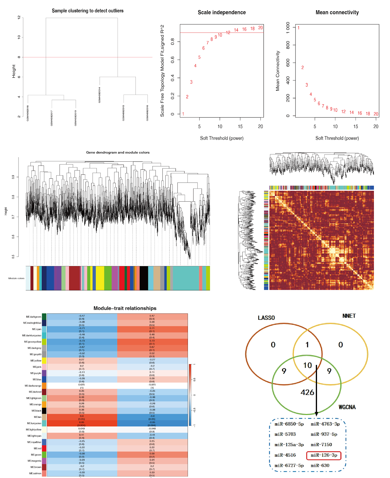
Xiao Ban Tong Mai Fang regulates autophagy via targeting miR-126-3p: bioinformatics analysis for prevention and treatment of atherosclerosisGraphical AbstractVerified
Chinese Journal of Tissue Engineering Research2026

Xiao Ban Tong Mai Fang regulates autophagy via targeting miR-126-3p: bioinformatics analysis for prevention and treatment of atherosclerosis

BACKGROUND: Studies have shown that miR-126-3p plays an important role in regulating autophagy and is closely related to the occurrence and development of atherosclerosis. Xiao Ban Tong Mai Fang can exert anti-atherosclerotic effects by exerting anti-inflammatory effects and inhibiting the proliferation of human aortic vascular smooth muscle cells. OBJECTIVE: To explore the mechanism of Xiao Ban Tong Mai Fang in the treatment of atherosclerosis by targeting microRNAs (miRNAs) to regulate autophagy using bioinformatics techniques. METHODS: Machine learning methods were used to screen the differentially expressed miRNAs in the atherosclerosis dataset GSE137580. The screened miRNAs were intersected with the key module genes screened by weighted gene co-expression network analysis. The potential regulatory genes of the intersected miRNAs were predicted based on the database, and then the predicted genes were intersected with the autophagy gene set. Protein-protein interaction analysis and enrichment analysis were performed on the intersection genes. Based on the enrichment analysis results, experimental validation was carried out. Human umbilical vein endothelial cells and RAW264.7 cells were cultured in vitro, and an atherosclerotic cell foam model was induced by oxidized low-density lipoprotein. qPCR was used to detect the differential expression of miR-126-3p. A miR-126-3p overexpression vector was constructed, and the transfection efficiency and the intervention effect of Xiao Ban Tong Mai Fang were detected by qPCR. Western blot was used to detect the regulation of Xiao Ban Tong Mai Fang on autophagy and mitogen-activated protein kinase pathway in human umbilical vein endothelial cells. RESULTS AND CONCLUSION: (1) Combining machine learning and weighted gene co-expression network analysis, 10 key miRNAs were identified. Based on literature review and previous foundation, miR-126-3p was selected for experimental validation. A total of 3,892 potential regulatory genes were predicted, and 257 autophagy-related genes were obtained by intersecting with the autophagy gene set. Enrichment analysis found that these genes were widely enriched in the mitogen-activated protein kinase signaling pathway, so this pathway was selected for experimental validation. (2) qPCR results showed that miR-126-3p was upregulated in oxidized low-density lipoprotein-induced foam cell models of human umbilical vein endothelial cells and RAW264.7 cells (P < 0.05). (3) After intervention with Xiao Ban Tong Mai Fang, the expression of miR-126-3p in the mimic group of human umbilical vein endothelial cells was significantly reduced (P < 0.05). (4) Western Blot results showed that Xiao Ban Tong Mai Fang intervention inhibited the expression of autophagy-related proteins microtubule-associated protein 1 light chain 3 lipidated/non-lipidated (LC3-II/LC3-I) and autophagy adaptor protein p62 in human umbilical vein endothelial cells (P < 0.05), and downregulated the expression of mitogen-activated protein kinase pathway proteins (P < 0.05), similar to the effect of autophagy inhibitor 3-methyladenine. These results indicate that miR-126-3p is upregulated in atherosclerosis models, and its abnormal expression may be involved in the pathogenesis of atherosclerosis. Xiao Ban Tong Mai Fang exerts its therapeutic effect on atherosclerosis by downregulating miR-126-3p, inhibiting the mitogen-activated protein kinase pathway, and inhibiting autophagy in human umbilical vein endothelial cells.

Read Full Abstract10.12307/2026.21610
Animal models of Parkinson's disease: research status and trend analysisGraphical AbstractVerified
Chinese Journal of Tissue Engineering Research2026

Animal models of Parkinson's disease: research status and trend analysis

BACKGROUND: To further elucidate the etiology and pathogenesis of Parkinson's disease, standardized animal models that more closely mimic clinical conditions have garnered significant attention and achieved notable progress. OBJECTIVE: To review and summarize the current status, development trajectory, and trends in Parkinson's disease animal model research. METHODS: A total of 10 170 relevant literature from two databases, CNKI and Web of Science, were used as data samples. CiteSpace bibliometric software was then used to draw and analyze knowledge graphs of co-occurrence, clustering, emergent terms of keywords, literature publication volume, national/regional publication volume, institutional cooperation network, and literature citation in Parkinson's disease animal model research, followed by analysis. RESULTS AND CONCLUSION: The overall publication volume of Parkinson's disease animal model research, both domestically and internationally, has shown a fluctuating upward trend, with significant growth observed in the Web of Science database. China and the United States were the top publishing countries, accounting for 52.6% of total publications and emerging as primary contributors to research outcomes in this field. The leading domestic institution was Shanghai University of Traditional Chinese Medicine (131 papers), while internationally, Harvard University was the institution with the most publications (263 papers). The distribution of journals and disciplines at home and abroad is concentrated in neuroscience, neurology, molecular biology, traditional Chinese medicine, basic medicine, pharmacology, and other fields. In particular, traditional Chinese medicine treatment of Parkinson's disease has received great attention from domestic scholars, and the potential for interdisciplinary cooperation is huge. Pathological mechanisms related to Parkinson's disease animal models, such as neuroinflammation, gut microbiota, oxidative stress, mitochondrial dysfunction, and α-synuclein, are research hotspots in this field, and ferroptosis has become an emerging theme in domestic research on the mechanism of Parkinson's disease. In terms of treatment of Parkinson's disease symptoms, in addition to the continuous deepening of research on the treatment of Parkinson's disease in traditional Chinese medicine and Western medicine through animal models, new methods such as gene therapy and stem cell transplantation therapy have provided innovative therapeutic strategies in animal model research of Parkinson's disease. With the advancement of the national 'Brain Science Project' and the development of gene editing, artificial intelligence, machine learning, and brain-computer interface technology, these new technologies also have certain exploration space in Parkinson's disease research in the future.

Read Full Abstract10.12307/2026.21609
A visualized analysis of vagus nerve stimulation in the field of stroke rehabilitationGraphical AbstractVerified
Chinese Journal of Tissue Engineering Research2026

A visualized analysis of vagus nerve stimulation in the field of stroke rehabilitation

BACKGROUND: The vagus nerve stimulation technique enhances cortical excitability and neuroplasticity by stimulating the vagus nerve. Its application in stroke rehabilitation has received widespread attention. Currently, there is no study analyzing the research status and development trend of vagus nerve stimulation in the field of stroke rehabilitation. OBJECTIVE: To analyze the current development status, research hotspots, and development trends of vagus nerve stimulation in the field of stroke rehabilitation, in order to provide a reference for future research and optimization of clinical rehabilitation plans for stroke. METHODS: We retrieved relevant literature on the application of vagus nerve stimulation in stroke rehabilitation from China National Knowledge Infrastructure, WanFang, PubMed, and Web of Science Core Collection databases from January 1, 2012 to February 1, 2025. Using CiteSpace 6.3.R1 software, we performed visualized analyses of publication volumes, countries/regions and institutions, authors, keyword co-occurrence, keyword clustering, and keyword bursts. RESULTS AND CONCLUSION: A total of 297 articles were included, including 86 in Chinese and 211 in English. The annual publication volume on vagus nerve stimulation in stroke rehabilitation has generally increased both domestically and internationally. China had the highest number of publications. The most prolific authors in Chinese and English were Zhao Jingjun and Hays Seth A, respectively. The institutions with the most publications in Chinese and English were Chongqing Medical University and the University of Texas System, respectively. Universities are the main contributors to research output. Domestic research teams and institutions are more dispersed than international ones, with less collaboration among teams. Keyword co-occurrence and clustering results showed that research mainly focuses on stroke, upper limb function, neuroplasticity, and neuroprotective effects. Burst keywords were mainly related to motor function and cortical plasticity. Overall, research directions primarily revolve around the effects of vagus nerve stimulation on motor, swallowing, cognitive functions after stroke, and the exploration of mechanisms for improving neurological function. Future research may focus on non-invasive vagus nerve stimulation in stroke rehabilitation and the combined application of vagus nerve stimulation with other rehabilitation techniques, as well as exploring related mechanisms.

Read Full Abstract10.12307/2026.21606
Mechanism by which magnesium implant-activated integrin α10β1 promotes osteogenic differentiation of periosteal stem cellsGraphical AbstractVerified
Chinese Journal of Tissue Engineering Research2026

Mechanism by which magnesium implant-activated integrin α10β1 promotes osteogenic differentiation of periosteal stem cells

BACKGROUND: Periosteal stem cells are the key cellular population in magnesium-induced osteogenesis. Integrin α10β1 is a magnesium-dependent heterodimeric adhesion molecule. Mg²⁺ binds to the MIDAS domain of integrin α10β1, functioning as a molecular switch to regulate downstream biological processes. OBJECTIVE: To elucidate the mechanism by which magnesium-based implants regulate osteogenic differentiation of periosteal stem cells by activating integrin α10β1. METHODS: Forty-two C57BL/6 mice were randomly divided into a titanium rod implantation group (n=21) and a magnesium rod implantation group (n=21). Titanium rods and magnesium rods were implanted into the medullary cavity of the intercondylar fossa of the left knee joint femur, respectively. Three days post-surgery, samples were collected for TUNEL staining to observe cell apoptosis around the implants. EdU staining was utilized to observe cell proliferation activity in the cortical bone thickening area. Fourteen days post-surgery, samples were collected for Micro-CT analysis of cortical bone thickening and osteogenesis. Hematoxylin-eosin staining was applied to observe the morphology of new bone in the thickened cortical bone area. Calcein double labeling was used to analyze osteogenic differentiation of periosteal stem cells. qPCR was performed to detect the expression of osteogenic marker genes Runx2, osterix, alkaline phosphatase, bone sialoprotein, integrin α10, and integrin β1. Western blot was used to detect the protein expression of integrin α10, integrin β1, focal adhesion kinase (FAK), phosphorylated FAK, and components of Wnt/β-catenin and mitogen-activated protein kinase (MAPK) signaling pathways. Transcriptome sequencing was conducted to analyze the correlation between integrin α10β1 and osteogenic gene expression. RESULTS AND CONCLUSION: (1) TUNEL and EdU staining showed that around the titanium rod, a large number of apoptotic cells accumulated, and only a few proliferating cells were observed in the periosteal region without osteogenic differentiation; around the magnesium rod, no apoptotic cells were detected, and proliferating cells in the thickened periosteal area were significantly increased. (2) Micro-CT analysis showed that the titanium rod was not degraded, while the magnesium rod degraded significantly; the magnesium rod group exhibited better cortical bone thickening and osteogenesis than the titanium rod group. Hematoxylin-eosin and calcein double labeling staining showed that the magnesium rod group had superior cortical bone thickening and osteogenesis compared to the titanium rod group. qPCR results showed that the mRNA expression of Runx2, osterix, alkaline phosphatase, bone sialoprotein, integrin α10, and integrin β1 was higher in the magnesium rod group than in the titanium rod group. Western blot results showed that the protein expression of integrin α10, integrin β1, FAK, phosphorylated FAK, and Wnt/β-catenin was higher in the magnesium rod group, while MAPK expression was lower. Transcriptome sequencing analysis revealed a significant positive correlation between integrin α10β1 and the expression of Runx2, osterix, alkaline phosphatase, and bone sialoprotein. (3) These findings indicate that magnesium-based implants promote osteogenic differentiation of periosteal stem cells by activating the integrin α10β1-FAK/p-FAK signaling pathway, upregulating Wnt/β-catenin signaling, and inhibiting part of the MAPK signaling.

Read Full Abstract10.12307/2026.21542
Lung tissue repair mechanisms and risk models for chronic obstructive pulmonary disease: an analysis based on computer simulation and experimental validationGraphical AbstractVerified
Chinese Journal of Tissue Engineering Research2026

Lung tissue repair mechanisms and risk models for chronic obstructive pulmonary disease: an analysis based on computer simulation and experimental validation

BACKGROUND: Understanding the characteristics of lung tissue repair and risk factors in patients with chronic obstructive pulmonary disease (COPD) is crucial for improving disease management and enhancing patient quality of life. Existing clinical indicators cannot accurately quantify the tissue repair potential and disease progression risk in patients with a history of frequent hospitalizations. OBJECTIVE: To conduct a risk prediction model analysis of lung tissue repair mechanisms in COPD based on computer simulation and case validation, thereby promoting lung tissue repair/regeneration in COPD patients, reducing the frequency of hospitalizations, and improving quality of life. METHODS: Medical records from 200 patients with COPD hospitalized at Beijing Jingmei Group General Hospital from 2022-10-01 to 2023-10-01 were collected. Patients were grouped based on the number of hospitalizations for COPD within 1 year after discharge: 100 patients with ≥2 hospitalizations were assigned to the frequent hospitalization group, and 100 patients with <2 hospitalizations were assigned to the non-frequent hospitalization group. General clinical data, pulmonary function, blood gas analysis, and hematological indicators were compared between the two groups. Variables with P < 0.05 were included in a multivariate logistic regression model to identify risk factors for rehospitalization within 1 year. The area under the receiver operating characteristic curve (AUC) was used to evaluate the predictive efficacy of the clinical risk factor model. RESULTS AND CONCLUSION: (1) Pulmonary function: There were significant differences between the two groups in FEV1% predicted, FVC, FVC% predicted, FEV1/FVC, residual volume/total lung capacity ratio, diffusing capacity of the lung for carbon monoxide (DLCO), and DLCO/alveolar volume (P < 0.05). (2) Blood gas analysis: Significant differences were found in oxygen partial pressure, carbon dioxide partial pressure, oxygen saturation, and respiratory failure type (P < 0.05). (3) Hematological indicators: Significant differences were observed in absolute neutrophil count, D-dimer, absolute lymphocyte count, neutrophil percentage, and red blood cell distribution width (RDW) (P < 0.05), while no significant differences were found in eosinophil percentage, fibrinogen, and absolute eosinophil count (P > 0.05). (4) Logistic regression analysis showed that FEV1% predicted (OR=1.01, 95%CI: 1.004-1.017, P=0.011), DLCO (OR=2.28, 95%CI: 1.270-3.025, P=0.004), type I respiratory failure (OR=3.15, 95%CI: 2.414-5.947, P=0.001), type II respiratory failure (OR=7.03, 95%CI: 1.688-8.604, P=0.001), and RDW (OR=1.50, 95%CI: 0.65-3.44, P < 0.0001) were risk factors for frequent hospitalizations in COPD patients. (5) ROC curve analysis showed that when FEV1% predicted was below 52.9%, DLCO below 4 mmol/(min·kPa), presence of type I respiratory failure, or RDW above 14.5%, the risk of frequent hospitalizations increased. (6) The logistic regression model and ROC curve analysis indicated that severely impaired diffusion capacity, combined respiratory failure, and elevated RDW are major risk factors for frequent hospitalizations in COPD patients. Early identification and intervention of these factors are important for improving lung tissue repair potential, predicting disease progression risk, and enhancing quality of life.

Read Full Abstract10.12307/2026.21612
Visualization analysis of literature on medication-related osteonecrosis of the jawGraphical AbstractVerified
Chinese Journal of Tissue Engineering Research2026

Visualization analysis of literature on medication-related osteonecrosis of the jaw

BACKGROUND: Bibliometric and visualization analyses of theme-related literature on medication-related osteonecrosis of the jaw are essential for gaining a comprehensive understanding of its research foundation and emerging trends. OBJECTIVE: To reveal research hotspots, current status, and trends in medication-related osteonecrosis of the jaw through bibliometrics, citation analysis, and visualization analysis. METHODS: The top 100 most-cited medication-related osteonecrosis of the jaw research articles published between 2004 and 2024 were retrieved and screened from the Web of Science Core Collection (SCI-Expanded). A bibliometric analysis was conducted to extract and analyze core characteristics such as annual publication volume, countries, institutions, authors, journal sources, and co-citation relationships, followed by the use of CiteSpace 6.4.R1 software to generate knowledge maps, thereby identifying research hotspots and emerging trends. RESULTS AND CONCLUSION: The included literature accumulated a total of 12 337 citations, with an average annual citation frequency of 6.17 per article. The international core collaboration network was stable and close, with the United States, University of London, and Professor Salvatore L. Ruggiero ranking first in high-cited paper output among countries, research institutions, and authors, respectively. Hot keywords mainly included risk factors, bisphosphonates, cancer, and osteoclasts; the burst keyword was predominantly 'Denosumab', and research directions were diversified. The research heat on medication-related osteonecrosis of the jaw generally showed an upward trend, with core focus on pathological mechanisms, risk factors, and treatment strategies. High-level evidence, such as large cohort studies (LOE 2a), high-quality randomized controlled trials (LOE 1b), GRADE systematic reviews and meta-analyses, and clinical practice guidelines, significantly promoted the field's development. Looking forward, tissue engineering and regenerative medicine, including 3D-printed scaffolds, stem cell therapy, novel biomaterials, and in vitro models, are expected to provide breakthrough strategies for bone repair, regeneration, and mechanistic research of medication-related osteonecrosis of the jaw.

Read Full Abstract10.12307/2026.21607
Motor imagery-based brain-computer interface rehabilitation training improves upper limb motor function in stroke patients: a meta-analysisGraphical AbstractVerified
Chinese Journal of Tissue Engineering Research2026

Motor imagery-based brain-computer interface rehabilitation training improves upper limb motor function in stroke patients: a meta-analysis

OBJECTIVE: To systematically evaluate the effects of motor imagery-based brain-computer training on upper limb motor function in patients with stroke, thereby providing evidence-based guidance for clinical practice. METHODS: The randomized controlled trials about the effects of motor imagery-based brain-computer interface training in patients with stroke were retrieved from databases (PubMed, Web of Science, Embase, Cochrane Library, CBM, CNKI, VIP, and WanFang Data) from the establishment of the databases to July 2025. Two researchers independently conducted literature screening and data extraction. The Cochrane bias risk was used to evaluate the level of evidence. Rev Man 5.4 software was used for meta-analysis. RESULTS: Eleven studies encompassing 543 stroke survivors were ultimately included. The results of the meta-analysis showed that the experimental group had better outcomes than the control group in terms of the Fugl Meyer assess...

Read Full Abstract10.12307/2026.21605
Epidemiological association between metabolic health obesity phenotype and chronic low back pain: a cross-sectional analysis based on the NHANES databaseGraphical AbstractVerified
Chinese Journal of Tissue Engineering Research2026

Epidemiological association between metabolic health obesity phenotype and chronic low back pain: a cross-sectional analysis based on the NHANES database

BACKGROUND: Currently, whether and how the metabolically healthy obesity phenotype influences the risk and pathological progression of chronic low back pain compared to metabolically unhealthy obese individuals remain unclear. Research is urgently needed to clarify their association and potential mechanisms. OBJECTIVE: To investigate the epidemiological association between the metabolically healthy obesity phenotype and chronic low back pain. METHODS: Using data from the US National Health and Nutrition Examination Survey (NHANES) 1999-2004 and 2009-2010 (n=4,956), a retrospective cross-sectional study was conducted. Obesity was defined by body mass index and waist circumference according to WHO standards, and metabolic phenotype was stratified using core metabolic syndrome indicators (fasting glucose, lipid profile, blood pressure, etc.), establishing metabolically healthy obesity and metabolically unhealthy obesity classification. Multivariable-adjusted logistic regression models and restricted cubic spline curves were used to systematically evaluate the nonlinear dose-response relationship between metabolic health obesity phenotype and chronic low back pain risk. Multivariable-adjusted models were established to analyze chronic low back pain risk, and subgroup analysis, threshold effect analysis, and multiple sensitivity analyses were used to verify model robustness. RESULTS AND CONCLUSION: (1) Metabolically unhealthy obesity significantly increased the risk of chronic low back pain [BMI standard: OR (95%CI)=1.44 (1.11, 1.85), P=0.0070; waist circumference standard: OR (95%CI)=1.57 (1.25, 1.97), P=0.0003]. Smoking [OR (95%CI)=1.44 (1.24, 1.67), P<0.0001] and arthritis [OR (95%CI)=2.44 (2.06, 2.88), P<0.0001] significantly amplified the risk effect of metabolically healthy obesity on chronic low back pain. BMI ≥39.73 kg/m² and waist circumference ≥125 cm were risk thresholds for chronic low back pain in metabolically unhealthy individuals. Low education level, low income, and widowed/divorced or separated status increased the risk of chronic low back pain. In the total population and metabolically unhealthy participants, there was a linear relationship between BMI and chronic low back pain (P<0.05). In metabolically healthy participants, the dose-response relationship between BMI and chronic low back pain was approximately linear (P>0.05); in the total population, metabolically healthy and metabolically unhealthy participants, the dose-response relationship between waist circumference and chronic low back pain was approximately linear (P>0.05). (2) Based on NHANES cross-sectional analysis, metabolically unhealthy obesity was significantly and positively independently associated with chronic low back pain, while metabolically healthy obesity had a weaker and non-statistically significant association, indicating that metabolic health status is an important factor influencing chronic low back pain in obese individuals. The model constructed from international research provides a key theoretical reference for exploring racial heterogeneity and localized mechanisms (such as Asian-specific metabolic thresholds) in the Chinese population. The results warn that China should pay attention to the risk of low back pain in metabolically healthy obese individuals, promote optimized clinical classification intervention strategies (such as avoiding excessive weight loss), and prospectively prevent the public health burden of obesity-related musculoskeletal diseases.

Read Full Abstract10.12307/2026.21611
Regulatory effects of Yuping Shen’an Granules on neuronal autophagy in a mouse model of insomniaGraphical AbstractVerified
Chinese Journal of Tissue Engineering Research2026

Regulatory effects of Yuping Shen’an Granules on neuronal autophagy in a mouse model of insomnia

BACKGROUND: Modern medicine often uses benzodiazepines for the treatment of insomnia complicated by anxiety; however, long-term use can lead to drug dependence and adverse reactions. Traditional Chinese medicine, based on syndrome differentiation and holistic treatment, demonstrates definite efficacy and high safety, offering multi-pathway and multi-target comprehensive effects, thereby providing new ideas and directions for the treatment of this condition. OBJECTIVE: To investigate the effects of Yuping Shen’an Granules on hippocampal neuronal autophagy in mice with insomnia and anxiety via the phosphatidylinositol 3-kinase/protein kinase B/mammalian target of rapamycin signaling pathway. METHODS: Ultra-high performance liquid chromatography-tandem mass spectrometry was used to identify the components of Yuping Shen’an Granules, and network pharmacology was used to predict key pathways. Ninety male C57BL/6J mice were randomly divided into normal group, model group, low-, medium-, and high-dose Yuping Shen’an Granules groups [3.125, 6.25, 12.5 g/(kg·d)], eszopiclone group [0.06 mg/(kg·d)], recovery-model group, recovery-model + PI3K inhibitor group [10 mg/(kg·d)], recovery-Yuping Shen’an [12.5 g/(kg·d)] + inhibitor group [10 mg/(kg·d)], with 10 mice per group. Except for the normal group, all groups were exposed to chronic unpredictable mild stress for 14 days and intraperitoneal injection of para-chlorophenylalanine for 4 days to establish a mouse model of insomnia with anxiety. Behavioral assessments included open field and pentobarbital sodium sleep tests; hematoxylin-eosin and Nissl staining were used to observe hippocampal pathological changes; enzyme-linked immunosorbent assay was used to detect levels of 5-hydroxytryptamine, γ-aminobutyric acid, dopamine, and norepinephrine; immunofluorescence was used to detect microtubule-associated protein light chain 3B expression; western blotting was used to detect PI3K/AKT/mTOR pathway-related proteins, autophagy-related proteins (P62, Beclin-1, microtubule-associated protein light chain 3B), and 5-hydroxytryptamine receptor 1A protein expression; transmission electron microscopy was used to observe the number of autophagosomes. RESULTS AND CONCLUSION: ① Ultra-high performance liquid chromatography-tandem mass spectrometry identified 2,276 components of Yuping Shen’an Granules, and 35 main components were screened out; ② Network pharmacology analysis showed that the PI3K/AKT/mTOR pathway was a potential target; ③ Compared with the normal group, the model group showed increased exploratory behavior, prolonged sleep latency (P < 0.01), significant hippocampal neuronal damage, decreased levels of 5-hydroxytryptamine and γ-aminobutyric acid, increased levels of dopamine and norepinephrine (P < 0.01), upregulated microtubule-associated protein light chain 3B fluorescence and protein expression (P < 0.01), downregulated phosphorylated PI3K/AKT/mTOR, P62, and 5-hydroxytryptamine receptor 1A expression, upregulated Beclin-1 and microtubule-associated protein light chain 3B expression (P < 0.05), and increased number of autophagosomes; ④ Compared with the model group, the recovery-model + inhibitor group showed aggravated damage, while the high-dose Yuping Shen’an Granules group and recovery-Yuping Shen’an + inhibitor group significantly reversed the above changes, improving behavioral indicators, alleviating neuronal damage, increasing 5-hydroxytryptamine and γ-aminobutyric acid levels, decreasing dopamine and norepinephrine levels (P < 0.05), and reversing autophagy-related protein expression (P < 0.05), with reduced autophagosomes. These results indicate that Yuping Shen’an Granules improve insomnia with anxiety symptoms by activating the PI3K/AKT/mTOR pathway, regulating neurotransmitter balance, and inhibiting excessive autophagy in hippocampal neurons.

Read Full Abstract10.12307/2026.21587
Efficacy and safety of romozumab in the treatment of osteoporosis in adults: a meta-analysisGraphical AbstractVerified
Chinese Journal of Tissue Engineering Research2026

Efficacy and safety of romozumab in the treatment of osteoporosis in adults: a meta-analysis

OBJECTIVE: Romosozumab is a novel biologic agent currently being used to treat osteoporosis in postmenopausal women at high risk of fracture. This meta-analysis aims to systematically evaluate the efficacy and safety of romosozumab compared with placebo, alendronate sodium, teriparatide, and denosumab in the treatment of osteoporosis in adults. METHODS: The medical keywords “anti-sclerostin antibody,” “romosozumab,” “AMG 78500,” and “osteoporosis” were used to search PubMed, CNKI, Web of Science, and the Cochrane Central Register of Controlled Trials (CENTRAL) for randomized controlled trials comparing the safety and efficacy of romosozumab with alendronate, teriparatide, denosumab, or placebo in adult patients with osteoporosis. Two researchers independently screened studies, assessed risk of bias, and extracted data. The Cochrane Collaboration's risk of bias tool was used for quality assessment, and meta-analysis was performed using RevMan 5.4. The primary outcome was the percentage change from baseline in bone mineral density (BMD) at 6 and 12 months; secondary outcomes were the incidence of adverse events and cardiovascular complications during treatment. RESULTS: A total of 10 randomized controlled trials involving 12,570 patients were included. Compared with placebo, alendronate, and teriparatide, romosozumab significantly increased BMD at the lumbar spine, total hip, and femoral neck at 6 and 12 months. Compared with denosumab, romosozumab significantly increased lumbar spine BMD at 6 and 12 months (MD=3.68, 95%CI: 0.34-7.01, P=0.03; MD=5.20, 95%CI: 3.19-7.21, P<0.00001), while no significant differences were found in total hip and femoral neck BMD. In terms of safety, romosozumab had a lower incidence of adverse events compared with alendronate (RR=0.96, 95%CI: 0.93-0.99, P=0.02) but a higher incidence compared with teriparatide (RR=1.13, 95%CI: 1.01-1.25, P=0.03). No significant differences were found versus placebo or denosumab (RR=0.98, 95%CI: 0.96-1.00, P=0.11; RR=2.64, 95%CI: 0.74-9.36, P=0.13). Importantly, romosozumab did not significantly increase the risk of cardiovascular complications compared with other treatments (RR=1.25, 95%CI: 0.94-1.67, P=0.12). CONCLUSION: Romosozumab rapidly improves lumbar spine BMD with an overall manageable safety profile, particularly suitable for adult osteoporosis patients at high fracture risk who require rapid bone mass increase and have no cardiovascular contraindications. This meta-analysis is based on limited data and has certain limitations; more high-quality, longer-duration follow-up studies are needed to confirm the results.

Read Full Abstract10.12307/2026.21599
Molecular mechanism of icariin in prevention and treatment of osteoporosisGraphical AbstractVerified
Chinese Journal of Tissue Engineering Research2026

Molecular mechanism of icariin in prevention and treatment of osteoporosis

BACKGROUND: Pharmacodynamic characteristics and mechanisms of action of icariin in combating osteoporosis gradually gain recognition within the academic community. Related basic research and clinical translation efforts are increasingly becoming the focal point of research. OBJECTIVE: To summarize the research progress of icariin on anti-osteoporosis. METHODS: China National Knowledge Infrastructure (CNKI) and PubMed databases were searched for relevant literature. Chinese and English search terms included “icariin, osteoporosis, Chinese medicine compound, pathogenesis, signal path, BMSCs, osteoblast, osteoclast.” Based on inclusion criteria, 90 articles were ultimately included in the review. RESULTS AND CONCLUSION: Icariin treatment increased alkaline phosphatase activity and induced the expression of core binding factor α1, bone morphogenetic protein 2, and bone morphogenetic protein 4 in bone marrow mesenchymal stem cells in a dose-dependent manner. Icariin promoted fracture healing by increasing serum levels of osteocalcin, bone-specific alkaline phosphatase, N-terminal peptide of type I collagen, C-terminal peptide of type I collagen, and tartrate-resistant acid phosphatase 5b, thereby enhancing osteocalcin secretion at the fracture site. Icariin promoted proliferation and osteogenic differentiation of bone marrow mesenchymal stem cells in ovariectomized osteoporotic rats by upregulating alkaline phosphatase and osteocalcin levels and inhibiting the expression of Notch-1, CBF1, and Jagged-1 proteins in the Notch pathway, thus achieving prevention and treatment of osteoporosis. Icariin regulates bone metabolism through multiple signaling axes including Wnt/β-catenin, mitogen-activated protein kinase, phosphatidylinositol 3-kinase/protein kinase B, osteoprotegerin/receptor activator of nuclear factor-κB ligand/receptor activator of nuclear factor-κB, and Notch. Among these, the Wnt/β-catenin pathway and the osteoprotegerin/receptor activator of nuclear factor-κB ligand/receptor activator of nuclear factor-κB axis constitute the core regulatory mechanism, modulating the osteoblast-osteoclast dynamic balance through synergistic interactions. Icariin can influence the biological behavior of osteoblasts, osteoclasts, and bone marrow mesenchymal stem cells through multidimensional interventions including regulation of mRNA expression modifications, inhibition of oxidative stress, and improvement of the inflammatory microenvironment.

Read Full Abstract10.12307/2026.21591
Effects of exercise intervention on cortical excitability and motor performance in healthy populations: a meta-analysis based on transcranial magnetic stimulation measurementsGraphical AbstractVerified
Chinese Journal of Tissue Engineering Research2026

Effects of exercise intervention on cortical excitability and motor performance in healthy populations: a meta-analysis based on transcranial magnetic stimulation measurements

OBJECTIVE: Multiple studies have confirmed that exercise interventions can induce changes in cortical excitability detectable by transcranial magnetic stimulation; however, the neural regulatory effects among different training types remain inconsistent, with no unified conclusions. Based on this, this study aims to systematically evaluate the effects of exercise interventions on cortical excitability and motor performance in healthy populations, exploring the underlying mechanisms at neurophysiological and motor functional levels. METHODS: A systematic search was conducted in PubMed, Web of Science, Embase, Cochrane Library, and Chinese databases including CNKI, VIP, and WanFang. Randomized controlled trials and crossover trials involving healthy adults undergoing exercise interventions were included, where the trial group underwent any form of exercise intervention, while the control group received sham intervention or no exercise intervention. Outcome measures included cortical excitability indexes and motor performance assessed via transcranial magnetic stimulation. Meta-analysis was performed using RevMan 5.4 software, with subgroup and sensitivity analyses conducted to explore sources of heterogeneity. RESULTS: A total of 15 studies involving 380 participants were included. Meta-analysis showed that exercise intervention had significant positive effects on both cortical excitability and motor performance. Exercise intervention significantly enhanced cortical excitability [SMD=0.38, 95%CI(0.05, 0.72), P=0.03], with a small-to-moderate effect size; and significantly improved motor performance [SMD=0.42, 95%CI(0.07, 0.76), P=0.02], with a moderate effect size. Subgroup analysis revealed that strength training significantly enhanced cortical excitability [SMD=0.53, 95%CI(0.12, 0.94), P=0.01], while motor skill training, high-intensity interval training, and balance training did not significantly enhance cortical excitability [SMD=-0.29, 95%CI(-1.13, 0.55), P=0.50; SMD=0.04, 95%CI(-0.44, 0.53), P=0.86; SMD=0.45, 95%CI(-0.37, 1.26), P=0.28]. Heterogeneity among studies was high, possibly due to differences in exercise intervention types, training duration, or measurement indicators. Sensitivity analysis confirmed the robustness of the results, and funnel plot analysis suggested low risk of publication bias. CONCLUSION: Exercise intervention can effectively enhance cortical excitability and motor performance in healthy populations, with strength training showing particularly significant effects. Future research should explore the mechanisms of different training types and optimize training program designs to further improve neuroplasticity and motor performance.

Read Full Abstract10.12307/2026.21601
Molecular mechanism by which the imbalance of the functional network of tissue inhibitors of metalloproteinases drives intervertebral disc degenerationGraphical AbstractVerified
Chinese Journal of Tissue Engineering Research2026

Molecular mechanism by which the imbalance of the functional network of tissue inhibitors of metalloproteinases drives intervertebral disc degeneration

BACKGROUND: Intervertebral disc degeneration is a core pathological mechanism of discogenic diseases, characterized by an imbalance in extracellular matrix metabolism. Tissue inhibitors of metalloproteinases, as endogenous antagonists of matrix metalloproteinases, play a crucial role in regulating extracellular matrix homeostasis, but the specific functions of subtypes, signaling pathway interactions, and epigenetic regulatory mechanisms have not been systematically clarified. OBJECTIVE: To review the expression changes, functional heterogeneity, and regulatory networks of tissue inhibitors of metalloproteinases in intervertebral disc degeneration, focusing on the molecular mechanisms and signaling pathways in oxidative stress, mechanical load, and inflammatory microenvironment, and to evaluate the translational potential of gene therapy strategies based on tissue inhibitors of metalloproteinases. METHODS: The first author searched PubMed, Web of Science, Embase, CNKI, Wanfang and other databases from inception to March 2025. Chinese search terms included '椎间盘退变,椎间盘退行性变,椎间盘退化,金属蛋白酶类组织抑制剂,信号通路', and English search terms included 'Tissue Inhibitor of Metalloproteinases, Tissue Inhibitor of Metalloproteinase, TIMPs, Intervertebral disc degeneration, Disc degeneration, Degenerative Disc Disease, Degenerative Intervertebral Discs'. Finally, 76 eligible articles were included for review. RESULTS AND CONCLUSION: (1) Subtype functions of tissue inhibitors of metalloproteinases: TIMP1 exhibits dual regulation (early protection/late depletion); TIMP2 maintains extracellular matrix homeostasis by inhibiting matrix metalloproteinase activity, and its abnormal expression can activate pro-apoptotic signaling pathways (e.g., miR-185-5p/MMP2 axis and inflammatory factor-mediated MMP/TIMP imbalance); TIMP3 exerts multi-dimensional protective effects by inhibiting matrix metalloproteinase activity, tumor necrosis factor-alpha converting enzyme/tumor necrosis factor-alpha axis, and angiogenesis; TIMP4 is regulated by miR-155-5p/fibroblast growth factor 2 and participates in extracellular matrix homeostasis. (2) Epigenetic reprogramming mechanisms: Abnormal mechanical stress degrades TIMP3 mRNA through the WTAP/YTHDF2-m6A axis, while miR-222 targets TIMP3 to synergistically accelerate extracellular matrix degradation. (3) Multi-modal therapeutic strategies: Photobiomodulation (wavelength-specific regulation of TIMP/MMP), stem cell exosomes (miR-199a/GREM1 axis), and irisin intervention can remodel matrix metabolic balance. This review summarizes the theoretical framework of 'functional network imbalance of tissue inhibitors of metalloproteinases', revealing its multi-level regulatory characteristics as a core driver of intervertebral disc degeneration, and providing a theoretical basis for developing precise therapies targeting epigenetic modifications and mechano-biological coupling interventions.

Read Full Abstract10.12307/2026.21593
Type VI collagen: a multifunctional regulator in bone homeostasis and tissue engineeringGraphical AbstractVerified
Chinese Journal of Tissue Engineering Research2026

Type VI collagen: a multifunctional regulator in bone homeostasis and tissue engineering

BACKGROUND: As a key structural protein in the extracellular matrix, type VI collagen plays a critical role in skeletal development, homeostasis, and repair through its unique tetrameric microfibrillar network. Recent studies have found that abnormal expression of type VI collagen is closely related to skeletal diseases such as osteoporosis, osteoarthritis, and bone tumors, but its multidimensional regulatory mechanisms and translational application potential still need systematic summary. OBJECTIVE: To review the structural characteristics and biological functions of type VI collagen in the skeletal system, clarify the mechanism of type VI collagen in skeletal diseases, and explore the translational application prospects of type VI collagen in biomarker development, tissue engineering material construction, and therapeutic target design. METHODS: A systematic search of PubMed, Web of Science, Elsevier, and CNKI databases from January 1982 to May 2025 was conducted, including research articles and reviews, excluding duplicates and low-quality literature. Finally, 69 articles (68 in English and 1 in Chinese) were included for systematic content integration and analysis. RESULTS AND CONCLUSION: Type VI collagen affects bone health through a triple regulatory network: (1) Bone formation and resorption balance: promotes osteoblast 'matrix bridge' connection and inhibits tumor necrosis factor α/nuclear factor κB p65 subunit signaling pathway-mediated osteoclast activation; (2) Disease mechanisms: in osteoporosis, the α2 chain of type VI collagen is epigenetically inhibited by miR-128-2-5p; in early osteoarthritis, pericellular matrix degradation occurs; in bone tumors, the α1 chain of type VI collagen is highly expressed; (3) Translational applications: type VI collagen can enhance the osteogenic efficacy of scaffold materials; specific serum degradation products of type VI collagen can serve as diagnostic markers for fibrotic diseases; antisense oligonucleotide technology has been successfully used to correct abnormal splicing caused by splice site mutations. Type VI collagen is a core regulatory hub for maintaining bone homeostasis, integrating the structural support and signal transduction functions of the extracellular matrix to regulate the dynamic balance of bone formation and resorption. Degradation products and tissue distribution patterns of type VI collagen can provide new diagnostic markers for skeletal diseases, and gene-targeted therapy and type VI collagen-enhanced biological scaffolds are expected to become innovative treatment strategies for osteoporosis and bone defects. Future research needs to break through the technical bottleneck of tissue-targeted delivery and deepen the dynamic expression atlas research based on multi-omics.

Read Full Abstract10.12307/2026.21589
Exercise improves neuropathic pain: precision exercise prescription and multimodal synergy advance clinical applicationsGraphical AbstractVerified
Chinese Journal of Tissue Engineering Research2026

Exercise improves neuropathic pain: precision exercise prescription and multimodal synergy advance clinical applications

BACKGROUND: Neuropathic pain is a chronic pain condition caused by direct damage or functional abnormalities in the somatic sensory nervous system. Its clinical manifestations include spontaneous pain and tactile hypersensitivity, which are difficult to control effectively with traditional drug therapies. The pathogenesis of neuropathic pain involves multiple physiological processes, including neuronal hyperexcitability, glial cell activation, neurotransmitter imbalance, immune responses, and oxidative stress. Existing medications and invasive treatments often carry side effects and exhibit significant limitations in efficacy. Therefore, exploring safe and effective non-pharmacological interventions, particularly exercise-based interventions for improving neuropathic pain, has become a critical research focus in the field of neuropathic pain. OBJECTIVE: To review advances in understanding the mechanisms of neuropathic pain, analyze the potential and mechanisms of exercise intervention in alleviating neuropathic pain, demonstrate the clinical application prospects of exercise as a non-pharmacological intervention strategy, and emphasize future research directions. METHODS: PubMed and CNKI databases were searched using Chinese and English keywords including neuropathic pain, nerve injury pain, exercise, physical activity, aerobic exercise, resistance training, yoga, pathogenesis, inflammation, neurotransmitter, neurotrophin, oxidative stress, and rehabilitation. A total of 139 articles were included. The core mechanisms of neuropathic pain were analyzed, focusing on the multi-target and multi-pathway synergistic effects of exercise in alleviating neuropathic pain. RESULTS AND CONCLUSION: The pathogenesis of neuropathic pain involves neuronal hyperexcitability, glial cell activation, neurotransmitter imbalance, immune-inflammatory responses, and oxidative stress-induced nerve damage. Exercise intervention alleviates neuropathic pain by regulating neurotransmitter release, promoting neurotrophic factor expression, inhibiting inflammatory responses, and reducing oxidative stress. Specifically, exercise upregulates the expression of neurotrophic factors such as brain-derived neurotrophic factor and nerve growth factor, inhibits the release of pro-inflammatory cytokines such as tumor necrosis factor-alpha, and further regulates core pathways in neuropathic pain development. Exercise also produces analgesic effects by modulating the endogenous opioid system. The specific mechanisms of different exercise types on neuropathic pain need further investigation, and personalized exercise prescription design and exercise parameter optimization face many challenges. Future research should focus on constructing and validating exercise prescriptions, clarifying the synergistic effects of exercise combined with drug therapy, to support the advancement of clinical treatment for neuropathic pain.

Read Full Abstract10.12307/2026.21595
Mechanism by which non-apoptotic regulated cell death induces neuronal injury in ischemic strokeGraphical AbstractVerified
Chinese Journal of Tissue Engineering Research2026

Mechanism by which non-apoptotic regulated cell death induces neuronal injury in ischemic stroke

BACKGROUND: In recent years, the involvement of non-apoptotic regulated cell death in the development of ischemic stroke has become a research hotspot. OBJECTIVE: To summarize the roles and action mechanisms of non-apoptotic regulated cell death subroutines such as autophagy, ferroptosis, cuproptosis, disulfidptosis, necroptosis, and pyroptosis in the neuronal damage caused by ischemic stroke. METHODS: Relevant literature on non-apoptotic regulated cell death and ischemic stroke was retrieved from the China National Knowledge Infrastructure and PubMed databases. The search terms included "ischemic stroke, regulated cell death, autophagy, ferroptosis, cuproptosis, disulfidptosis, necroptosis, pyroptosis, alkaliptosis, oxeiptosis, parthanatos, mitochondrial permeability transition-driven necrosis, neutrophil extracellular trap-related death, lysosome-dependent cell death, immunogenic cell death, anoikis" in English and corresponding Chinese terms. Based on inclusion criteria, 176 articles were finally included for analysis and summary. RESULTS AND CONCLUSION: The regulatory mechanisms of non-apoptotic regulated cell death mainly include autophagy, ferroptosis, anoikis, cuproptosis, disulfidptosis, necroptosis, pyroptosis, oxeiptosis, alkaliptosis, parthanatos, mitochondrial permeability transition-driven necrosis, neutrophil extracellular trap-related death, lysosome-dependent cell death, and immunogenic cell death. Autophagy plays a dual regulatory role in neuronal injury after ischemic stroke: under ischemic conditions, autophagy exerts a neuroprotective effect, whereas excessive autophagy during reperfusion can lead to neuronal death. Ferroptosis can aggravate neuronal injury in ischemic stroke through iron overload and lipid peroxidation. Cuproptosis can regulate glutathione-induced ferroptosis by modulating the protein ferredoxin 1. There is partial crosstalk between disulfidptosis and ferroptosis; under glucose deprivation, upregulation of solute carrier family 7 member 11 consumes NADPH, leading to abnormal accumulation of disulfide compounds and promoting disulfidptosis in neurons. Mixed lineage kinase domain-like pseudokinase, a key participant in necroptosis, is also associated with activation of the pyroptosis-related protein NLRP3 inflammasome, further promoting neuronal pyroptosis during necroptosis in ischemic stroke. Neutrophil extracellular trap-related death in ischemic stroke is mainly caused by citrullination, stress-triggered neutrophil extracellular trap formation, and inflammatory responses mediated by release of various cytotoxic proteases. Other emerging subtypes such as immunogenic cell death cause neuronal damage in ischemic stroke through various specific mechanisms.

Read Full Abstract10.12307/2026.21598
Applications and advances of tissue/organ perfusionGraphical AbstractVerified
Chinese Journal of Tissue Engineering Research2026

Applications and advances of tissue/organ perfusion

BACKGROUND: As critical physiological and functional units, tissue/organ have been utilized in experimental perfusion research for over two centuries. Today, perfusion technology is widely applied in various fields, including the screening of bioactive components in traditional Chinese medicine, pharmacodynamics and mechanisms, pharmacokinetics, pathological mechanisms, local cancer therapy, tissue/organ transplantation, tissue/organ fixation, cell preparation, and metabolite production. OBJECTIVE: To summarize the experimental methods, influencing factors, and recent advances in tissue/organ perfusion technology, providing a reference for its practical application. METHODS: Relevant articles published from inception to September 2025 were retrieved and analyzed from the CNKI and PubMed databases using Chinese search terms “cardiac perfusion, cerebral perfusion, liver perfusion, kidney perfusion, intestinal perfusion, lung perfusion, neural perfusion, limb perfusion” and English search terms “heart perfusion/cardiac perfusion, brain perfusion, liver perfusion, kidney perfusion, intestinal perfusion, lung perfusion, neural perfusion/nerves perfusion, limb perfusion, forelimb perfusion, hindlimb perfusion, rat, rabbit”. After excluding clinical trials, duplicate publications, and irrelevant articles, 86 articles were included for review. RESULTS AND CONCLUSION: (1) Tissue/organ perfusion technology serves as an intermediate bridge between cell experiments and whole-animal experiments, providing a research platform that allows precise control of experimental conditions and avoids interference from complex in vivo factors, thus playing an important role in basic and applied life science research. (2) Although perfusion procedures share commonalities across different tissues/organs, successful application highly depends on parameter optimization (e.g., perfusate composition, flow rate) tailored to the specific physiological functions and anatomical structures of the target organ, characterized by the same basic principles but different specific applications. (3) Perfusion technology has broad application fields, covering multiple levels from new drug development to pathological mechanism exploration, and its application is highly targeted; for example, intestinal perfusion for drug absorption, liver perfusion for metabolism studies, and kidney perfusion for excretion mechanisms, fully leveraging the unique physiological functions of different target organs. (4) Perfusion technology itself is continuously innovating, from the classic Langendorff model to the more physiological “working heart” model. Currently, it is being combined with cutting-edge technologies such as transgenic animal models, organ-on-a-chip, and organoids, moving towards a more precise and humane direction.

Read Full Abstract10.12307/2026.21588
MicroRNA-23a-3p improves neurological function in mice with traumatic brain injury by regulating microglial polarizationGraphical AbstractVerified
Chinese Journal of Tissue Engineering Research2026

MicroRNA-23a-3p improves neurological function in mice with traumatic brain injury by regulating microglial polarization

BACKGROUND: Previous studies have demonstrated neuroprotective potential of microRNA-23a-3p in traumatic brain injury. However, direct evidence is still lacking regarding whether this protective effect stems from its precise regulation of the M1/M2 polarization balance of microglia. OBJECTIVE: To clarify the expression changes of microRNA-23a-3p in mouse brain tissue after traumatic brain injury and to explore the specific mechanism by which it affects neurological function through regulating microglial polarization. METHODS: Eighty C57BL/6J mice were randomly assigned to four groups: a sham operation group, a traumatic brain injury group, a traumatic brain injury + agomir-NC group, and a traumatic brain injury + agomir-MicroRNA-23a-3p group. The traumatic brain injury model was established using the cortical impact method. The sham group did not receive cortical impact. The intervention groups received intracerebroventricular injection of agomir-NC or agomir-MicroRNA-23a-3p after modeling. Six mice from the sham and traumatic brain injury groups were analyzed at 1, 3, 7, and 14 days post-injury, and six mice from the other two groups were analyzed at 14 days post-injury. Neurological deficits were assessed using the modified neurological severity score (mNSS). Hematoxylin-eosin staining and Nissl staining were used to observe pathological changes in brain tissue and neurons. qRT-PCR and western blot were used to detect the expression levels of MicroRNA-23a-3p, M1 markers (CD16, CD86), M2 markers (CD206, arginase-1), and inflammatory cytokines (tumor necrosis factor-α and interleukin-10). Immunohistochemistry was used to evaluate microglial M1/M2 polarization and the aggregation of F4/80-positive cells in the injured area. RESULTS AND CONCLUSION: Compared with the sham group, the expression of MicroRNA-23a-3p in the traumatic brain injury group showed a "V"-shaped curve, with downregulation in the early phase and upregulation starting at 7 days post-injury. Upregulation of MicroRNA-23a-3p reduced the mNSS score in traumatic brain injury mice. Morphological results showed that upregulation of MicroRNA-23a-3p alleviated brain edema and neuronal damage. Molecular biology results showed that upregulation of MicroRNA-23a-3p promoted microglial polarization from M1 to M2 phenotype. These findings indicate that MicroRNA-23a-3p can promote neurological function recovery after traumatic brain injury in mice by regulating microglial polarization.

Read Full Abstract10.12307/2026.21585