🧬 SinoBioData Academic Portal
📚 Peer-Reviewed Translated Literature

All Biomedical & Clinical Articles (Page 17)

Browse complete peer-reviewed translations from top Chinese biomedical, oncology, and genomics journals. Read verified previews and download full authentic clinical reports.

Published Research Papers

Showing 24 of 1542 peer-reviewed translated articles (Page 17 of 65)

Lentivirus-mediated gene therapy in a beta-thalassemia mouse modelGraphical AbstractVerified
Chinese Journal of Tissue Engineering Research2026

Lentivirus-mediated gene therapy in a beta-thalassemia mouse model

BACKGROUND: Lentiviral vector (LV)-mediated autologous hematopoietic stem cell gene therapy is expected to be a novel curative treatment for β-thalassemia. The LV serves as a core agent of gene therapy, directly influencing future clinical efficacy and treatment costs. Therefore, the primary task is to develop high-performance lentiviral vectors. OBJECTIVE: To explore the feasibility of an ex vivo gene therapy and assess the activity and functionality of the β-globin-LV in thalassemic mice. METHODS: A novel lentiviral vector, HS40-LV, carrying the human β-globin gene cassette, was constructed. 7.5 Gy-conditioned Hbbth3/+ mice were subjected to HS40-LV-modified hematopoietic stem cell transplantation. Normal mice and untreated thalassemic mice served as controls. Peripheral blood samples were collected from mice at 2, 4, 6, 8, and 10 months post-treatment. The integrated proviral DNA in the individual sample was detected by using qPCR. The proportion of red blood cells expressing human β-globin was detected by fluorescence-activated cell sorting. Fresh whole blood was collected for blood smears, which were used for Giemsa staining, reticulocyte staining, and fully automated blood cell analysis. At 10 months post-treatment, the liver, spleen, and bone marrow tissues were sampled from all three groups to prepare single-cell suspensions and extract genomic DNA for qPCR detection of vector marking; flow cytometry was used to detect cells expressing transgenic β-globin; portions of spleen and liver were subjected to hematoxylin-eosin staining and Prussian blue staining. RESULTS AND CONCLUSION: (1) The HS40-LV vector achieved a transduction efficiency of 50% in hematopoietic stem cells. (2) During the 10-month follow-up, the proportion of vector-marked cells and β-globin-positive red blood cells in peripheral blood of treated mice steadily increased, reaching an average of 50% vector marking and 70% β-globin-positive red blood cells at 10 months post-transplantation. (3) Biological distribution of the lentiviral vector and expression of transgenic β-globin were also detected in liver, spleen, and bone marrow hematopoietic tissues. (4) Gene therapy corrected hematological parameters in thalassemic mice, such as significant reductions in poikilocytes, reticulocytes, and cell fragments, and a significant increase in overall hemoglobin levels. (5) Histopathological improvements were also observed, with significant reductions in iron deposition in spleen and liver, and improved extramedullary hematopoiesis. These results indicate that the novel HS40-LV vector achieved stable expression in vivo, and modified cells corrected some symptoms in thalassemic mice.


Read Full Abstract10.12307/2026.21491
Mechanisms by which mangiferin alleviates pain in osteoarthritis: integration of microarray data analysis, network pharmacology, and experimental validation in a rat modelGraphical AbstractVerified
Chinese Journal of Tissue Engineering Research2026

Mechanisms by which mangiferin alleviates pain in osteoarthritis: integration of microarray data analysis, network pharmacology, and experimental validation in a rat model

BACKGROUND: Mangiferin, a major bioactive compound derived from mango trees, is widely present in various traditional Chinese medicinal herbs and exhibits multiple biological functions including antibacterial, cholesterol-lowering, and anti-allergic effects. Existing studies have suggested that mangiferin may prevent and treat osteoarthritis pain. However, its specific mechanism of action remains unclear to date. OBJECTIVE: To systematically investigate the key targets and potential mechanisms of mangiferin in the treatment of osteoarthritis by integrating gene expression omnibus (GEO) microarray data analysis, network pharmacology, and molecular docking techniques, and to validate the findings in a rat model. METHODS: First, GEO microarray data were mined to identify potential therapeutic targets for osteoarthritis. Next, professional databases were integrated to predict the targets of mangiferin, and target information related to osteoarthritis was collected. A Venn diagram was generated using the Weishengxin platform, a protein-protein interaction network was constructed based on the STRING database, and Gene Ontology and Kyoto Encyclopedia of Genes and Genomes pathway enrichment analyses were performed. Cytoscape 3.8.0 software was used to construct a drug-target-pathway-disease network, and molecular docking analysis and visualization were performed using the CBDOCK2 online docking platform. A rat model of osteoarthritis was established by anterior cruciate ligament transection of the left knee joint, and different concentrations of mangiferin were administered to observe and record the therapeutic effects. RESULTS AND CONCLUSION: A total of 144 potential targets of mangiferin were identified from multiple databases. Protein-protein interaction network analysis revealed important targets including interleukin-6, tumor necrosis factor, and nuclear factor kappa B1. Kyoto Encyclopedia of Genes and Genomes pathway enrichment analysis showed that 235 signaling pathways might be involved, including lipid and atherosclerosis-related pathways, advanced glycation end products-receptor, hypoxia-inducible factor 1, and estrogen, which are closely related to inflammation. In animal experiments, after 4 weeks of intervention with 40 μmol/L mangiferin, there was no significant difference in hindlimb weight-bearing compared with the sham-operated group. These findings suggest that mangiferin may exert therapeutic effects on osteoarthritis through a multi-target, multi-pathway mode of action, providing a new strategy and theoretical support for the treatment of osteoarthritis.

Read Full Abstract10.12307/2026.21488
Mazdutide improves cognitive function in APP/PS1/Tau triple transgenic miceGraphical AbstractVerified
Chinese Journal of Tissue Engineering Research2026

Mazdutide improves cognitive function in APP/PS1/Tau triple transgenic mice

BACKGROUND: The pathological process of Alzheimer's disease is closely related to β-amyloid/Tau deposition and cerebrovascular dysfunction, and existing therapies are difficult to effectively intervene. In recent years, research on glucagon-like peptide-1 receptor agonists for the treatment of Alzheimer's disease has gained increasing popularity. As a novel dual agonist of the glucagon-like peptide-1 receptor and glucagon receptor, the therapeutic mechanism of Mazdutide in Alzheimer’s disease remains to be elucidated. OBJECTIVE: To systematically explore the mechanism by which Mazdutide improves cognitive function in Alzheimer's disease using network pharmacology and experimental validation, and to identify potential core molecular targets, providing a theoretical basis for new therapeutic strategies. METHODS: A multi-omics integration strategy was employed. Co-localization targets of Alzheimer's disease and Mazdutide were screened using DisGeNET and SEA databases. A protein-protein interaction network was constructed via STRING, and hub genes were identified by Cytoscape topological analysis. In APP/PS1/Tau triple transgenic Alzheimer's disease mice, Mazdutide (30 nmol/kg) was administered intraperitoneally. Cognitive function was assessed using Morris water maze, novel object recognition, and Y maze tests. Western blot was used to detect hippocampal β-amyloid (6E10), p-Tau181, and endothelin receptor A expression. RESULTS AND CONCLUSION: ① Fifty-two co-localized targets were identified, with endothelin receptor A (EDNRA) as the top hub gene. ② Mazdutide significantly improved cognition in 3xTg mice: shortened spatial memory latency, increased novel object recognition index, increased spontaneous alternation in Y maze, and reduced hippocampal pathological burden, with decreased p-Tau181 and EDNRA overexpression. ③ Gene Ontology and KEGG enrichment revealed core pathways: biological processes included blood pressure regulation, amine transport regulation, and amine transport; cellular components included symmetric synapses, sperm head, and pseudopodia; molecular functions included G protein-coupled peptide receptor activity, peptide receptor activity, and neuropeptide receptor activity. KEGG pathways included neuroactive ligand-receptor interaction, calcium signaling, and hormone signaling. ④ This study confirms that Mazdutide may improve cognitive deficits in Alzheimer's disease by targeting and inhibiting EDNRA overexpression, reducing neuronal pathological damage, and acting through multiple dimensions. The discovery of EDNRA provides a novel therapeutic target for Alzheimer's disease.

Read Full Abstract10.12307/2026.21489
Establishment and identification of a patient-derived organoid model for esophagogastric junction adenocarcinomaGraphical AbstractVerified
Chinese Journal of Tissue Engineering Research2026

Establishment and identification of a patient-derived organoid model for esophagogastric junction adenocarcinoma

BACKGROUND: Traditional cell culture models have substantial limitations in accurately simulating the biological characteristics of tumors. Organoid technology, which retains key biological features of primary tumors, provides technical support for in-depth exploration of the pathogenesis of esophagogastric junction adenocarcinoma (EGJA) and for precision diagnosis and treatment research. OBJECTIVE: To construct a patient-derived organoid model of EGJA, evaluate its consistency with the biological characteristics of the primary tumor, and provide a standardized in vitro model for studying the pathogenesis and precision diagnosis and treatment of EGJA. METHODS: Surgical resection specimens from 50 patients with EGJA were collected for tissue dissociation, culture, and organoid model construction. Cell viability in organoid models was analyzed by fluorescence co-localization staining. Structural features of organoid models and primary tumor tissues were analyzed by hematoxylin-eosin staining. Mucin secretion phenotype in organoid models was detected by periodic acid-Schiff staining. Immunohistochemical staining was used to evaluate the consistency of organoid models with primary tumor histopathology. Chi-square test and univariate logistic regression were used to analyze the correlation between organoid model establishment success rate and patient clinicopathological features and tissue characteristics. RESULTS AND CONCLUSION: A patient-derived EGJA organoid model culture system was successfully established, with a primary culture success rate of 86.0% (43/50). Organoids exhibited typical three-dimensional structures, gradually developing from cell clusters into glandular-like three-dimensional solid spheres. After 7-10 days of primary culture, they could be stably passaged, and no significant phenotypic changes were observed after multiple passages. After cryopreservation and resuscitation, organoids still proliferated stably. Fluorescence co-localization staining showed that organoid models maintained high viability at different culture stages. Hematoxylin-eosin staining results showed that both organoid models and primary tumor tissues exhibited atypical glandular arrangement and high nuclear-to-cytoplasmic ratio. Periodic acid-Schiff staining showed positive mucin secretion. Immunohistochemical staining showed high consistency in the positive expression rates of Ki67, CEA, CK7, and Cadherin17 between organoid models and primary tumor tissues. Furthermore, chi-square test and logistic regression analysis showed that poorly differentiated tumor tissue (P=0.02), major pathological response to neoadjuvant chemotherapy (P=0.007), tumor tissue ex vivo time (P=0.006), and tumor mass (P=0.006) significantly affected the success rate of patient-derived EGJA organoid model establishment.

Read Full Abstract10.12307/2026.21490
Immunological mechanisms of Xuling Jiangu Formula in intervening osteoporosis model ratsGraphical AbstractVerified
Chinese Journal of Tissue Engineering Research2026

Immunological mechanisms of Xuling Jiangu Formula in intervening osteoporosis model rats

BACKGROUND: Osteoporosis is not only a metabolic disease, but also an inflammatory or autoimmune disease. Immune cells Treg and Th17 have a mutually antagonistic effect on the differentiation and maturation of osteoclasts, and the maintenance of cell balance between the two is the basis for ensuring normal bone metabolism. OBJECTIVE: To explore the immunomodulatory mechanism of Xuling Jiangu Formula on osteoporosis and its relationship with the balance of Treg/Th17 cells. METHODS: Sprague-Dawley rats were randomly divided into a sham operation group, a model group, and a drug group, with eight rats in each group. The latter two groups underwent bilateral ovariectomy to establish the animal model; the sham operation group underwent the same procedure except for ovary removal. Four weeks after modeling, bone mineral density was measured using dual-energy X-ray absorptiometry to evaluate whether the modeling was successful. Drug intervention started 4 weeks after surgery: the Xuling Jiangu Formula group was given 10 mL/kg Xuling Jiangu Formula by gavage once daily for 16 weeks; the model and sham operation groups were given the same volume of normal saline. After 16 weeks of drug administration, bone mineral density and blood routine parameters were measured, bone tissue morphology was observed by hematoxylin-eosin staining, serum immune factor levels were detected by ELISA, and the gene and protein expressions of osteoprotegerin, receptor activator of nuclear factor-κB ligand, retinoic acid-related orphan receptor γt, and forkhead box P3 were detected by quantitative PCR and western blot. The numbers of Treg and Th17 cells were detected by flow cytometry. RESULTS AND CONCLUSION: After 16 weeks of administration, compared with the model group, the bone mineral density of rats in the Xuling Jiangu Formula group was significantly increased (P < 0.05), serum levels of interleukin-10 and transforming growth factor-β were increased but without significant difference, while levels of interleukin-17 and interleukin-6 were significantly decreased (P < 0.05). After 16 weeks of administration, the gene and protein expressions of osteoprotegerin and Treg marker gene forkhead box P3 were significantly increased (P < 0.05), while the gene and protein expressions of Th17 marker gene retinoic acid-related orphan receptor γt were significantly decreased (P < 0.05), and the protein expression of receptor activator of nuclear factor-κB ligand was significantly decreased (P < 0.05), but the gene expression showed a decreasing trend without significant difference. Flow cytometry results showed that the number of Treg cells was significantly increased (P < 0.05), while the number of Th17 cells was decreased but not statistically significant. These findings suggest that the immune mechanism of Xuling Jiangu Formula in treating osteoporosis is to affect the Treg/Th17 balance by increasing the number of Treg cells, then reduce the secretion of interleukin-17 and interleukin-6, promote the upregulation of forkhead box P3 and downregulation of retinoic acid-related orphan receptor γt, thereby changing the osteoprotegerin/receptor activator of nuclear factor-κB ligand ratio, and subsequently inhibit osteoclast differentiation.

Read Full Abstract10.12307/2026.21487
Effects of different frequency electroacupuncture on mitochondrial function and oxidative stress injury in quadriceps femoris muscle of rabbits with anterior cruciate ligament injuryGraphical AbstractVerified
Chinese Journal of Tissue Engineering Research2026

Effects of different frequency electroacupuncture on mitochondrial function and oxidative stress injury in quadriceps femoris muscle of rabbits with anterior cruciate ligament injury

BACKGROUND: Oxidative stress is one of the potential factors contributing to muscle atrophy following anterior cruciate ligament injury. Alleviating skeletal muscle fatigue facilitates proprioceptive recovery, thereby accelerating rehabilitation after anterior cruciate ligament injury. Improving mitochondrial function helps mitigate skeletal muscle fatigue-related damage. OBJECTIVE: To verify that electroacupuncture at different frequencies alleviates skeletal muscle oxidative stress damage and improves mitochondrial function in rabbits, thereby reducing skeletal muscle fatigue, restoring proprioceptive function, and accelerating rehabilitation following anterior cruciate ligament injury. METHODS: Twenty-four healthy New Zealand rabbits were randomly divided into blank group, model group, low-frequency electroacupuncture group, and high-frequency electroacupuncture group, with 6 rabbits in each group. The model group, low-frequency electroacupuncture group, and high-frequency electroacupuncture group were used to construct a knee anterior cruciate ligament injury model. In the low-frequency and high-frequency electroacupuncture groups, electroacupuncture was applied to the acupoints Xuehai and Liangqiu on the affected knee joint 7 days after modeling. The blank and model groups were only grasped and fixed without electroacupuncture intervention, once daily for 21 consecutive days. After intervention, ELISA was used to detect the levels of superoxide dismutase, succinate dehydrogenase, and malondialdehyde in the quadriceps femoris; western blot was used to detect the protein expression levels of silent information regulator 2-related enzyme 1, peroxisome proliferator-activated receptor gamma coactivator 1 alpha, and mitochondrial transcription factor A in skeletal muscle tissue, as well as the mRNA expression of silent information regulator 2-related enzyme 1, peroxisome proliferator-activated receptor gamma coactivator 1 alpha, nuclear respiratory factor 1, and mitochondrial transcription factor A. RESULTS AND CONCLUSION: After anterior cruciate ligament injury, the level of superoxide dismutase in the quadriceps femoris of rabbits increased, the level of succinate dehydrogenase decreased, and the concentration of malondialdehyde increased; the protein expressions of silent information regulator 2-related enzyme 1, peroxisome proliferator-activated receptor gamma coactivator 1 alpha, and mitochondrial transcription factor A decreased; the mRNA expressions of silent information regulator 2-related enzyme 1, peroxisome proliferator-activated receptor gamma coactivator 1 alpha, nuclear respiratory factor 1, and mitochondrial transcription factor A were significantly downregulated. After electroacupuncture intervention, the concentration of malondialdehyde in the affected quadriceps femoris decreased, the activities of superoxide dismutase and succinate dehydrogenase increased, and the protein expressions of silent information regulator 2-related enzyme 1, peroxisome proliferator-activated receptor gamma coactivator 1 alpha, and mitochondrial transcription factor A increased; the mRNA expressions of mitochondrial biogenesis-related genes such as silent information regulator 2-related enzyme 1, peroxisome proliferator-activated receptor gamma coactivator 1 alpha, nuclear respiratory factor 1, and mitochondrial transcription factor A increased, and the low-frequency electroacupuncture group was superior to the high-frequency electroacupuncture group. These results indicate that electroacupuncture can reduce oxidative stress damage in skeletal muscle after anterior cruciate ligament injury by increasing the contents of superoxide dismutase and succinate dehydrogenase and decreasing the content of malondialdehyde; and improve mitochondrial function by regulating the expression of proteins related to the silent information regulator 2-related enzyme 1/peroxisome proliferator-activated receptor gamma coactivator 1 alpha signaling pathway and mitochondrial biogenesis-related genes, thereby accelerating rehabilitation after anterior cruciate ligament injury.

Read Full Abstract10.12307/2026.21483
Effects of Gushukang Granules on the expression of myogenic and osteogenic factors in the muscles and bones of sarcopenic osteoporosis ratsGraphical AbstractVerified
Chinese Journal of Tissue Engineering Research2026

Effects of Gushukang Granules on the expression of myogenic and osteogenic factors in the muscles and bones of sarcopenic osteoporosis rats

BACKGROUND: In sarcopenic osteoporosis, muscle loss and osteoporosis often coexist, leading to a significant increase in the risk of falls and fractures. Gushukang Granules are clinically used for the treatment of osteoporosis; however, the mechanism of action on myogenic and osteogenic factors in muscle and bone remains unclear. OBJECTIVE: To investigate the effects of Gushukang Granules on myogenic and osteogenic factors in the muscles and bones of rats. METHODS: Thirty-six healthy Sprague-Dawley rats were randomly divided into control, model, and Gushukang groups (n=12 per group). Osteoporosis was induced in the latter two groups by ovariectomy. Four weeks after surgery, the Gushukang group received 1.05 mL/kg Gushukang Granules solution by gavage, while the other groups received an equal volume of saline, once daily for 12 weeks. General conditions were observed. Hematoxylin-eosin staining was used to assess morphological changes in muscle and bone tissues. RT-qPCR was performed to detect mRNA expression of interleukin-6 (IL-6), tumor necrosis factor-alpha (TNF-alpha) in muscle, and osteocalcin (OCN) and insulin-like growth factor 1 (IGF-1) in bone. Immunohistochemistry was used to observe the expression of IL-6, TNF-alpha, IGF-1, and OCN in muscle and bone tissues. Western blot was used to detect protein expression levels of these factors. RESULTS AND CONCLUSION: Compared with the control group, the model group exhibited typical sarcopenic osteoporosis phenotype: muscle fibers were sparse, disordered, and atrophic; bone trabeculae were reduced, sparse, and disconnected. mRNA and protein expression of IL-6 and TNF-alpha were significantly increased (P < 0.01), while IGF-1 and OCN were significantly decreased (P < 0.01). Compared with the model group, the Gushukang group showed significant improvement: muscle fiber arrangement became more orderly with interstitial fibrosis; bone trabeculae increased and connectivity improved (though still loose). IL-6 and TNF-alpha expression were significantly decreased (P < 0.01), while IGF-1 and OCN were significantly increased (P < 0.01). Western blot results were consistent with immunohistochemistry. These findings suggest that Gushukang Granules can improve muscle atrophy and bone trabecular thinning in ovariectomized rats by downregulating the abnormal high expression of IL-6 and TNF-alpha, and upregulating IGF-1 and OCN, possibly through regulating muscle-bone crosstalk signaling pathways and balancing inflammatory and growth factor levels, providing experimental support for the clinical treatment of sarcopenic osteoporosis.

Read Full Abstract10.12307/2026.21484
Application of tissue clearing technology in a rat model of chronic spinal cord injuryGraphical AbstractVerified
Chinese Journal of Tissue Engineering Research2026

Application of tissue clearing technology in a rat model of chronic spinal cord injury

BACKGROUND: Studies have shown that tissue clearing technology enables the three-dimensional (3D) visualization of neurons in the spinal cord injury area, clearly presenting morphological changes of neurons, including soma atrophy, dendrite fragmentation, and axonal degeneration. OBJECTIVE: To systematically evaluate the application potential of tissue clearing technology in a rat model of chronic spinal cord injury. METHODS: Thirty-six female Sprague-Dawley rats were randomly and equally divided into a normal group (n=12), a sham surgery group (n=12), and a surgery group (n=12). The normal group received no treatment. The sham group underwent implantation and immediate removal of a poly(vinyl alcohol)/polyacrylamide interpenetrating network hydrogel into the C5-C7 spinal canal. The surgery group received implantation of the hydrogel to compress the spinal cord at C5-C7 to establish a chronic spinal cord injury model. At postoperative days 1, 3, 7, and 14, motor function was assessed using the Basso, Beattie, and Bresnahan (BBB) score and the modified Rivlin inclined plane test. At day 14, spinal cord tissue was harvested for hematoxylin-eosin staining to observe morphology, and tissue clearing combined with neuron-specific nuclear protein immunofluorescence labeling was used for three-dimensional reconstruction and cross-sectional view analysis. RESULTS AND CONCLUSION: (1) The BBB scores and inclined plane test angles in the surgery group were significantly lower than those in the normal and sham groups at all time points (P < 0.001). (2) Hematoxylin-eosin staining showed significant spinal cord injury in the surgery group, with swelling and destruction of nerve cells in the gray matter, loss of uniformity in white matter structure, disappearance of some nuclei, reduced cell number, massive glial cell proliferation and aggregation in the compression area, disordered white matter structure, and formation of numerous cavities. (3) Three-dimensional reconstruction and cross-sectional analysis of the spinal cord showed that in the normal and sham groups, the spinal cord appeared continuous and full, with uniform distribution of neuron-specific nuclear protein red fluorescence, dense layered arrangement of neurons in the anterior horn of the gray matter, and intact white matter fiber tracts. In the surgery group, the spinal cord appeared depressed or even interrupted, with significantly reduced fluorescence intensity of neuron-specific nuclear protein in the compressed segment, disrupted gray matter neuronal layer structure, and regional fluorescence interruption. These results indicate that tissue clearing technology can effectively display structural changes after spinal cord injury, providing strong support for studying the pathological mechanisms of spinal cord injury.

Read Full Abstract10.12307/2026.21486
Mechanism of epothilone B improving spinal cord microcirculation after spinal cord injury in ratsGraphical AbstractVerified
Chinese Journal of Tissue Engineering Research2026

Mechanism of epothilone B improving spinal cord microcirculation after spinal cord injury in rats

BACKGROUND: Animal experiments have found that epothilone B can remodel blood microcirculation and reduce tissue scar formation after spinal cord injury, but the specific mechanism remains unclear. OBJECTIVE: To clarify the mechanism by which epothilone B improves spinal cord microcirculation after spinal cord injury. METHODS: Fifty Sprague-Dawley rats were randomly divided into a sham-operated group (n=10), a spinal cord injury group (n=20), and an epothilone B group (n=20). The sham-operated group underwent only laminectomy at T10, while the other two groups were subjected to laminectomy at T10 followed by spinal cord contusion. Immediately after modeling, the epothilone B group received intraperitoneal injections of epothilone B solution, while the other two groups received corresponding solvents. At corresponding time points after modeling, motor function was assessed using Basso-Beattie-Bresnahan (BBB) score, inclined plane test, and open field test; blood flow recovery in the posterior median spinal cord vessels was detected by laser speckle flow imaging; hematoxylin-eosin staining was used to evaluate the overall spinal cord tissue; Western blot was used to detect Toll-like receptor 4 and nuclear factor κB protein expression; immunofluorescence staining was used to detect vascular endothelial growth factor receptor 2 and Toll-like receptor 4 expression in spinal cord injury tissue. RESULTS AND CONCLUSION: (1) The BBB score in the epothilone B group was higher than that in the spinal cord injury group at 14 and 28 days after modeling (P < 0.05), the inclined plane test angle was greater than that in the spinal cord injury group at 28 days after modeling (P < 0.05), and the open field test moving distance was greater than that in the spinal cord injury group at 14 and 28 days after modeling (P < 0.05), indicating that epothilone B improved motor function in rats with spinal cord injury. Laser speckle flow imaging at 28 days after modeling showed that epothilone B increased blood flow recovery in spinal cord injury rats. Hematoxylin-eosin staining at 28 days after modeling showed that the spinal cord cavity area in the epothilone B group was smaller than that in the spinal cord injury group (P < 0.05). Western blot at 5 days after modeling showed that the expression of Toll-like receptor 4 and nuclear factor κB protein in the spinal cord injury group was higher than that in the sham-operated group and epothilone B group (P < 0.05). Immunofluorescence staining at 5 days after modeling showed that the expression of vascular endothelial growth factor receptor 2 in the spinal cord injury group was lower than that in the sham-operated group and epothilone B group (P < 0.05), while the expression of Toll-like receptor 4 was higher than that in the sham-operated group and epothilone B group (P < 0.05). (2) These results indicate that epothilone B may reduce local inflammation after spinal cord injury by regulating the Toll-like receptor 4 and nuclear factor κB pathway, ensuring the regeneration of vascular endothelial cells in spinal cord tissue, thereby promoting the reconstruction of blood microcirculation.

Read Full Abstract10.12307/2026.21485
Therapeutic targets for knee osteoarthritis: identification via a bioinformatics approachGraphical AbstractVerified
Chinese Journal of Tissue Engineering Research2026

Therapeutic targets for knee osteoarthritis: identification via a bioinformatics approach

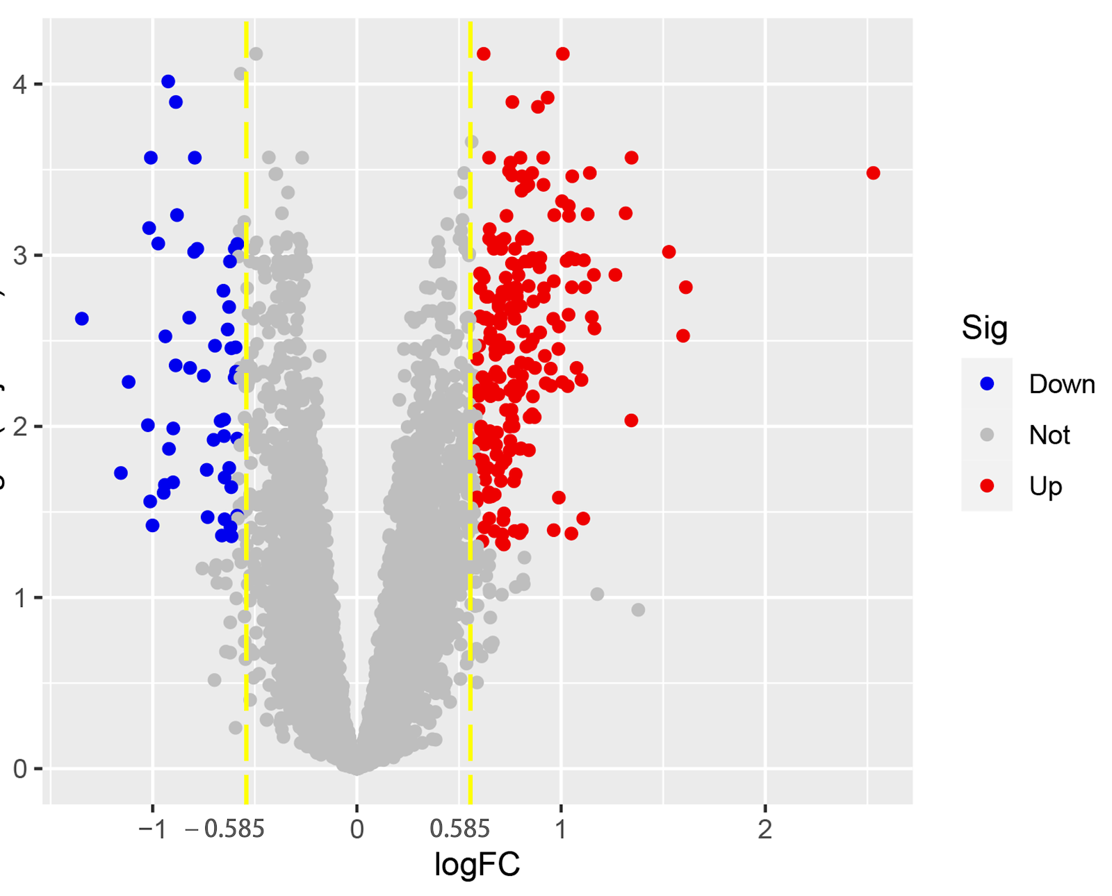
BACKGROUND: The etiology of knee osteoarthritis is complex and its mechanisms are not fully understood. Research on candidate target genes for knee osteoarthritis will help further clarify the pathogenesis of the disease and provide a basis for precision treatment. OBJECTIVE: To identify therapeutic targets for knee osteoarthritis based on summary data using Mendelian randomization combined with bioinformatics methods, followed by cellular validation. METHODS: Gene expression profiles GSE46750, GSE55235, GSE82107, and GSE206848 were downloaded from the Gene Expression Omnibus database. Differentially expressed genes were obtained using R software with screening criteria of |log2FC| > 0.585 and adjusted P < 0.05. Module genes with the highest correlation were acquired using the Weighted Gene Co-expression Network Analysis algorithm and intersected with differentially expressed genes. Gene Ontology and Kyoto Encyclopedia of Genes and Genomes enrichment analyses were performed on the intersection genes. In the eQTLGen database, summary-data-based Mendelian randomization analysis was used to identify genetic genes significantly associated with knee osteoarthritis, and genes commonly identified by bioinformatics and summary-data-based Mendelian randomization were considered core genes. Molecular docking and dynamics simulations were used to evaluate the binding of celecoxib to core genes, and immune infiltration analysis was performed using the CIBERSORT algorithm. Human chondrocytes were divided into normal and experimental groups (interleukin-1β-induced osteoarthritis cell model), and mRNA expression of adrenomedullin, osteopontin, and lysosomal protein transmembrane 5 was detected by qPCR. RESULTS AND CONCLUSION: Bioinformatics identified 229 differentially expressed genes. GO enrichment analysis showed that differentially expressed genes were mainly related to inflammatory response, positive regulation of response to external stimulus, regulation of cell activation, chemotaxis, and other biological functions. KEGG enrichment analysis showed that differentially expressed genes were mainly enriched in phagosome, osteoclast differentiation, complement and coagulation cascades, and interleukin-17 signaling pathway. Summary-data-based Mendelian randomization identified 76 significantly associated genetic genes (P < 0.05, FDR < 0.05, HEIDI test P > 0.05). Adrenomedullin, osteopontin, and lysosomal protein transmembrane 5 were core genes, among which osteopontin and lysosomal protein transmembrane 5 were negatively correlated with knee osteoarthritis development, while adrenomedullin was positively correlated. Molecular docking and dynamics simulations confirmed good structure-activity relationships between core genes and celecoxib. Immune infiltration analysis suggested that adrenomedullin, osteopontin, and lysosomal protein transmembrane 5 were correlated with multiple immune cells. qPCR showed that mRNA expression of osteopontin and lysosomal protein transmembrane 5 in the experimental group was lower than that in the normal group (P < 0.001), while adrenomedullin mRNA expression was higher (P < 0.001). These results indicate that adrenomedullin, osteopontin, and lysosomal protein transmembrane 5 are key genes in knee osteoarthritis development and may serve as new targets for prevention and treatment.

Read Full Abstract10.12307/2026.21479
Bibliometric analysis of development of precision, minimally invasive and intelligent robot-assisted spinal surgeryGraphical AbstractVerified
Chinese Journal of Tissue Engineering Research2026

Bibliometric analysis of development of precision, minimally invasive and intelligent robot-assisted spinal surgery

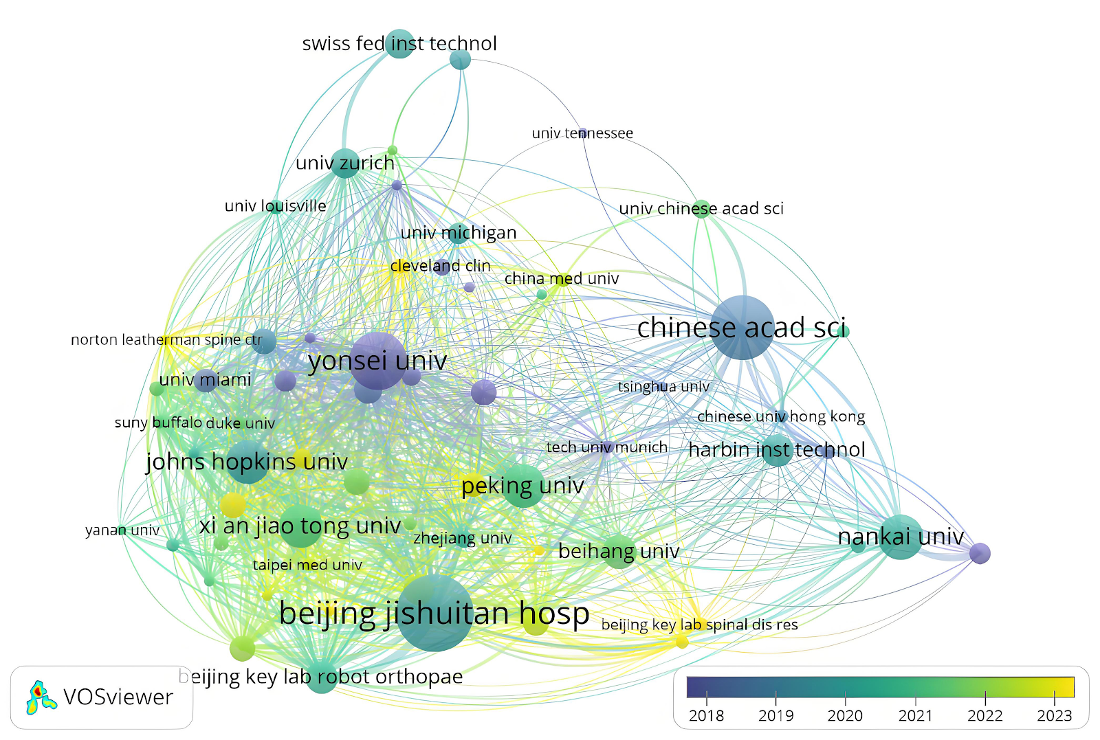
BACKGROUND: In recent years, the application of robot-assisted surgery technology in the field of spinal surgery has become increasingly widespread, significantly enhancing the precision and minimally invasive level of surgeries and promoting the rapid development of this field. However, with the explosive growth of related literature, researchers urgently need to conduct systematic bibliometric analysis to sort out the research hotspots, cooperation networks and development trends in this field, so as to grasp the future research directions and promote academic exchanges and cooperation. OBJECTIVE: To analyze the research hotspots, development trends, and domestic and international cooperation characteristics of robot-assisted surgical techniques in the field of spinal surgeries and explore their promoting effects on the precision and minimally invasive nature of spinal surgeries using bibliometric methods. METHODS: Original research papers on robot-assisted spinal surgery published in the Science Citation Index Expanded (SCI-E) database of the Web of Science (WOS) from 2005-01-01 to 2025-03-31 were retrieved. Bibliometric analysis was performed using the Web of Science online analysis platform, Bibliometric platform, and VOSviewer software. RESULTS AND CONCLUSION: A total of 772 original research articles were included. Research on robot-assisted spinal surgery has shown rapid growth, with annual publications exceeding 50 since 2019 and reaching a historical peak of 136 in 2024. China (279 articles) and the United States (255 articles) were the top publishing countries, while the United Kingdom had the highest average citations per article (69.57). The core journals in this field were the International Journal of Medical Robotics and Computer Assisted Surgery (84 articles) and Spine (33 articles, 539 total citations). Research hotspots focused on the accuracy and safety of robot-assisted pedicle screw placement, with high-frequency keywords including 'accuracy', 'navigation', and 'pedicle screw'. International collaboration networks showed close cooperation among China, the United States, and Germany. In China, Beijing Jishuitan Hospital was the core research institution, and Professor He Da was the most prolific author (21 articles). Highly cited articles often compared robot-assisted versus conventional techniques in terms of screw placement accuracy, safety, and radiation dose. Robot-assisted surgical technology is driving spinal surgery toward precision, minimally invasive, and intelligent development. Future efforts should strengthen domestic and international interdisciplinary collaboration to promote further advancement in this field.

Read Full Abstract10.12307/2026.21701
Functional characterization of Caspr2 in a mouse model of sciatic nerve injuryGraphical AbstractVerified
Chinese Journal of Tissue Engineering Research2026

Functional characterization of Caspr2 in a mouse model of sciatic nerve injury

BACKGROUND: The intrinsic molecular mechanisms and regulatory networks of peripheral nervous system injury remain to be systematically analyzed. Caspr2, as a cell adhesion molecule specifically expressed on the surface of neuronal axons, plays an important role in the myelination of nerve fibers, but the dynamic regulatory mechanism of Caspr2 in peripheral nerve regeneration has not been clarified. OBJECTIVE: To focus on the functional characterization of Caspr2 in a sciatic nerve injury model, reveal the molecular mechanism by which Caspr2 regulates axonal regeneration of dorsal root ganglion neurons, and provide a new perspective for developing precise repair strategies for peripheral nerve injury. METHODS: An ICR mouse model of sciatic nerve crush injury was established. The transcriptional and protein expression characteristics of Caspr2 in dorsal root ganglion tissue were analyzed by qRT-PCR and western blot, and the expression of Caspr2 in dorsal root ganglion neurons was analyzed by immunofluorescence staining. An intervention model was constructed using primary dorsal root ganglion neuron culture system: Caspr2 siRNA negative control group, Caspr2 siRNA group; Caspr2 empty vector control group, Caspr2 overexpression group. Axonal regeneration dynamics were quantitatively evaluated by Tuj1 immunofluorescence staining and AxioVision image analysis system. RESULTS AND CONCLUSION: (1) Injury response characteristics: On day 3 after sciatic nerve injury, the mRNA and protein expression levels of Caspr2 in dorsal root ganglion tissue decreased, and further decreased on day 7 after injury. In addition, the expression of Caspr2 in dorsal root ganglion neurons also significantly decreased on day 7 after injury. (2) Loss-of-function effect: Caspr2 siRNA treatment significantly promoted neuronal axon growth. (3) Overexpression effect: Caspr2 overexpression significantly inhibited neuronal axon extension. These results indicate that Caspr2, as a key inhibitory factor in peripheral nerve regeneration, participates in the nerve repair process by bidirectionally regulating axonal regeneration ability.

Read Full Abstract10.12307/2026.21481
Interleukin-10 alleviates inflammatory responses after acute tendon injuryGraphical AbstractVerified
Chinese Journal of Tissue Engineering Research2026

Interleukin-10 alleviates inflammatory responses after acute tendon injury

BACKGROUND: During the repair process following tendon injury, an excessive inflammatory response can cause tendon cell apoptosis, thereby leading to a reduction in the biomechanical properties of the tendon. Meanwhile, a persistent inflammatory response can also trigger tissue fibrosis and adhesion. Studies have confirmed that interleukin-10 exerts an inflammatory regulatory role in connective tissue cells such as fibroblasts and can block inflammatory responses produced in various models. OBJECTIVE: To explore the effect of interleukin-10 against inflammatory responses following acute tendon injury. METHODS: Forty-two Sprague-Dawley rats were randomly divided into a normal group (n=6), model group (n=12), control group (n=12), and intervention group (n=12). Except for the normal group, the other three groups underwent acute Achilles tendon injury modeling via intra-tendinous injection of type I collagenase solution (the model was successfully established after 3 days). On the day of modeling, the control and intervention groups were subjected to daily injections of PBS and interleukin-10 protein solution, respectively, at the 1 cm points on both sides of the hind limb midline and abdominal midline intersection. Injections were given once daily for 4 consecutive days. On day 3 after successful modeling, ultrasound examination of the Achilles tendon was performed. On days 3 and 7 after successful modeling, tissue samples were collected for hematoxylin-eosin staining to observe pathological changes, immunohistochemical staining to detect phosphorylated nuclear factor kappa B (p-NF-κB) and tumor necrosis factor alpha (TNF-α) protein expression, RT-PCR to detect NF-κB, TNF-α, and cyclooxygenase-2 (COX-2) mRNA expression, and western blot to detect p-NF-κB, TNF-α, and COX-2 protein expression. RESULTS AND CONCLUSION: Ultrasound showed that the model group had blurred tendon boundaries and increased thickness, while the intervention group had clearer boundaries and thickness close to normal. Hematoxylin-eosin staining on day 3 showed that the model and control groups had disordered collagen fibers, massive inflammatory cell infiltration, and round nuclei concentrated; the intervention group had reduced fiber disorder, fewer inflammatory cells, and more elongated spindle-shaped tenocytes. On day 7, the model and control groups showed significantly improved collagen fiber arrangement, reduced inflammatory infiltration, and tenocytes transitioning from round to spindle shape with mostly aligned nuclei; the intervention group showed parallel and orderly collagen fibers approaching normal, further reduced inflammatory infiltration, and mostly elongated spindle-shaped tenocytes. Immunohistochemistry showed that on days 3 and 7, the model group had higher p-NF-κB and TNF-α protein expression than the normal and intervention groups (P < 0.05). RT-PCR showed that on days 3 and 7, the model group had higher NF-κB, TNF-α, and COX-2 mRNA expression than the normal and intervention groups (P < 0.05). Western blot showed that on days 3 and 7, the model group had higher p-NF-κB, TNF-α, and COX-2 protein expression than the normal group (P < 0.05); on day 3, the intervention group had lower p-NF-κB, TNF-α, and COX-2 protein expression than the model group (P < 0.05). These findings indicate that interleukin-10 can alleviate inflammatory responses during acute tendon injury repair.

Read Full Abstract10.12307/2026.21482
Mechanism by which Hernandezine alleviates osteoporosis through macrophage polarization and osteoclast activationGraphical AbstractVerified
Chinese Journal of Tissue Engineering Research2026

Mechanism by which Hernandezine alleviates osteoporosis through macrophage polarization and osteoclast activation

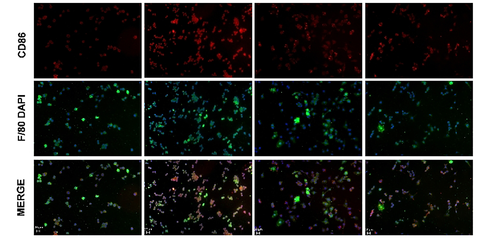
BACKGROUND: Hernandezine has shown promising therapeutic effects due to its anti-inflammatory bioactivity in diseases such as suppression of tumors, antiplatelet agglutination and diabetes. However, there are no basic studies on the effects and molecular mechanism of Hernandezine on macrophage phenotype and osteoclast activation. OBJECTIVE: To investigate the role of Hernandezine on the regulation of macrophage polarization, osteoclast activation and osteoporosis. METHODS: (1) Cellular experiments: RAW264.7 was used as macrophage model and divided into four groups: Control group, lipopolysaccharide group, lipopolysaccharide + 2.5 μmol/L Hernandezine group, lipopolysaccharide + 5 μmol/L Hernandezine group. Macrophage polarization was induced in the latter three groups using a complete medium supplemented with lipopolysaccharide. The two drug-treated groups received 2.5 and 5 μmol/L Hernandezine, respectively. RAW264.7 cells were induced toward osteoclast differentiation using a complete medium supplemented with nuclear factor κB receptor activator ligand. Macrophage polarization was assessed via qRT-PCR and immunofluorescence for inflammatory cytokine expression. The effects of Hernandezine on osteoclast differentiation were evaluated using qRT-PCR, tartrate-resistant acid phosphatase staining, and F-actin staining. (2) In vivo experiments: Twenty-four female C57BL/6J mice were randomly divided into four groups: sham operation, ovariectomy, ovariectomy + 5 mg/kg Hernandezine, and ovariectomy + 10 mg/kg Hernandezine. The latter three groups underwent bilateral ovariectomy to establish an osteoporosis model. The two drug-treated groups received intraperitoneal injections of Hernandezine at 5 or 10 mg/kg every two days post-surgery. After 8 weeks, femurs were collected for Micro-CT scanning, bone parameter analysis, and hematoxylin-eosin staining to evaluate bone loss. RESULTS AND CONCLUSION: Hernandezine inhibited lipopolysaccharide-induced pro-inflammatory gene expression in macrophages by downregulating the transcription of Toll-like receptor 4/nuclear factor κB signaling pathway-related genes, exhibiting a concentration-dependent effect, with 5 μmol/L showing more significant inhibition. Hernandezine also inhibited the expression of genes related to osteoclast activation and bone resorption, and suppressed osteoclast activation in vitro in a concentration-dependent manner. In vivo, Hernandezine reduced bone loss in estrogen-deficient osteoporotic mice, with the 10 mg/kg group showing better recovery. CONCLUSION: This study confirms that Hernandezine inhibits macrophage pro-inflammatory phenotype transformation and osteoclast activation by downregulating the Toll-like receptor 4/nuclear factor κB signaling pathway, and alleviates excessive bone loss in estrogen-deficient osteoporosis.

Read Full Abstract10.12307/2026.21477
SinoBioData ResearchChinese Journal of Tissue Engineering Research
Chinese Journal of Tissue Engineering Research2026

Mechanism of glucocorticoid-induced mitochondrial dysfunction in osteoblasts in steroid-induced osteonecrosis of the femoral head

BACKGROUND: The pathogenesis of steroid-induced osteonecrosis of the femoral head remains unclear; however, it is closely associated with mitochondrial damage in osteoblasts. OBJECTIVE: To explore the impact of dexamethasone on mitochondrial dysfunction in osteoblasts following steroid-induced osteonecrosis of the femoral head and to analyze its regulatory roles in osteoblast apoptosis and autophagy. METHODS: MC3T3-E1 cells were cultured in vitro and divided into control group (no treatment) and dexamethasone group (1 μmol/L dexamethasone treatment for 24 hours). Osteoblast differentiation capacity was assessed by alizarin red staining and qRT-PCR. Mitochondrial morphology was examined using transmission electron microscopy, MitoTracker Red fluorescence staining, and flow cytometry. Mitochondrial membrane potential and energy metabolism were evaluated by JC-1 fluorescence staining and ATP content detection. Mitochondrial superoxide levels were measured using MitoSOX fluorescence probe and flow cytometry. Intracellular total reactive oxygen species and glutathione content were also measured to assess oxidative stress status. Additionally, Western blot and qRT-PCR were used to detect the expression of apoptosis-related proteins (Bax, Bcl-2) and autophagy markers (LC3B, p62), flow cytometry was used to analyze apoptosis rate, and autophagy flux was observed via mRFP-GFP-LC3 adenovirus transfection combined with confocal microscopy. RESULTS AND CONCLUSION: Compared with the control group, the dexamethasone group showed significantly reduced osteogenic differentiation capacity of MC3T3-E1 cells, abnormal mitochondrial structure (swelling, cristae disruption), decreased mitochondrial membrane potential, reduced ATP synthesis, increased mitochondrial superoxide and total reactive oxygen species levels, and increased glutathione consumption (P < 0.05). The dexamethasone group showed significantly upregulated pro-apoptotic protein Bax (P < 0.01), significantly downregulated anti-apoptotic protein Bcl-2 (P < 0.01), increased LC3B-II/I ratio (P < 0.01), and decreased p62 levels (P < 0.01); dexamethasone treatment significantly increased the apoptosis rate (P < 0.01). mRFP-GFP-LC3 adenovirus tracing revealed increased formation of autophagosomes and autolysosomes. These results indicate that dexamethasone induces mitochondrial dysfunction and oxidative stress, synergistically regulating apoptosis and autophagy in MC3T3-E1 cells, thereby impairing bone formation and repair function. This mechanism may be a key pathological basis for the pathogenesis of steroid-induced osteonecrosis of the femoral head.

Read Full Abstract10.12307/2026.21475
WGCNA and machine learning identify autophagy and senescence signature genes in osteoarthritis chondrocytesGraphical AbstractVerified
Chinese Journal of Tissue Engineering Research2026

WGCNA and machine learning identify autophagy and senescence signature genes in osteoarthritis chondrocytes

BACKGROUND: Autophagy and senescence are considered important factors in the pathogenesis of osteoarthritis, but their specific regulatory mechanisms remain unclear. OBJECTIVE: To screen autophagy- and senescence-related genes in osteoarthritis through bioinformatics analysis combined with machine learning methods, providing new molecular targets for early diagnosis and treatment of osteoarthritis. METHODS: Osteoarthritis-related datasets (including GSE51588, GSE169077, and GSE114007) were downloaded from the GEO database. Differential expression analysis, weighted gene co-expression network analysis, and functional enrichment analysis were performed to screen autophagy- and senescence-related genes in osteoarthritis. LASSO regression, random forest (RF), and support vector machine (SVM) were used to further screen potential core genes, and receiver operating characteristic curve analysis was used to evaluate the diagnostic value of core genes. Based on the GSE51588 dataset, the CIBERSORT algorithm was used to analyze the proportions of immune cell types such as T cell subsets, B cells, and macrophages in osteoarthritis and healthy control knee cartilage specimens. The expression of ubiquitin-conjugating enzyme E2I, ribosomal protein S6 kinase 1, interleukin-2 receptor beta chain, YEATS protein family member 4, histone H4 variant, and Toll-like receptor 3 was detected in the external validation set GSE114007. Clinical knee cartilage specimens from 5 osteoarthritis patients and 5 healthy controls were collected, and RT-qPCR was used to detect the mRNA expression of these genes. RESULTS AND CONCLUSION: (1) A total of 26 autophagy- and senescence-related differentially expressed genes were obtained. Functional enrichment analysis showed that these genes were mainly involved in biological processes such as cellular homeostasis, immune regulation, and cell death, and played important roles in multiple signaling pathways. Six key genes were screened by machine learning: ubiquitin-conjugating enzyme E2I, ribosomal protein S6 kinase 1, interleukin-2 receptor beta chain, YEATS protein family member 4, histone H4 variant, and Toll-like receptor 3. The area under the receiver operating characteristic curve (AUC) values of these genes were all greater than 0.8, indicating high diagnostic performance. Immune infiltration analysis showed that the infiltration of plasma cells, resting CD4 memory T cells, resting NK cells, monocytes, M2 macrophages, eosinophils, and neutrophils was significantly decreased in the osteoarthritis group, while the infiltration of follicular helper T cells, gamma delta T cells, activated NK cells, M1 macrophages, and resting dendritic cells was significantly increased. (2) In the external validation set, the expression of ubiquitin-conjugating enzyme E2I, interleukin-2 receptor beta chain, and Toll-like receptor 3 was higher in the osteoarthritis group than in the healthy control group (P < 0.05), while there was no significant difference in the expression of histone H4 variant, YEATS protein family member 4, and ribosomal protein S6 kinase 1 between the two groups (P > 0.05). In clinical samples, the mRNA expression of ribosomal protein S6 kinase 1, interleukin-2 receptor beta chain, YEATS protein family member 4, histone H4 variant, and Toll-like receptor 3 was higher in the osteoarthritis group than in the healthy control group (P < 0.05), while there was no significant difference in the expression of ubiquitin-conjugating enzyme E2I mRNA between the two groups (P > 0.05). (3) These results indicate that Toll-like receptor 3 and interleukin-2 receptor beta chain can serve as key genes for autophagy and senescence in osteoarthritis chondrocytes, and may become diagnostic molecular markers and potential therapeutic targets for osteoarthritis.

Read Full Abstract10.12307/2026.21480
Mechanism by which luteolin regulates macrophage polarization in the treatment of knee osteoarthritisGraphical AbstractVerified
Chinese Journal of Tissue Engineering Research2026

Mechanism by which luteolin regulates macrophage polarization in the treatment of knee osteoarthritis

BACKGROUND: The polarization state of macrophages is closely related to the occurrence and development of knee osteoarthritis. Luteolin can regulate the nuclear transcription factor κB (NF-κB) signaling pathway to affect the polarization process of macrophages, but the specific mechanism remains unclear. OBJECTIVE: To investigate the mechanism of luteolin in the treatment of knee osteoarthritis. METHODS: (1) Intersectional targets of luteolin, macrophage polarization, and knee osteoarthritis were screened by network pharmacology, a protein-protein interaction network was constructed, and GO, KEGG enrichment analysis and molecular docking were performed. (2) After knocking out NF-κB p65, RAW264.7 cells were induced to polarize to M1 and M2 types, and flow cytometry was used to detect cell polarization tendency. Different concentrations of luteolin were used to intervene in RAW264.7 cells, and the appropriate concentration of luteolin was screened by CCK-8 method for subsequent experiments. RAW264.7 cells were induced to polarize to M1 and M2 types, and luteolin was added for intervention, and flow cytometry was used to detect cell polarization tendency. RAW264.7 cells were induced to polarize to M1 type, and luteolin was added for intervention. ELISA was used to detect the levels of tumor necrosis factor α, interleukin-6, and interleukin-10 in the cell supernatant, immunofluorescence staining was used to detect the nuclear translocation of NF-κB p65/p50, and western blot was used to detect the expression and phosphorylation of NF-κB p65 and NF-κB inhibitor protein α. RESULTS AND CONCLUSION: (1) A total of 137 intersection genes were screened, of which 135 target genes were involved in the construction of the protein-protein interaction network. GO and KEGG analysis showed that the NF-κB complex was enriched in the protein-protein interaction network. Molecular docking showed that luteolin docked well with NF-κB inhibitor protein α and NF-κB p50/p65 proteins. (2) After NF-κB p65 knockout, the polarization tendency of RAW264.7 cells to M1 type was inhibited, and spontaneous M2 polarization tendency appeared. Luteolin intervention could inhibit the polarization of RAW264.7 cells to M1 type and promote the polarization to M2 type. During the induction of RAW264.7 cells to M1 polarization, luteolin intervention could inhibit the release of pro-inflammatory factors, promote the release of anti-inflammatory factors, inhibit the nuclear translocation of NF-κB p65/p50, promote the expression of NF-κB inhibitor protein α and inhibit its phosphorylation, and inhibit the expression and phosphorylation of NF-κB p65. The results indicate that luteolin can regulate macrophage polarization through the NF-κB signaling pathway and has a potential therapeutic effect on knee osteoarthritis.

Read Full Abstract10.12307/2026.21478
Role of myeloid-derived suppressor cells in osteoclast differentiation in primary osteoporosisGraphical AbstractVerified
Chinese Journal of Tissue Engineering Research2026

Role of myeloid-derived suppressor cells in osteoclast differentiation in primary osteoporosis

BACKGROUND: Recent studies have found that immune cells play an important role in bone metabolism. Myeloid-derived suppressor cells, as a type of immunosuppressive cell, play a significant role in tumor development, but their role in primary osteoporosis remains unclear. OBJECTIVE: To investigate the osteoclastogenic potential of myeloid-derived suppressor cells in naturally aged and ovariectomy-induced osteoporosis mouse models. METHODS: (1) Myeloid-derived suppressor cells and bone marrow-derived macrophages were isolated from 6-8-week-old female C57BL/6 mice. Both cell types were induced for osteoclast differentiation. After 5 days of induction, osteoclast formation was detected by tartrate-resistant acid phosphatase staining. After 3 days of induction, mRNA expression of nuclear factor of activated T-cells 1 and osteoclast-associated immunoglobulin-like receptor was detected by qRT-PCR. (2) 6-8-week-old female C57BL/6 mice (young group, n=6) and 18-month-old female C57BL/6 mice (naturally aged group, n=6) were taken. Bone microstructure of the distal femur was analyzed by Micro-CT. Bone marrow cells were collected from both groups, and the proportion of myeloid-derived suppressor cells was detected by flow cytometry. Myeloid-derived suppressor cells were isolated and induced for osteoclast differentiation. After 5 days of induction, osteoclast formation was detected by tartrate-resistant acid phosphatase staining; after 3 days of induction, mRNA expression of nuclear factor of activated T-cells 1 and osteoclast-associated immunoglobulin-like receptor was detected by qRT-PCR. (3) 6-8-week-old female C57BL/6 mice were randomly divided into sham-operated group (n=6) and ovariectomy group (n=6). Eight weeks after ovariectomy, bone microstructure of the distal femur was analyzed by Micro-CT. Bone marrow cells were collected from both groups, and the proportion of myeloid-derived suppressor cells was detected by flow cytometry. Serum levels of tumor necrosis factor-alpha and interleukin-6 were measured by ELISA. Myeloid-derived suppressor cells were isolated and induced for osteoclast differentiation. After 5 days of induction, osteoclast formation was detected by tartrate-resistant acid phosphatase staining; after 3 days of induction, mRNA expression of nuclear factor of activated T-cells 1 and osteoclast-associated immunoglobulin-like receptor was detected by qRT-PCR. RESULTS AND CONCLUSION: (1) Tartrate-resistant acid phosphatase staining and qRT-PCR showed that the osteoclast differentiation ability of myeloid-derived suppressor cells was stronger than that of bone marrow-derived macrophages. (2) Micro-CT analysis showed that compared with the young group, the naturally aged group had lower bone mineral density, bone volume fraction, and trabecular number (P < 0.05), and increased trabecular separation (P < 0.05). The proportion of myeloid-derived suppressor cells in the naturally aged group was higher than that in the young group (P < 0.05). Tartrate-resistant acid phosphatase staining and qRT-PCR showed that the osteoclast differentiation ability of myeloid-derived suppressor cells in the naturally aged group was stronger than that in the young group. (3) Micro-CT analysis showed that compared with the sham-operated group, the ovariectomy group had lower bone mineral density, bone volume fraction, and trabecular number (P < 0.05), and increased trabecular separation (P < 0.05). The proportion of myeloid-derived suppressor cells and serum levels of tumor necrosis factor-alpha and interleukin-6 in the ovariectomy group were higher than those in the sham-operated group (P < 0.05). Tartrate-resistant acid phosphatase staining and qRT-PCR showed that the osteoclast differentiation ability of myeloid-derived suppressor cells in the ovariectomy group was stronger than that in the sham-operated group. (4) These results indicate that the proportion and osteoclastogenic ability of myeloid-derived suppressor cells increase under conditions of natural aging and estrogen deficiency, which may participate in the occurrence and development of osteoporosis.

Read Full Abstract10.12307/2026.21476
Total knee arthroplasty alignment technology: visual analysis of the latest research hotspotsGraphical AbstractVerified
Chinese Journal of Tissue Engineering Research2026

Total knee arthroplasty alignment technology: visual analysis of the latest research hotspots

BACKGROUND: Some patients report dissatisfaction after total knee arthroplasty due to persistent issues like limited mobility, joint instability, and residual pain. To address these challenges, various osteotomy methods and alignment strategies have been proposed and clinically implemented. However, the optimal approach remains debated, with no clear consensus. OBJECTIVE: To comprehensively analyze global trends in alignment techniques for total knee arthroplasty and identify research hotspots through bibliometric and visual analysis, offering evidence-based insights to guide clinical decision-making. METHODS: We retrieved publications (articles and reviews) on total knee arthroplasty alignment techniques published between January 1, 2015 and December 31, 2024, from the Web of Science database (https://www.webofscience.com, developed by the Institute for Scientific Information, USA). Data on countries, institutions, publication years, authors, journals, mean citation rates, H-index, titles, keywords, and the top 25 most-cited articles were extracted and analyzed. VOSviewer and Citespace software were used to analyze keyword co-occurrence and predict research hotspots. RESULTS AND CONCLUSION: A total of 866 publications were identified, including 792 articles and 74 reviews. The United States had the highest number of publications, with the University of California being the most prolific institution and Howell, Stephen M being the most prolific author. The journal 'Knee Surgery Sports Traumatology Arthroscopy' had the highest publication and citation counts. Common keywords included total knee arthroplasty, alignment technique, arthroplasty, clinical outcomes, and survival rate. Keyword and reference burst detection indicated that kinematic alignment and robotic navigation are future research hotspots. The number of publications on total knee arthroplasty alignment techniques is increasing annually. The United States is currently the leading contributor, followed by the United Kingdom. Increasingly precise tools, such as computer-assisted navigation and patient-specific instrumentation, are being developed and applied to achieve alignment goals, but no optimal solution has yet been established. Future research frontiers may include 'robot-assisted navigation', 'patient satisfaction', 'long-term survival', 'indication range', and 'patient-specific alignment'.

Read Full Abstract10.12307/2026.21700
Local administration of liposomal bupivacaine reduces postoperative pain and complications in spinal surgery: a meta-analysisGraphical AbstractVerified
Chinese Journal of Tissue Engineering Research2026

Local administration of liposomal bupivacaine reduces postoperative pain and complications in spinal surgery: a meta-analysis

OBJECTIVE: The purpose of this meta-analysis was to assess whether the local application of liposomal bupivacaine in spinal surgery effectively reduces postoperative pain, opioid consumption, and the incidence of related complications. METHODS: The study was conducted strictly in accordance to the methodological guidelines proposed by Cochrane Handbook. The protocol of the present work was registered with PROSPERO. Two researchers independently conducted literature searches in electronic databases including CNKI, Wanfang, VIP, PubMed, Web of Science, Embase, Cochrane Library, and Google Scholar, collecting trials published from January 2015 to December 2024 that met the inclusion criteria. Data extraction and synthesis were performed. Risk of bias or quality of included studies was independently assessed by two reviewers. Statistical analysis was conducted using STATA software. RESULTS: A total of 13 studies involving 2,962 patients were included. The risk of bias in randomized controlled trials was considered low to moderate, and the quality scores of cohort studies were rated as high. The results showed that the total postoperative opioid consumption in the liposomal bupivacaine group was significantly lower than that in the control group [SMD=-0.36, 95%CI(-0.71, -0.02)], but sensitivity analysis indicated that the robustness of this result was not high. In addition, pain scores at each follow-up point, opioid consumption, and the incidence of treatment-related complications were not significantly different between the liposomal bupivacaine group and the control group (P > 0.05). CONCLUSION: Local application of liposomal bupivacaine in spinal surgery does not increase treatment-related complications and can effectively reduce total opioid consumption; however, liposomal bupivacaine did not show a significant advantage in improving postoperative pain after spinal surgery.

Read Full Abstract10.12307/2026.21683
Integration of CD4+ T cell dynamic expression of quantitative trait loci reveals immunotherapeutic targets for sarcopeniaGraphical AbstractVerified
Chinese Journal of Tissue Engineering Research2026

Integration of CD4+ T cell dynamic expression of quantitative trait loci reveals immunotherapeutic targets for sarcopenia

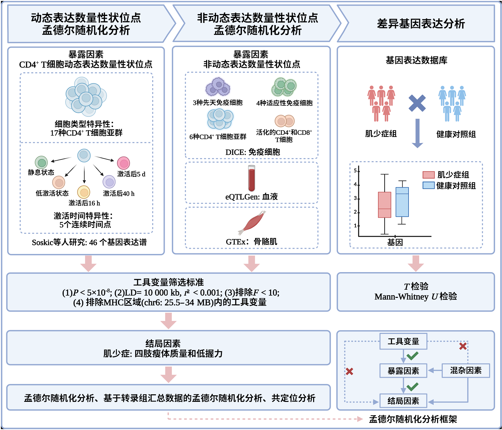
BACKGROUND: Sarcopenia is a degenerative illness in the elderly, and there is currently a dearth of particular therapeutic medications. Abnormalities in immune cells are risk factors for sarcopenia. CD4+ T lymphocytes play a vital role in skeletal muscle repair and regeneration. Previous research generally used expression quantitative trait loci data from whole tissues or blood to identify pharmacological targets, making it difficult to uncover the regulatory effects of gene expression on distinct cell subpopulations and their dynamic activation states. This study integrates dynamic expression quantitative trait locus data of CD4+ T cells to evaluate the immune cell specificity and activation time-dependent impacts of gene expression on sarcopenia, providing a platform for the development of precise immune intervention techniques. OBJECTIVE: To reveal the specific causal relationship between gene expression in different activation phases of CD4+ T cell subpopulations and sarcopenia. METHODS: Based on the dynamic eQTL data of CD4+ T cells from SOSKIC et al. (covering 46 cell-activation states in European populations), the Database of Immune Cell Expression, eQTLs and Epigenomics (DICE), eQTLGen, Genotype-Tissue Expression (GTEx), and GWAS Catalog, a two-sample Mendelian randomization analysis was systematically conducted. First, using CD4+ T cell dynamic eQTLs as exposure and sarcopenia phenotypes as outcomes, candidate genes were screened. Then, eQTL data from immune cells, whole blood, and skeletal muscle tissue were used for validation. Additionally, summary-data-based Mendelian randomization (SMR), heterogeneity tests, colocalization analysis, and differential gene expression analysis (using GEO dataset GSE111016) were performed to verify reliability. All data were publicly available summary statistics and met ethical requirements. All analyses strictly selected instrumental variables and followed the Strengthening the Reporting of Observational Studies in Epidemiology (STROBE) guidelines. RESULTS AND CONCLUSION: (1) SMR and heterogeneity tests showed that RAB29, NDUFS3, and MMP24OS had specific causal associations with sarcopenia. Specifically, expression of RAB29 in CD4+ naive T cells activated for 5 days and MMP24OS in CD4+ memory T cells activated for 5 days were positively associated with sarcopenia risk, while NDUFS3 expression in naive T cells activated for 16 and 40 hours was negatively associated with sarcopenia risk. (2) Colocalization analysis further confirmed that eQTLs for RAB29, NDUFS3, and MMP24OS shared potential causal variants with sarcopenia GWAS signals. (3) Differential expression analysis showed that NDUFS3 was significantly downregulated in sarcopenia patients compared with healthy controls, while RAB29 and MMP24OS showed no significant difference. NDUFS3 was identified as a potential gene therapy target with temporal regulatory characteristics in CD4+ T cells. This analysis was based on European population data; future studies should introduce dynamic eQTL Mendelian randomization frameworks to develop precise T-cell functional timing intervention strategies for the Chinese population.

Read Full Abstract10.12307/2026.21694
Healing characteristics and influencing factors of large-segment infectious bone defect of tibia repaired by bone transferGraphical AbstractVerified
Chinese Journal of Tissue Engineering Research2026

Healing characteristics and influencing factors of large-segment infectious bone defect of tibia repaired by bone transfer

BACKGROUND: Severe segmental infectious bone defects of the tibia pose a significant challenge in orthopedic treatment due to persistent infection, poor local blood supply, and poor soft tissue conditions. Although bone transplantation techniques are the main repair method, the healing process is complex and influenced by multiple factors. The underlying mechanisms are not yet fully understood. Therefore, it is necessary to conduct in-depth research on the healing characteristics and influencing factors to optimize the treatment. OBJECTIVE: To explore the healing characteristics and influencing factors of large-segment infectious bone defect of tibia repaired by bone transfer and analyze the influencing factors. METHODS: A total of 98 patients with large-segment infectious bone defect of the tibia treated by Ilizarov bone transfer in The First Affiliated Hospital of Xinjiang Medical University from May 2020 to October 2022 were selected as the study subjects. According to the clinical criteria of delayed or nonunion and union of fractures, they were divided into delayed or nonunion group (n=48) and union group (n=50), and the general data of the two groups were compared. A combined model was constructed. Cox regression analysis was used to evaluate the relationship between fluctuations in serum bone turnover markers (bone-specific alkaline phosphatase, N-terminal midfragment of osteocalcin, and N-terminal propeptide of type I procollagen) and bone healing. Least absolute shrinkage and selection operator regression and multivariate logistic regression were used to analyze risk factors affecting healing. After adjusting for confounders, Cox proportional hazards model analyzed the association of these markers with poor healing. A regression equation y=1-1/(1+e-z) was established for prediction and validated. RESULTS AND CONCLUSION: (1) Significant differences were found between the delayed/nonunion and union groups in long-term smoking history, diabetes, soft tissue injury, fibula fracture, wound infection, weight-bearing within 6 weeks postoperatively, transport direction, transport distance, and levels of bone-specific alkaline phosphatase, N-terminal midfragment of osteocalcin, and N-terminal propeptide of type I procollagen (P < 0.05). (2) The combined model showed that each unit decrease in these markers increased the risk of poor healing by 3%, 2%, and 4%, respectively. (3) LASSO and multivariate logistic regression identified soft tissue injury, weight-bearing within 6 weeks, wound infection, fibula fracture, and decreased levels of these markers as independent risk factors (P < 0.05). (4) Adjusted Cox model showed that bone-specific alkaline phosphatase (HR=0.67, 95%CI: 0.54-0.87, P < 0.001), N-terminal midfragment of osteocalcin (HR=0.80, 95%CI: 0.55-0.99, P < 0.001), and N-terminal propeptide of type I procollagen (HR=0.85, 95%CI: 0.43-0.97, P < 0.001) were significant factors for poor healing. (5) As levels decreased (Q2-Q4), the association increased, with significant trend tests (P trend < 0.05). Bootstrap validation showed good discrimination and accuracy of the prediction model. (6) These findings suggest that decreased serum bone turnover markers are closely related to poor healing after Ilizarov bone transfer, and soft tissue injury, weight-bearing within 6 weeks, wound infection, fibula fracture, and these markers are important factors affecting healing.

Read Full Abstract10.12307/2026.21640
Construction and validation of a temperature prediction model for cortical bone during orthopedic surgeryGraphical AbstractVerified
Chinese Journal of Tissue Engineering Research2026

Construction and validation of a temperature prediction model for cortical bone during orthopedic surgery

BACKGROUND: Cortical bone drilling, cutting, friction, and heat generation can easily cause local temperature rise. If it exceeds the bone tissue tolerance threshold and continues to act, it can lead to complications such as bone necrosis, delayed healing, or prosthesis loosening. The coupling of commonly used clinical parameters such as rotational speed, feed rate, and irrigation significantly affects the degree of thermal accumulation, and there is an urgent need to establish a quantitative tool that can predict the temperature field and heat affected zone to define the safe operating window. OBJECTIVE: To establish a temperature prediction model for cortical bone in orthopedic surgery by analyzing the temperature distribution of cortical bone at different depths and radial directions. METHODS: A three-dimensional transient heat transfer control equation was established to describe the cortical bone drilling process. The moving/distributed heat source method was introduced to characterize the interface heat input caused by the shear of the anterior cutting surface and the friction of the posterior cutting surface, and the temperature field evolution at different radial and depth positions was calculated. Using the inverse heat transfer method, the distribution ratio of heat flux and heat between the tool chip bone interface was inverted under the constraint of a finite temperature sequence of measurement points. The model prediction was further validated through experimental comparison. RESULTS AND CONCLUSION: (1) Inverse heat transfer inversion showed that approximately 11.7% of the total heat entered the cortical bone under given conditions; (2) The established temperature prediction model showed good consistency with fresh porcine bone drilling experiments in terms of peak temperature, onset of temperature rise, and temperature-time curve shape, confirming the reliability of the model for spatiotemporal temperature distribution; (3) In terms of spatial distribution, the closer to the hole wall (radius approaching 2.0 mm), the earlier the temperature rise and the higher the peak; along the depth direction (z=0–5 mm), temperature rise first occurred near the surface and gradually extended to deeper parts; (4) The heat affected zone increased with rotational speed: under conditions of drill diameter 4 mm and feed rate 60 mm/min, the heat affected zone was approximately 0.71 mm at 800 r/min and approximately 0.86 mm at 1,000 r/min; (5) These results indicate that under the premise of controlling drill diameter and feed rate, increasing rotational speed increases bone thermal load and heat affected zone thickness; therefore, clinically, rotational speed and cooling/irrigation strategies need to be optimized synergistically to reduce the risk of thermal bone injury; this model can be used for preoperative parameter screening and intraoperative risk assessment, providing quantitative basis for formulating a 'safe parameter window', improving tool and irrigation protocols, and enhancing patient prognosis.

Read Full Abstract10.12307/2026.21650
Effects of Fascia Iliaca Compartment Block and Femoral Nerve Block on Pain, Inflammation, and Stress Response in Hip Fracture SurgeryGraphical AbstractVerified
Chinese Journal of Tissue Engineering Research2026

Effects of Fascia Iliaca Compartment Block and Femoral Nerve Block on Pain, Inflammation, and Stress Response in Hip Fracture Surgery

BACKGROUND: Hip fracture is a common and serious injury in the elderly population, with postoperative pain management directly impacting rehabilitation quality and functional recovery. While peripheral nerve blocks have become an integral component of multimodal analgesia, systematic comparison of different blocking techniques across various types of hip fractures remains limited. OBJECTIVE: To compare the analgesic effects, safety profiles, and postoperative recovery impacts of fascia iliaca compartment block and femoral nerve block in different types of hip fracture surgeries. METHODS: A total of 180 patients scheduled for hip fracture surgery between January 2022 and June 2024 were enrolled and divided according to fracture type into femoral neck fracture group (n=60), intertrochanteric fracture group (n=60), and subtrochanteric fracture group (n=60). Patients in each group were randomly assigned to either fascia iliaca compartment block group or femoral nerve block group using a random number table, with 30 patients in each subgroup. Corresponding nerve blocks were performed preoperatively. Numeric pain scores were recorded immediately and at 6, 12, and 24 hours postoperatively; 24-hour morphine consumption, time to first ambulation, hospital stay, inflammatory markers (interleukin-6, tumor necrosis factor-α), stress response markers (cortisol, glucose), and adverse events were also assessed. Multivariate regression analysis identified factors influencing analgesic effect, and a nomogram prediction model was constructed. RESULTS AND CONCLUSION: In femoral neck and intertrochanteric fracture groups, the fascia iliaca compartment block subgroup had significantly lower numeric pain scores immediately and at 6 and 12 hours postoperatively compared with the femoral nerve block subgroup (P < 0.01). In the femoral neck fracture group, mean differences in pain scores were 0.86 (95%CI: 0.47-1.25), 0.90 (95%CI: 0.45-1.35), and 0.79 (95%CI: 0.30-1.28) at immediate, 6, and 12 hours, respectively; in the intertrochanteric fracture group, differences were 0.83 (95%CI: 0.43-1.23), 0.87 (95%CI: 0.44-1.30), and 0.70 (95%CI: 0.31-1.09). In these two groups, fascia iliaca compartment block significantly reduced 24-hour morphine consumption (P < 0.001), shortened time to first ambulation (P < 0.05), and reduced hospital stay (P < 0.05). In the subtrochanteric fracture group, no significant differences were observed between the two block techniques for any outcome (P > 0.05). Fascia iliaca compartment block significantly lowered inflammatory markers (interleukin-6, tumor necrosis factor-α) and stress markers (cortisol, glucose) compared with femoral nerve block (P < 0.001). Adverse event rates were comparable (P=0.825). Multivariate regression identified block type (β=-3.76, P < 0.001), fracture type (subtrochanteric β=5.47, P < 0.001), age ≥75 years (β=-1.75, P=0.022), and ASA grade III (β=2.32, P=0.016) as independent predictors of analgesic effect. The nomogram showed good calibration. Interleukin-6 levels positively correlated with hospital stay (r=0.42, P < 0.001) and time to first ambulation (r=0.38, P < 0.001). In conclusion, compared with femoral nerve block, fascia iliaca compartment block provides superior postoperative analgesia for femoral neck and intertrochanteric fractures, reduces inflammation and stress responses, and promotes early recovery; however, in subtrochanteric fractures, the two techniques are equivalent. Block type, fracture type, age, and ASA grade are independent factors influencing analgesic effect. The nomogram may guide individualized analgesic strategies.

Read Full Abstract10.12307/2026.21643