🧬 SinoBioData Academic Portal
📚 Peer-Reviewed Translated Literature

All Biomedical & Clinical Articles (Page 16)

Browse complete peer-reviewed translations from top Chinese biomedical, oncology, and genomics journals. Read verified previews and download full authentic clinical reports.

Published Research Papers

Showing 24 of 1542 peer-reviewed translated articles (Page 16 of 65)

Fabrication and characterization of hydrogels with both antibacterial and osteogenic functionsGraphical AbstractVerified
Chinese Journal of Tissue Engineering Research2026

Fabrication and characterization of hydrogels with both antibacterial and osteogenic functions

BACKGROUND: Hydrogel materials have garnered significant attention in tissue repair due to their good biocompatibility and degradability, but single hydrogels lack antibacterial and osteogenic functions, limiting clinical application. OBJECTIVE: To prepare hydrogels with both antibacterial and osteogenic functions for bone tissue repair. METHODS: Copper and zinc co-doped hydroxyapatite (Cu/Zn HA) was synthesized by chemical precipitation. Cu/Zn HA, epigallocatechin gallate (EGCG), and Cu/Zn HA+EGCG were separately added to photoinitiators, and methacrylated gelatin (GelMA) was added to the photoinitiator solutions. After UV irradiation at 405 nm for 20 s, four hydrogels were prepared: GelMA (G), Cu/Zn HA/GelMA (G-Cu/Zn HA), EGCG-modified GelMA (G-E), and EGCG-modified Cu/Zn HA/GelMA (G-E-Cu/Zn HA). The microstructure, compressive mechanical properties, swelling, degradation, and release kinetics of metal ions and EGCG were characterized. Antibacterial properties were evaluated against Staphylococcus aureus and Escherichia coli using agar plate coating, live/dead staining, and scanning electron microscopy. Cytocompatibility was assessed with MC3T3-E1 cells via live/dead staining and CCK-8 assay. Osteogenic activity was evaluated after osteogenic induction using alkaline phosphatase staining, alizarin red S staining, and osteogenic-related gene expression. RESULTS AND CONCLUSION: Scanning electron microscopy showed porous internal structures in all hydrogels, with G and G-E having relatively smooth surfaces, while G-Cu/Zn HA and G-E-Cu/Zn HA had increased surface roughness. Compressive stresses were 10.48, 12.91, 23.64, and 41.03 kPa for G, G-E, G-Cu/Zn HA, and G-E-Cu/Zn HA, respectively. Compared with G, the swelling time and equilibrium swelling ratio decreased in G-E, G-Cu/Zn HA, and G-E-Cu/Zn HA. G-E-Cu/Zn HA exhibited prolonged degradation. G-Cu/Zn HA and G-E-Cu/Zn HA released Cu2+, Zn2+, and Ca2+ over 30 days, with G-Cu/Zn HA releasing more Cu2+ and Zn2+ than G-E-Cu/Zn HA. G-E-Cu/Zn HA significantly inhibited the burst release of EGCG compared with G-E. Antibacterial assays showed that all modified hydrogels inhibited bacteria, with G-E-Cu/Zn HA showing the strongest effect. All hydrogels were cytocompatible. Osteogenic assays showed that G had the weakest osteogenic ability, while G-E-Cu/Zn HA had the strongest. CONCLUSION: The EGCG-modified Cu/Zn HA/GelMA composite hydrogel exhibits excellent antibacterial and osteogenic properties.


Read Full Abstract10.12307/2026.21451
Preparation and characterization of beta-tricalcium phosphate/polyvinyl alcohol and hydroxyapatite/polyvinyl alcohol bone substitute materialsGraphical AbstractVerified
Chinese Journal of Tissue Engineering Research2026

Preparation and characterization of beta-tricalcium phosphate/polyvinyl alcohol and hydroxyapatite/polyvinyl alcohol bone substitute materials

BACKGROUND: Currently, many oral periodontal bone substitute materials suffer from drawbacks such as poor biomechanical properties and degradation characteristics. Using composite periodontal bone substitute materials can compensate for the shortcomings of single materials and better achieve the construction of 'periodontal bone tissue engineering constructs'. OBJECTIVE: To prepare beta-tricalcium phosphate/polyvinyl alcohol and hydroxyapatite/polyvinyl alcohol composite materials by freeze-drying method and characterize their properties. METHODS: Polyvinyl alcohol hydrogels with a concentration of 15% were prepared. Different masses of beta-tricalcium phosphate were added to the polyvinyl alcohol hydrogels, and after freeze-drying, 10%, 20%, and 30% beta-tricalcium phosphate/polyvinyl alcohol composite materials were obtained. Similarly, different masses of hydroxyapatite were added to polyvinyl alcohol hydrogels, and after freeze-drying, 10%, 20%, and 30% hydroxyapatite/polyvinyl alcohol composite materials were obtained. The properties of each composite material were characterized using scanning electron microscopy, X-ray energy dispersive spectroscopy, X-ray diffraction, X-ray photoelectron spectroscopy, and Raman spectroscopy. RESULTS AND CONCLUSION: (1) Scanning electron microscopy and X-ray energy dispersive spectroscopy analysis showed that beta-tricalcium phosphate and hydroxyapatite particles were successfully combined with polyvinyl alcohol hydrogel to form stable composite materials. With increasing concentration of beta-tricalcium phosphate or hydroxyapatite, the aggregation degree of particles in the composite materials increased and the porosity decreased. The main elements in each composite material included Ca, P, C, and O. (2) X-ray photoelectron spectroscopy, X-ray diffraction, and Raman spectroscopy results showed that no new bending vibration peaks, stretching vibration peaks, or characteristic peaks appeared in the beta-tricalcium phosphate/polyvinyl alcohol and hydroxyapatite/polyvinyl alcohol composite materials. These results indicate that the beta-tricalcium phosphate/polyvinyl alcohol and hydroxyapatite/polyvinyl alcohol composite materials prepared by freeze-drying have stable properties.

Read Full Abstract10.12307/2026.21446
Multi-objective optimization of coronary artery stent design in ensemble surrogate modelGraphical AbstractVerified
Chinese Journal of Tissue Engineering Research2026

Multi-objective optimization of coronary artery stent design in ensemble surrogate model

BACKGROUND: Percutaneous coronary intervention stent implantation is primarily used to treat coronary artery stenosis. However, current multi-objective stent optimization methods are limited by sample size constraints, resulting in insufficient prediction accuracy when balancing key performance indicators such as support and compliance, hindering the effectiveness of stent optimization design. OBJECTIVE: To establish an innovative optimization framework for coronary stents based on a ensemble surrogate model. METHODS: A three-dimensional parametric model of the vascular stent was constructed, and a mechanical response database was established through finite element simulation. A dynamic weight fusion strategy was adopted to integrate the global optimization characteristics of the Kriging model and the local nonlinear representation advantages of the radial basis function model. A ensemble surrogate model was constructed based on 20 groups of initial samples, and the non-dominated sorting genetic algorithm-II was used to optimize the parameter space. RESULTS AND CONCLUSION: Experimental results demonstrated that the ensemble surrogate model exhibited significant advantages in the finite sample setting. The coefficient of determination for the inverse prediction of the radial stiffness of the stent reached 0.974 2, a 4.4% improvement compared to the single model, validating the efficient modeling capability of the ensemble surrogate model in the finite sample setting. The prediction accuracy of the stent's bending stiffness also improved by 4.4% compared to the single radial basis function surrogate model. After optimization, the stent performance achieved dual-objective synergistic optimization. The inverse radial stiffness of the stent in the ensemble surrogate model group was reduced by 13.92% and 9.57% compared to the Kriging model group and the single radial basis function surrogate model group, respectively. The bending stiffness of the stent was optimized by 0.38% and 2.56% compared to the Kriging model group and the single radial basis function surrogate model group, respectively. The proposed ensemble surrogate model breaks through the performance limitations of traditional single models, providing a low-cost, high-precision solution for the 'rigid-flexible' synergistic optimization of coronary stents.

Read Full Abstract10.12307/2026.21449
Ginsenoside Rg3-loaded liposome hydrogel promotes chondrogenic differentiation of stem cellsGraphical AbstractVerified
Chinese Journal of Tissue Engineering Research2026

Ginsenoside Rg3-loaded liposome hydrogel promotes chondrogenic differentiation of stem cells

BACKGROUND: Ginsenoside Rg3 has potential value in cartilage protection and repair, but its application is limited by poor water solubility, short half-life, and low bioavailability. To improve the pharmacokinetic properties of drugs, embedding drug-loaded liposomes into methylated gelatin, polysaccharide, or silk fibroin-based hydrogels has become a research hotspot in cartilage tissue engineering. OBJECTIVE: To prepare ginsenoside Rg3-loaded liposome methacrylated silk fibroin hydrogel and further analyze its effect on chondrogenic differentiation of mouse bone marrow mesenchymal stem cells. METHODS: (1) Ginsenoside Rg3 liposomes were prepared by thin-film dispersion method. Dil-labeled liposomes were co-cultured with mouse bone marrow mesenchymal stem cells, and phalloidin staining was used to detect cellular uptake of liposomes. Methacrylated silk fibroin (SilMA) was prepared. Ginsenoside Rg3 liposomes were mixed with SilMA and crosslinked by light to prepare composite hydrogel (SilMA@Lipo-Rg3). The microstructure, mechanical properties, rheological properties, swelling properties, and drug release properties of the hydrogel were evaluated. (2) Mouse bone marrow mesenchymal stem cells were cultured with different concentrations of SilMA hydrogel extract or SilMA@Lipo-Rg3 hydrogel extract. CCK-8 assay and live/dead cell staining were used to evaluate cytocompatibility. Cells were cultured with 1/8 concentration of SilMA hydrogel extract or SilMA@Lipo-Rg3 hydrogel extract. After chondrogenic induction, qPCR was used to detect mRNA expression of collagen type II, SOX9, and aggrecan. Alcian blue and safranin O staining were used to detect the expression of proteoglycans and glycosaminoglycans. RESULTS AND CONCLUSION: (1) Phalloidin staining showed that Dil-labeled liposomes could be successfully taken up by mouse bone marrow mesenchymal stem cells. Scanning electron microscopy showed that SilMA@Lipo-Rg3 hydrogel had a loose porous network structure. Compression and rheological tests showed that the compressive stiffness of SilMA@Lipo-Rg3 hydrogel was slightly lower than that of SilMA hydrogel; there was no significant difference in swelling properties between the two hydrogels. SilMA@Lipo-Rg3 hydrogel had good sustained release performance, releasing ginsenoside Rg3 for more than 14 days. (2) CCK-8 assay and live/dead cell staining showed that SilMA and SilMA@Lipo-Rg3 hydrogels had good cytocompatibility. qPCR showed that the mRNA expression of collagen type II, SOX9, and aggrecan in the SilMA@Lipo-Rg3 group was higher than that in the SilMA group (P < 0.05). Alcian blue and safranin O staining showed that the expression of proteoglycans and glycosaminoglycans in the SilMA@Lipo-Rg3 group was higher than that in the SilMA group. These results indicate that SilMA@Lipo-Rg3 hydrogel can promote chondrogenic differentiation of mouse bone marrow mesenchymal stem cells.

Read Full Abstract10.12307/2026.21450
Comparison of different 3D-printed allogeneic bone and artificial polymer composite porous scaffold materials for repairing cranial bone defects in ratsGraphical AbstractVerified
Chinese Journal of Tissue Engineering Research2026

Comparison of different 3D-printed allogeneic bone and artificial polymer composite porous scaffold materials for repairing cranial bone defects in rats

BACKGROUND: Allogeneic bone repair materials have good effect on bone defect repair, but they have drawbacks such as immune rejection, high cost, and low rigidity. OBJECTIVE: To evaluate the bone repair ability, tissue response and degradation performance of three kinds of 3D-printed allogeneic bone and artificial polymer composite porous scaffold materials. METHODS: Three kinds of bone repair materials were prepared: (1) Sample A: A bone repair material was prepared by mixing polylactic acid-glycolic acid copolymer with allogeneic bone powder at a mass ratio of 1:4, using low-temperature deposition 3D printing. (2) Sample B: A bone repair material was prepared by mixing polycaprolactone with allogeneic bone powder at a mass ratio of 1:4, using low-temperature deposition 3D printing. (3) Sample C: A bone repair material was prepared by mixing polycaprolactone with allogeneic bone powder at a mass ratio of 3:7, using high-temperature melt 3D printing. In 35 SD rats, one 5 mm diameter circular bone defect was drilled on each side of the sagittal suture. Ten defects received no intervention (blank control group), 15 defects were implanted with allogeneic bone material (positive control group), 15 defects were implanted with sample A (sample A group), 15 defects with sample B (sample B group), and 15 defects with sample C (sample C group). At 2, 4, 8, 12, and 26 weeks postoperatively, samples were harvested. Hematoxylin-eosin staining was used to observe material degradation, tissue response, and new bone formation. Masson staining was used to observe collagen fiber formation. Immunohistochemical staining was used to observe the expression of RUNT-related transcription factor 2 and type I collagen. RESULTS AND CONCLUSION: (1) Hematoxylin-eosin staining: During the experimental period, all implanted materials degraded to varying degrees. Samples A and B degraded faster, followed by the positive control, and sample C degraded slower. Over time, the inflammatory response in each implanted material group tended to decrease, but fibrous tissue proliferation and neovascularization were still obvious. At 26 weeks, the inflammatory response in sample A group was significantly reduced, followed by sample B group and positive control group, while sample C group showed no obvious reduction. In sample B group, a small amount of new bone formation was visible at 8 weeks; in sample A and positive control groups, new bone formation was visible at 12 weeks; these three groups showed a large amount of new bone formation at 26 weeks. Sample C group never showed obvious new bone formation. (2) Masson staining: At 2 weeks, a small amount of disordered collagen deposition was seen in each implanted material group; at 26 weeks, a large amount of regularly arranged collagen deposition was seen. (3) Immunohistochemical staining: Over time, the expression of RUNT-related transcription factor 2 and type I collagen increased in each implanted material group. At 12 and 26 weeks, the expression in positive control, sample A, and sample B groups was higher than that in sample C group. (4) These results indicate that samples prepared by low-temperature deposition 3D printing of polylactic acid-glycolic acid copolymer or polycaprolactone with allogeneic bone powder degraded faster, reduced inflammatory response, and showed more obvious expression of osteogenic factors and new bone formation than samples prepared by high-temperature melt printing of high-content polycaprolactone with allogeneic bone powder.

Read Full Abstract10.12307/2026.21448
3D printed neobavaisoflavone-coated scaffolds promote bone regeneration by regulating osteoblast/osteoclast activitiesGraphical AbstractVerified
Chinese Journal of Tissue Engineering Research2026

3D printed neobavaisoflavone-coated scaffolds promote bone regeneration by regulating osteoblast/osteoclast activities

BACKGROUND: Neobavaisoflavone could promote bone formation and may be a potential small molecule drug for bone regeneration. The use of 3D printed bone tissue engineering scaffolds as drug delivery carriers for neobavaisoflavone is expected to enhance the potential application of bone regeneration. OBJECTIVE: To explore the effects of polylactic acid/polydopamine/neobavaisoflavone bone scaffold on osteoclast and osteoblast activity. METHODS: (1) Fused deposition modeling technology was used to manufacture a 3D printed polylactic acid scaffold. These polylactic acid scaffolds were immersed in a dopamine solution containing or without neobavaisoflavone to produce polylactic acid/polydopamine/neobavaisoflavone scaffolds and polylactic acid/polydopamine scaffolds, respectively. The surface morphology, surface hardness, and compressive strength of the three groups of scaffolds were characterized, and the drug release properties of the polylactic acid/polydopamine/neobavaisoflavone scaffolds were investigated. (2) Mouse embryonic osteoblast MC3T3-E1 cells were co-cultured with the three groups of scaffolds. CCK-8 assay and live/dead staining were used to evaluate the cytocompatibility of the scaffolds. Transwell assay was used to evaluate the effect of scaffolds on osteoblast migration. Alkaline phosphatase quantitative assay and alizarin red staining were used to evaluate the effect of scaffolds on osteoblast differentiation. RAW264.7 cells were co-cultured with the three groups of scaffolds. After osteoclast induction, tartrate-resistant acid phosphatase staining was used to evaluate the effect of scaffolds on osteoclast differentiation. RESULTS AND CONCLUSION: (1) Scanning electron microscopy showed that all three groups of scaffolds had three-dimensional structure and regular interconnected porous structure with an average pore size of 400 µm. The surface hardness and compressive strength of polylactic acid/polydopamine scaffolds and polylactic acid/polydopamine/neobavaisoflavone scaffolds were higher than those of polylactic acid scaffolds (P < 0.05). Polylactic acid/polydopamine/neobavaisoflavone scaffolds had good drug release behavior and could continuously release drugs for more than 14 days in vitro. (2) CCK-8 assay and live/dead staining showed that all three groups of scaffolds had good cytocompatibility, and polylactic acid/polydopamine scaffolds and polylactic acid/polydopamine/neobavaisoflavone scaffolds could promote MC3T3-E1 cell proliferation. Transwell assay showed that compared with polylactic acid scaffolds, polylactic acid/polydopamine scaffolds and polylactic acid/polydopamine/neobavaisoflavone scaffolds could promote MC3T3-E1 cell migration. Alkaline phosphatase quantitative assay and alizarin red staining showed that compared with the other two groups, polylactic acid/polydopamine scaffolds and polylactic acid/polydopamine/neobavaisoflavone scaffolds could promote osteogenic differentiation of MC3T3-E1 cells. Tartrate-resistant acid phosphatase staining showed that polylactic acid/polydopamine/neobavaisoflavone scaffolds could inhibit osteoclast differentiation of RAW264.7 cells. (3) These results indicate that polylactic acid/polydopamine/neobavaisoflavone scaffolds have good biosafety and can promote bone regeneration by regulating osteoblast and osteoclast activities.

Read Full Abstract10.12307/2026.21447
Osteogenic and antibacterial properties of polyether ether ketone modified by multifunctional composite coatingGraphical AbstractVerified
Chinese Journal of Tissue Engineering Research2026

Osteogenic and antibacterial properties of polyether ether ketone modified by multifunctional composite coating

BACKGROUND: Polyether ether ketone (PEEK) has insufficient inherent bioactivity, and as a bone implant material, it carries risks of poor osseointegration and implant-related infection. Developing surface modification strategies with both osteogenic and antibacterial functions is of great clinical significance for improving the implantation performance of PEEK. OBJECTIVE: To analyze the biocompatibility, osteogenic and antibacterial effects of PEEK loaded with polydopamine-modified strontium-doped hydroxyapatite-silver composite coating. METHODS: (1) A polydopamine-hydroxyapatite composite coating was prepared on the PEEK surface, denoted as PEEK@PDA-HA. According to the ratios of Sr2+/(Sr2++Ca2+) of 0%, 5%, 10%, and 20%, polydopamine-strontium-doped hydroxyapatite-silver composite coatings were prepared on the PEEK surface, denoted as PEEK@PDA-HA-Ag, PEEK@PDA-5Sr/HA-Ag, PEEK@PDA-10Sr/HA-Ag, and PEEK@PDA-20Sr/HA-Ag, respectively. MC3T3-E1 cells were co-cultured with PEEK, PEEK@PDA-HA-Ag, PEEK@PDA-5Sr/HA-Ag, PEEK@PDA-10Sr/HA-Ag, and PEEK@PDA-20Sr/HA-Ag. The best material was selected for subsequent experiments through cell proliferation, live/dead staining, and adhesion assays. The surface morphology and water contact angle of PEEK@PDA-10Sr/HA-Ag were characterized. (2) MC3T3-E1 cells were seeded on PEEK, PEEK@PDA-HA, PEEK@PDA-HA-Ag, and PEEK@PDA-10Sr/HA-Ag surfaces. After osteogenic induction, alkaline phosphatase staining, alizarin red staining, and osteocalcin immunofluorescence staining were performed to evaluate the osteogenic differentiation performance of the materials. (3) Escherichia coli (or Staphylococcus aureus) were co-cultured with PEEK, PEEK@PDA-HA, PEEK@PDA-HA-Ag, and PEEK@PDA-10Sr/HA-Ag. The antibacterial properties were evaluated by agar plate counting and bacterial live/dead staining. RESULTS AND CONCLUSION: (1) Cell proliferation, live/dead staining, and adhesion assays showed that PEEK@PDA-10Sr/HA-Ag had the best effect on promoting MC3T3-E1 cell proliferation, and the cells adhered on the material surface exhibited good morphology with many filopodia, making it suitable for subsequent experiments. Scanning electron microscopy revealed a rough and uneven surface of PEEK@PDA-10Sr/HA-Ag with numerous spherical nanoparticle aggregates. Compared with PEEK, the water contact angle of PEEK@PDA-10Sr/HA-Ag decreased, indicating enhanced hydrophilicity. (2) Alkaline phosphatase staining, alizarin red staining, and osteocalcin immunofluorescence staining showed that PEEK@PDA-10Sr/HA-Ag had the strongest osteogenic effect. (3) Bacterial plate counting and live/dead staining showed that compared with the other three groups, PEEK@PDA-10Sr/HA-Ag effectively inhibited the growth of Escherichia coli and Staphylococcus aureus. (4) These results indicate that PEEK loaded with polydopamine-modified strontium-doped hydroxyapatite-silver composite coating has good biocompatibility, osteogenic and antibacterial effects.

Read Full Abstract10.12307/2026.21445
Three-dimensional printed isoniazid liposome photothermal composite scaffolds and their performance evaluationGraphical AbstractVerified
Chinese Journal of Tissue Engineering Research2026

Three-dimensional printed isoniazid liposome photothermal composite scaffolds and their performance evaluation

BACKGROUND: Drug-loaded bone scaffolds for tuberculosis treatment suffer from problems such as drug burst release in the early stages of treatment and insufficient drug release in the later stages, which leads to low drug concentration at the lesion, therefore, it is imperative to build a drug controlled-release system on the tissue-engineered bone scaffolds. OBJECTIVE: To prepare photothermal composite scaffolds with different isoniazid liposome doping ratios and characterize their mechanical and biological properties. METHODS: Isoniazid liposomes were prepared using the thin-film hydration method. The microstructure, encapsulation efficiency, drug loading capacity, particle size, and Zeta potential of liposomes were characterized. 15 mg of isoniazid was selected as the total drug amount for a single drug-loaded scaffold. Isoniazid drug powder, isoniazid liposome powder at 2%, 5%, and 8% of the total drug amount, respectively, were mixed uniformly with the scaffold base material (hydroxyapatite and β-tricalcium phosphate in a mass ratio of 6:4) and 3% of the photothermal agent polypyrrole nanoparticles (by mass fraction of the scaffold base material) to prepare extrusion printing powders. 13% polyvinyl alcohol gel was added at a mass ratio of 1:1 to the extrusion printing powder, and pure isoniazid scaffolds and three types of isoniazid liposome photothermal scaffolds were fabricated by extrusion-based 3D printing. The microstructure, porosity, mechanical properties, and drug release performance of the photothermal scaffolds were characterized. Mouse embryonic osteoblast MC3T3 cells were cultured with the extracts of the four scaffolds, and the cytotoxicity of the scaffolds was evaluated by MTT assay. RESULTS AND CONCLUSION: (1) Transmission electron microscopy showed that isoniazid liposomes were spherical vesicles with regular shape; the encapsulation efficiency was (28.67±0.62)%, drug loading was (3.54±0.19)%, average particle size was (363.63±10.42) nm, and average Zeta potential was (-4.68±0.72) mV. (2) Scanning electron microscopy showed that compared with pure isoniazid scaffolds, isoniazid liposome photothermal scaffolds had more internal pores; with the increase of isoniazid liposome content, the porosity of photothermal scaffolds increased, while the compressive strength and elastic modulus decreased, but still met the minimum compressive strength required for animal in vivo tissue implantation experiments. The incorporation of isoniazid liposomes effectively solved the drug burst release in the early stage, and the cumulative release rate in the early stage was inversely proportional to the doping amount of isoniazid liposomes. After irradiation with 808 nm near-infrared laser, the cumulative release rate of isoniazid liposome photothermal scaffolds increased compared with that without near-infrared laser irradiation, and with the increase of isoniazid liposome doping amount, the photothermal controlled-release performance in the later stage became more significant. MTT assay showed that the relative proliferation rates of MC3T3 cells cultured with the extracts of the four scaffolds for 24, 72, and 120 h were all greater than 70%, indicating no obvious cytotoxicity. The results indicate that isoniazid liposome photothermal scaffolds have good mechanical properties, drug controlled-release performance, and cytocompatibility.

Read Full Abstract10.12307/2026.21443
Triangular fibrocartilage complex injuries: a visualization analysis of treatment hotspots and frontiersGraphical AbstractVerified
Chinese Journal of Tissue Engineering Research2026

Triangular fibrocartilage complex injuries: a visualization analysis of treatment hotspots and frontiers

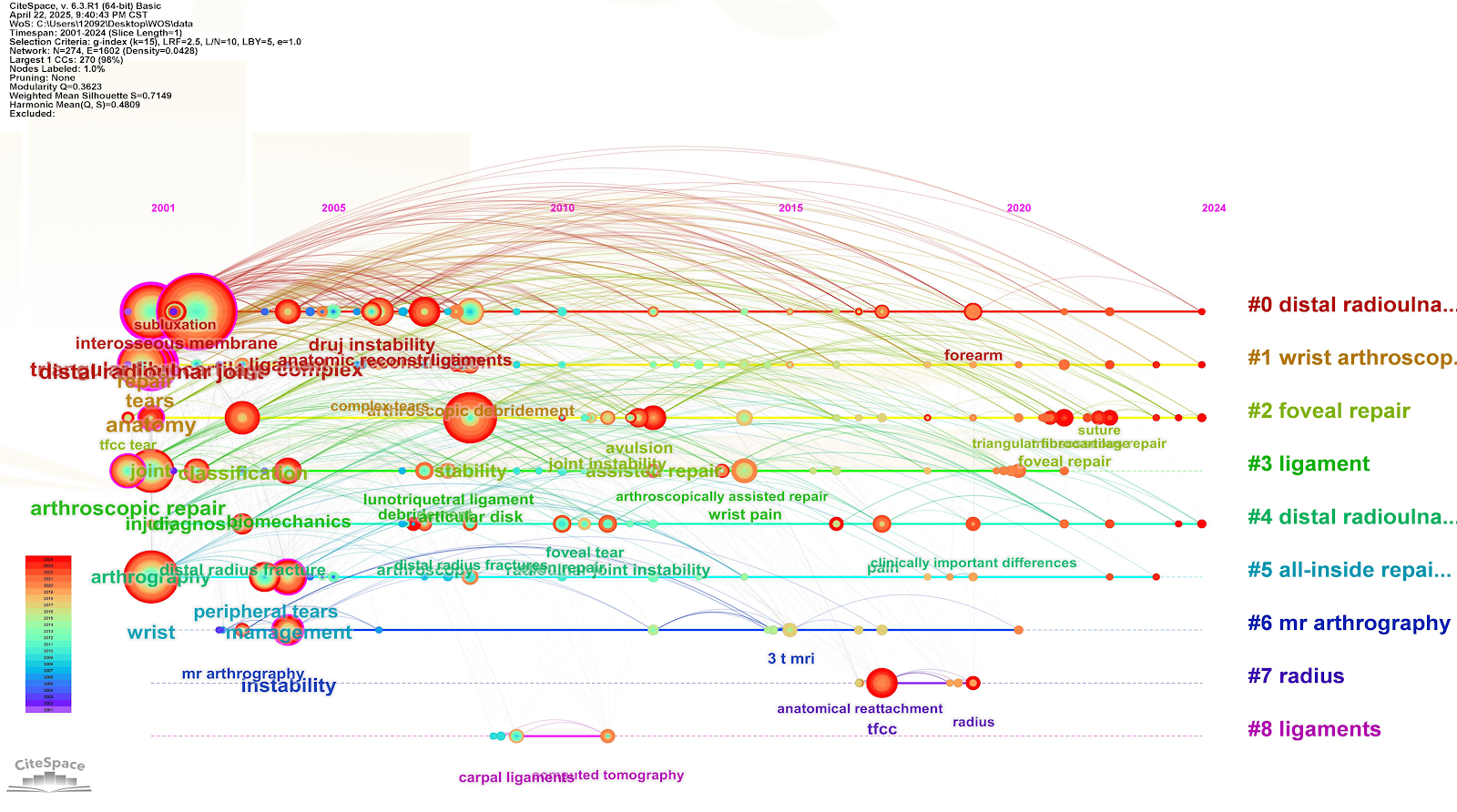
BACKGROUND: As a common condition of the musculoskeletal system, triangular fibrocartilage complex injury necessitates effective treatment strategies to improve patients' quality of life and functional recovery. However, there is a lack of systematic integration of global research trends and core hotspots. OBJECTIVE: To conduct a bibliometric analysis of the literature on triangular fibrocartilage complex injury treatment, aiming to explore the current research status, hotspots, and trends in this field. METHODS: Relevant literature in the field of triangular fibrocartilage complex injury treatment published between 2001 and 2024 was retrieved from the Web of Science Core Collection. Citespace software was utilized for visual analysis of countries, authors, institutions, co-cited references, and keywords. RESULTS AND CONCLUSION: (1) After screening, 352 articles were included, with an increasing trend in the annual publication volume. The United States was the leading contributing country, and Mayo Clinic (USA) was the most prolific institution. (2) The top five most frequent keywords were triangular fibrocartilage complex, wrist, distal radioulnar joint, anatomy, and tear, with 'anatomy' having the highest centrality. (3) Temporal analysis revealed that early research focused on anatomical foundations and diagnostic methods for wrist injuries; from 2010 onward, the focus shifted to complications and long-term outcomes after arthroscopic surgery. In recent years, research has increasingly emphasized precision, standardization, and interdisciplinary integration. (4) Research on triangular fibrocartilage complex injury treatment has entered an innovative phase of multidisciplinary convergence, with vigorous development in areas such as precision anatomy, intelligent diagnosis and treatment, and biomaterials.

Read Full Abstract10.12307/2026.21507
In vitro osteogenic and anti-inflammatory properties of icariin sustained-release microsphere three-dimensional scaffoldsGraphical AbstractVerified
Chinese Journal of Tissue Engineering Research2026

In vitro osteogenic and anti-inflammatory properties of icariin sustained-release microsphere three-dimensional scaffolds

BACKGROUND: Icariin has the dual activity of promoting bone formation and inhibiting bone resorption, but its clinical application is plagued by low bioavailability, difficulty in controlling dosage, and a high risk of adverse reactions. OBJECTIVE: To prepare a three-dimensional scaffold containing icariin sustained-release microspheres and characterize their osteogenic activity in vitro. METHODS: A silk fibroin/chitosan/nanohydroxyapatite three-dimensional scaffold (SF/CS/nHA scaffold), icariin sustained-release microspheres, and a silk fibroin/chitosan/nanohydroxyapatite three-dimensional scaffold loaded with icariin sustained-release microspheres (SF/CS/nHA-ICA scaffold) were prepared. The drug loading efficiency, encapsulation efficiency, and in vitro drug release of the icariin sustained-release microspheres were characterized. The pore size, porosity, water absorption expansion rate, and hot water dissolution rate of the two scaffolds were measured. Rabbit bone marrow mesenchymal stem cells (or human rheumatoid arthritis fibroblast-like synoviocytes) were seeded on SF/CS/nHA and SF/CS/nHA-ICA scaffolds, with cells cultured alone as controls. Cell adhesion was observed by scanning electron microscopy. Cell proliferation and viability were assessed by CCK-8 assay, live/dead staining, and F-actin staining. The mRNA and protein expression of Runx-2, osteocalcin, and type I collagen in bone marrow mesenchymal stem cells were detected by RT-qPCR and western blot. RESULTS AND CONCLUSION: (1) The drug loading efficiency and encapsulation efficiency of icariin sustained-release microspheres were (29.38±0.04)% and (52.01±0.09)%, respectively, and the microspheres could sustainably release icariin for more than 90 days in vitro. (2) Scanning electron microscopy showed a honeycomb-like porous structure with interconnected pores in both scaffolds. There were no significant differences in pore size, porosity, water absorption expansion rate, or total hot water dissolution rate between the two groups (P > 0.05). (3) Scanning electron microscopy showed that both cell types adhered tightly to the scaffold surface and pores, with more extended pseudopodia on the SF/CS/nHA-ICA scaffold. CCK-8 assay, live/dead staining, and F-actin staining showed that compared with the control and SF/CS/nHA groups, the SF/CS/nHA-ICA scaffold promoted the proliferation and viability of rabbit bone marrow mesenchymal stem cells, while inhibiting the proliferation and viability of human rheumatoid arthritis fibroblast-like synoviocytes. (4) RT-qPCR and western blot showed that compared with the control and SF/CS/nHA groups, the mRNA and protein expression of Runx-2, osteocalcin, and type I collagen were increased in the SF/CS/nHA-ICA group (P < 0.05). (5) These results indicate that the icariin sustained-release microsphere three-dimensional scaffold has good cytocompatibility, and in vitro osteogenic and anti-inflammatory effects.

Read Full Abstract10.12307/2026.21444
Global analysis and hotspot exploration of acne scarring: a bibliometric visualization approachGraphical AbstractVerified
Chinese Journal of Tissue Engineering Research2026

Global analysis and hotspot exploration of acne scarring: a bibliometric visualization approach

BACKGROUND: As societal attention to acne scarring has increased, the number of emerging studies in this field has risen markedly. However, traditional reviews and meta-analyses are limited in their ability to accurately predict future development trends. Therefore, bibliometric methods are needed to clarify the research landscape and trajectory within this domain. OBJECTIVE: To analyze the research status and future trends of acne scars from 2000 to 2025 using bibliometric methods. METHODS: The relevant papers and reviews on the research of acne scars from 2000 to 2025 were retrieved using the Web of Science Core Collection. And Excel, CiteSpace, VOSviewer and BibliomeTools (R-Tool of R-Studio) were used to analyze the citation frequency, countries/institutions, authors/co-cited authors, journals/co-cited journals, keywords and co-cited references. RESULTS AND CONCLUSION: From January 1, 2000 to February 28, 2025, both the annual publication volume and the number of citations in the field of acne scars showed an upward trend. Among them, the annual publication volume reached a peak of 114 articles in 2022; the number of citations reached the highest value of 3 933 times in 2024. The analysis of countries/regions shows that the United States is in a leading position globally in this field, with the highest publication volume and citation count. French author Dreno, Brigitte and American author Alster, Ts rank first in publication volume and co-citation count, respectively, and are authoritative authors in this field. The institution with the highest number of publications is Tehran University of Medical Sciences in Iran, while more than half of the top-cited institutions are from the United States, represented by Harvard University. Dermatologic Surgery published the most articles in this field and is also the most co-cited journal. The most common keyword is 'acne scar', appearing 275 times. Keywords such as 'microneedling', 'atrophic scar', and 'subcision' have received increasing attention in recent years. The most cited article is 'Acne vulgaris' by Williams, HC et al., with 814 citations as of February 28, 2025. This bibliometric analysis reveals that the treatment of acne scars, especially laser therapy, has been a research hotspot and will continue to be a future development trend.

Read Full Abstract10.12307/2026.21508
Correlation between skeletal muscle mass and Chinese healthy eating index in older adultsGraphical AbstractVerified
Chinese Journal of Tissue Engineering Research2026

Correlation between skeletal muscle mass and Chinese healthy eating index in older adults

BACKGROUND: With the deepening of China's aging population, low skeletal muscle mass has become a serious public health problem. Diet is closely linked to muscle mass; however, existing research has mostly focused on the effects of a single nutrient, and there is still a lack of systematic exploration of the relationship between China's overall dietary pattern and skeletal muscle quality. OBJECTIVE: To analyze the correlation between Chinese Healthy Eating Index and skeletal muscle mass in older adults, providing scientific basis for dietary optimization and prevention of low skeletal muscle mass. METHODS: Based on the 2018 China Longitudinal Healthy Longevity Survey (CLHLS), 8,114 participants were included. Dietary quality was assessed using the Chinese Healthy Eating Index, and skeletal muscle mass was evaluated using the skeletal muscle mass index. Low skeletal muscle mass was diagnosed according to the 2019 Asian Working Group for Sarcopenia criteria. Multivariate logistic regression was used to explore the association between Chinese Healthy Eating Index and low skeletal muscle mass. RESULTS AND CONCLUSION: (1) Univariate analysis showed that the highest quartile (Q4) of overall Chinese Healthy Eating Index (OR=0.52, 95%CI: 0.46-0.59), animal-based Chinese Healthy Eating Index (OR=0.85, 95%CI: 0.75-0.97), and plant-based Chinese Healthy Eating Index (OR=0.43, 95%CI: 0.38-0.49) were significantly associated with low skeletal muscle mass (P < 0.001). (2) Multivariate logistic regression showed that compared with the lowest quartile (Q1), the highest quartile (Q4) of overall, animal-based, and plant-based Chinese Healthy Eating Index were associated with 30% (OR=0.70, 95%CI: 0.60-0.83, P < 0.05), 20% (OR=0.80, 95%CI: 0.68-0.94, P < 0.05), and 20% (OR=0.80, 95%CI: 0.69-0.94, P < 0.05) lower risk of low skeletal muscle mass, respectively. Trend tests further indicated a significant association between higher dietary quality and lower risk of low skeletal muscle mass (P_trend < 0.001). (3) Subgroup analysis showed that in female participants, compared with Q1, the highest quartile (Q4) of overall, animal-based, and plant-based Chinese Healthy Eating Index were associated with 43% (OR=0.57, 95%CI: 0.45-0.72, P < 0.05), 29% (OR=0.71, 95%CI: 0.56-0.88, P < 0.001), and 33% (OR=0.67, 95%CI: 0.54-0.83, P < 0.001) lower risk of low skeletal muscle mass, respectively. No significant association was found in males. The results indicate that higher dietary quality is significantly associated with lower risk of low skeletal muscle mass, and adopting a healthy diet is an effective intervention to prevent skeletal muscle loss.

Read Full Abstract10.12307/2026.21504
Different doses of aquatic exercise for improving muscle strength in older adults: a meta-analysisGraphical AbstractVerified
Chinese Journal of Tissue Engineering Research2026

Different doses of aquatic exercise for improving muscle strength in older adults: a meta-analysis

OBJECTIVE: To systematically evaluate and quantify the effects of different doses of aquatic exercise on muscle strength in healthy older adults using a Bayesian model-based dose-response meta-analysis, thereby determining the optimal exercise regimen and providing evidence-based recommendations for precise exercise prescription. METHODS: A systematic search was conducted in both English and Chinese databases, including PubMed, Embase, Web of Science, CNKI, and WanFang, to identify randomized controlled trials published up to March 2025 that investigated the effects of aquatic exercise on muscle strength in older adults. Data analysis was performed using R 4.5.0. A conventional meta-analysis was first conducted to estimate the overall effect, followed by a Bayesian model-based dose-response meta-analysis to quantify the nonlinear relationships between different exercise dose dimensions (frequency, session duration, weekly total duration, period, and intensity) and muscle strength improvement. The standardized mean difference (SMD) with 95% confidence interval (CI) was used as the effect size. RESULTS: A total of 13 randomized controlled trials involving 531 participants were included. The overall meta-analysis showed that aquatic exercise significantly improved muscle strength in older adults compared with control (SMD=0.56, 95%CI 0.39-0.74, P < 0.0001). Dose-response analysis revealed cumulative trends for training period, frequency, and weekly total duration: the effect peaked at 24 weeks (SMD=0.65, 95%CI 0.40-0.66); significant gains were achieved at a frequency of twice per week (SMD=0.56, 95%CI 0.22-0.58), with a slight increase at three times (SMD=0.62, 95%CI 0.24-0.62); weekly cumulative duration showed significant effects at 100 minutes (SMD=0.58, 95%CI 0.34-0.60) and plateaued after 200 minutes. In contrast, session duration and intensity exhibited an inverted U-shaped relationship: the effect peaked at 40 minutes per session (SMD=0.62, 95%CI 0.32-0.82), with an optimal range of 30-45 minutes; the optimal intensity was Borg RPE 10-12 (SMD=0.45, 95%CI 0.18-0.46), with diminishing returns beyond this range. CONCLUSION: Aquatic exercise is an effective strategy for improving muscle strength in healthy older adults. It is recommended that older adults engage in aquatic exercise two to three times per week, with each session lasting approximately 40 minutes, at a moderate-to-vigorous intensity (Borg RPE 10-12), and as a long-term strategy.

Read Full Abstract10.12307/2026.21503
Future medical research on brain organoids: interdisciplinary training, bioengineering technologies, and optimized model maturityGraphical AbstractVerified
Chinese Journal of Tissue Engineering Research2026

Future medical research on brain organoids: interdisciplinary training, bioengineering technologies, and optimized model maturity

BACKGROUND: In recent years, brain organoids have rapidly emerged as a hotspot in neuroscience research; however, no comprehensive analysis of knowledge graphs related to medical research on brain organoid has been conducted. OBJECTIVE: To conduct a bibliometric analysis of brain organoid medical research and systematically collate research hotspots, emerging trends, and developmental trajectories in this field, thereby providing scholars with a rapid and systematic overview of the current landscape. METHODS: Relevant publications on brain organoids from January 1, 2014 to December 31, 2024 were retrieved and analyzed using the Web of Science Core Collection. CiteSpace, Microsoft Office, and Origin software were used to systematically analyze publication volume, country distribution, institutional contributions, authors, highly cited references, and keyword hotspots. RESULTS AND CONCLUSION: The number of publications in the field of brain organoid medical research has shown a continuous rapid growth trend, with the United States leading in publication volume and influence. The University of California system is the highest-producing institution, and most institutions have frequent cooperation and exchange, forming a broad cooperation network. Research teams are closely connected internally, but cooperation between teams is limited. The most prolific author is Knoblich J. After 2021, research hotspots entered a period of concentrated outbreak. Long-term research hotspots include pluripotent stem cells, in vitro models, and culture. Recent hotspot research directions include choroid plexus, human cortical organoids, and microglia. The results indicate that the field of brain organoid medical research has entered a new stage of rapid development in recent years, with high thematic diversity. In vitro three-dimensional model culture has always been a research hotspot. Future research will focus on interdisciplinary integration of brain organoid culture and bioengineering technology to optimize model maturity, providing a precise in vitro research platform for regenerative medicine, brain disease treatment, and exploration of neurological mechanisms.

Read Full Abstract10.12307/2026.21506
Medication patterns for traditional Chinese medicine in children with cerebral palsy: an analysis based on medical records and literatureGraphical AbstractVerified
Chinese Journal of Tissue Engineering Research2026

Medication patterns for traditional Chinese medicine in children with cerebral palsy: an analysis based on medical records and literature

BACKGROUND: The Affiliated Hospital of Jiangxi University of Chinese Medicine has used traditional Chinese medicine (TCM) to treat children with cerebral palsy (CP) for over 20 years, but no analysis of medication patterns has been conducted. OBJECTIVE: To analyze TCM syndrome types and explore medication patterns for CP in children based on medical records and literature. METHODS: An evidence-based retrieval strategy was used to search and manage literature and medical records on TCM treatment for CP in children. Bibliometric methods were applied to mine and analyze data characteristics. VOSviewer software was used to create visual knowledge maps. IBM SPSS Modeler software was used for association rule analysis of TCM drugs. Radar chart method was used to analyze the four natures and five flavors of drugs. RESULTS AND CONCLUSION: A total of 503 medical records and 90 articles were included. Syndrome analysis showed that the main TCM syndrome type of CP in children was liver-kidney deficiency. Intervention analysis showed that external therapy was most frequently used, and among oral medications, drugs for nourishing liver and kidney were most common. Medication pattern analysis showed that among the top 20 drugs by frequency in both medical records and literature, 12 (60%) were the same. The drug pairs Shanyao (Rhizoma Dioscoreae) and Fuling (Poria), and Shudi (Radix Rehmanniae Preparata) and Fuling (Poria) had high support and confidence above 82.50%, indicating significant association. Among all TCM drugs for CP in children, the nature was mainly warm, followed by neutral and cold; the flavor was mainly sweet, followed by bitter and pungent; the meridian tropism was mainly liver and kidney meridians, followed by spleen, heart, and lung meridians. Most drugs in these formulas were non-toxic. The results indicate that CP in children is mainly characterized by liver-kidney deficiency, and the formulas used in clinical practice and related clinical research are mostly for nourishing liver and kidney, among which Liuwei Dihuang Pill and its derivative formulas appear most frequently. The drug pairs with the highest frequency and reliability are Shudi and Fuling, and Shanyao and Fuling. The nature and flavor of drugs are mainly warm, sweet, and attributed to liver and kidney meridians.

Read Full Abstract10.12307/2026.21505
Synovial fluid exosome-mediated crosstalk between synoviocytes and chondrocytes in development and progression of knee osteoarthritisGraphical AbstractVerified
Chinese Journal of Tissue Engineering Research2026

Synovial fluid exosome-mediated crosstalk between synoviocytes and chondrocytes in development and progression of knee osteoarthritis

BACKGROUND: Increasing evidence has demonstrated the crucial role of synovial cell-chondrocyte signaling crosstalk mediated by synovial fluid exosomes in knee osteoarthritis. The signaling interaction between synovial cells and chondrocytes reveals, at the molecular level, the bidirectional regulatory role of synovial fluid exosomes in the progression of knee osteoarthritis. OBJECTIVE: To comprehensively interpret the interactions between synoviocytes and chondrocytes mediated by synovial fluid exosomes from multiple perspectives and levels to provide new insights and directions for research and clinical applications in the related field. METHODS: A literature search was conducted in PubMed for publications from January 2001 to June 2025 using the keywords “osteoarthritis*, exosomes, synovial cells, chondrocytes.” All relevant original studies, reviews, and clinical trials published from January 2001 to June 2025 were included, while irrelevant mechanisms and low-relevance studies were excluded. Finally, 93 articles were selected for comprehensive analysis. RESULTS AND CONCLUSION: During the development and progression of knee osteoarthritis, synovial cells and chondrocytes establish close signal communication via synovial fluid exosomes. This signaling interaction significantly affects the progression of knee osteoarthritis, potentially exacerbating cartilage degeneration and synovial inflammation, while also exerting protective regulatory effects under specific conditions. Therefore, the dual nature of this mechanism highlights the necessity for precise intervention, and targeting key molecules in exosome-mediated intercellular signaling pathways may offer new therapeutic strategies and targets for knee osteoarthritis.

Read Full Abstract10.12307/2026.21499
Mitochondrial kinetic mechanism by which triptolide alleviates hydrogen peroxide-induced apoptosis in SH-SY5Y cellsGraphical AbstractVerified
Chinese Journal of Tissue Engineering Research2026

Mitochondrial kinetic mechanism by which triptolide alleviates hydrogen peroxide-induced apoptosis in SH-SY5Y cells

BACKGROUND: Previous studies from our group have shown that triptolide exerts protective effects on nerve cells and alleviates symptoms of neurodegenerative diseases. However, whether it acts by improving mitochondrial dynamic abnormalities requires further investigation. OBJECTIVE: To explore the effect and mechanism of triptolide in regulating the mitochondrial fusion-fission balance to mitigate hydrogen peroxide (H₂O₂)-induced apoptosis in SH-SY5Y cells. METHODS: Human neuroblastoma SH-SY5Y cells were cultured and divided into three groups: control group, model group (200 μmol/L H₂O₂), and triptolide group (2.5 nmol/L triptolide + 200 μmol/L H₂O₂). After 24 hours of intervention, oxidative stress markers (superoxide dismutase activity and malondialdehyde levels), mitochondrial membrane potential, and apoptosis levels were measured. Western blot was used to detect the expression of apoptosis-related proteins, mitochondrial dynamics-related proteins, and respiratory chain-related proteins. Immunofluorescence staining was used to detect the expression of phosphorylated dynamin-related protein 1, optic atrophy protein 1, cytochrome C oxidase 1, and ATP synthase F1 subunit alpha. RESULTS AND CONCLUSION: Compared with the control group, the model group showed significantly decreased superoxide dismutase activity, mitochondrial membrane potential, anti-apoptotic protein Bcl-2, mitochondrial fusion proteins 1 and 2, optic atrophy protein 1, and oxidative phosphorylation complex proteins (NADH dehydrogenase [ubiquinone] iron-sulfur protein 8, ubiquinol-cytochrome c reductase core protein 2, cytochrome c oxidase 1, succinate dehydrogenase B, ATP synthase F1 subunit alpha) (P < 0.05). Meanwhile, malondialdehyde levels, pro-apoptotic proteins Bax and Caspase-3, mitochondrial fission protein 1, phosphorylated dynamin-related protein 1 expression, and apoptosis rate were significantly increased (P < 0.05). Compared with the model group, triptolide intervention reduced malondialdehyde levels, increased superoxide dismutase activity and mitochondrial membrane potential, promoted fusion protein expression, inhibited fission protein expression, increased oxidative phosphorylation complex protein levels, and decreased apoptosis rate (P < 0.05). These results confirm that triptolide can regulate mitochondrial dynamic imbalance to alleviate H₂O₂-induced apoptosis in SH-SY5Y cells.

Read Full Abstract10.12307/2026.21492
Key role of m6A methylation in sarcopeniaGraphical AbstractVerified
Chinese Journal of Tissue Engineering Research2026

Key role of m6A methylation in sarcopenia

BACKGROUND: The role of epigenetic regulatory mechanisms, especially N6-methyladenine (m6A) RNA modification, in muscle cell proliferation, differentiation, and disease development is increasingly being studied. However, the multidimensional mechanism of m6A methylation in sarcopenia still needs to be systematically integrated. OBJECTIVE: To explore the key role of m6A methylation in the occurrence and development of sarcopenia, and to review the latest research progress on the involvement of m6A related regulatory factors in the pathological process of sarcopenia. METHODS: Using "m6A methylation, N6 methyladenine, sarcopenia, muscle atrophy, muscle regeneration, muscle, skeletal muscle" as Chinese keywords, and "m6A RNA methylation, sarcopenia, skeletal muscle, muscle mass loss" as English keywords, CNKI and PubMed were searched to screen high-quality literature in recent years, and the mechanism of action and related signaling pathways of m6A methylation in sarcopenia were summarized. RESULTS AND CONCLUSION: m6A methylation participates in the pathological process of sarcopenia through a dynamic and reversible regulatory network (methyltransferases METTL3/METTL14, demethylases FTO/ALKBH5, reader proteins YTHDF1/YTHDF2, etc.). m6A methylation affects the proliferation and differentiation of skeletal muscle cells by regulating satellite cells, ubiquitin-proteasome system, non-coding RNAs, etc. Most studies are based on cell models or animal experiments, with few clinical sample validation and translational application studies. The feasibility of m6A-related factors as diagnostic markers or therapeutic targets for sarcopenia needs further verification.

Read Full Abstract10.12307/2026.21500
Eight-week aerobic exercise and aerobic exercise combined with full-body vibration training improve the microcirculation function of obese college studentsGraphical AbstractVerified
Chinese Journal of Tissue Engineering Research2026

Eight-week aerobic exercise and aerobic exercise combined with full-body vibration training improve the microcirculation function of obese college students

BACKGROUND: The occurrence and development of obesity are closely related to microcirculation dysfunction. Improving microcirculation dysfunction is of great significance to obese people. Studies have shown that both aerobic exercise and full-body vibration training can improve microcirculation dysfunction. However, most existing research focuses on single exercise modalities, with insufficient exploration of combined intervention studies. OBJECTIVE: To compare the effects of 8-week aerobic exercise and aerobic exercise combined with full-body vibration training on the microcirculation function of obese college students. METHODS: Sixty obese college students (with a body fat percentage of ≥ 28% for females and ≥ 20% for males) were recruited and divided into three groups using a matched grouping method: control group (n=20) did not undergo systematic exercise training; aerobic exercise group (n=20) performed 8 weeks of aerobic jogging (4 times per week, 60 min each); combined exercise group (n=20) performed 8 weeks of aerobic exercise combined with full-body vibration training [4 times per week, 60 min each (51 min aerobic jogging + 9 min full-body vibration training)]. Before and after the exercise intervention, microvascular reactivity, transcutaneous oxygen pressure, muscle oxygen saturation, and serum levels of nitric oxide, endothelial nitric oxide synthase, and endothelin-1 were measured in all three groups. RESULTS AND CONCLUSION: (1) After the intervention, the heated values of microvascular blood perfusion, blood cell concentration, blood cell velocity, transcutaneous oxygen pressure, and muscle oxygen saturation in the aerobic exercise and combined exercise groups were higher than those in the control group (P < 0.05). The combined exercise group showed higher values than the aerobic exercise group (P < 0.05). In the combined exercise group, the baseline values of microvascular blood perfusion and blood cell velocity after intervention were higher than before (P < 0.01). In both exercise groups, the heated values of microvascular blood perfusion, blood cell concentration, blood cell velocity, transcutaneous oxygen pressure, and muscle oxygen saturation were higher after intervention than before (P < 0.05, P < 0.01). (2) After the intervention, serum levels of nitric oxide, endothelial nitric oxide synthase, and nitric oxide/endothelin-1 ratio in the aerobic exercise and combined exercise groups were higher than those in the control group (P < 0.05), while serum endothelin-1 levels were lower (P < 0.05). The combined exercise group had higher nitric oxide, endothelial nitric oxide synthase, and nitric oxide/endothelin-1 ratio than the aerobic exercise group (P < 0.05), and lower endothelin-1 levels (P < 0.05). In both exercise groups, serum nitric oxide, endothelial nitric oxide synthase, and nitric oxide/endothelin-1 ratio increased after intervention (P < 0.05, P < 0.01), while endothelin-1 decreased (P < 0.01). (3) These results indicate that 8 weeks of aerobic exercise and aerobic exercise combined with full-body vibration training can improve microcirculation function in obese subjects, with the combined intervention being more effective. Promoting endogenous nitric oxide release and increasing the nitric oxide/endothelin-1 ratio may be the potential mechanism by which exercise improves microcirculation function.

Read Full Abstract10.12307/2026.21493
Regional specificity of brain organoids and their application in ischemic stroke modeling and drug developmentGraphical AbstractVerified
Chinese Journal of Tissue Engineering Research2026

Regional specificity of brain organoids and their application in ischemic stroke modeling and drug development

BACKGROUND: The construction of brain organoid technology and its application in ischemic stroke are new research hotspots in recent years. The anatomy and functional organization of the brain of rodents are significantly different from those of the human brain, which determines that they cannot fully mimic the physiological, pathological and anatomical characteristics of the human brain. At the same time, there are ethical issues in obtaining brain tissue samples from patients in clinical practice, so the importance of brain organoids in ischemic stroke research lies in their ability to replace clinical brain tissue and animal models to construct in vitro research models that are more closely related to human brain tissue, and they are of great value in disease mechanism modeling and drug development. OBJECTIVE: To review and summarize the current research on regional specificity of brain organoids, and to provide new technical means and research strategies for the application of brain organoids in ischemic stroke modeling and drug development. METHODS: Literature search on organoids and ischemic stroke was conducted in CNKI and PubMed databases, with the search time limit from database inception to May 2025. Chinese search terms were "ischemic stroke, stroke, organoids, ischemia-reperfusion injury, oxygen-glucose deprivation/reoxygenation, forebrain organoids, hippocampal organoids, thalamic organoids, midbrain organoids", and English search terms were "ischemic stroke, stroke, organoids, oxygen-glucose deprivation/reoxygenation, middle cerebral artery occlusion, forebrain organoids, hippocampal organoids, thalamic organoids, midbrain organoids". All retrieved literature included original research and related reviews, and finally 98 articles were screened for analysis and summary. RESULTS AND CONCLUSION: (1) Dorsal forebrain organoids mimic cortical neuroepithelium and differentiate into neural cells such as astrocytes, excitatory neurons, and oligodendrocytes; ventral forebrain organoids mimic ganglionic eminences and develop into basal ganglia and striatum, exhibiting extensive neuronal migration and maintaining GABAergic properties. (2) Human induced pluripotent stem cell-derived hippocampal organoids can integrate functionally with mouse hippocampal neural circuits, receive excitatory input, and form synaptic-connected neuronal networks. (3) Thalamic/hypothalamic organoids mimic the development and cellular diversity of the human hypothalamic arcuate nucleus and explore dopaminergic neuron specificity. (4) Midbrain organoids are widely used in modeling Parkinson's disease, gene editing, and drug development. (5) Oxygen-glucose deprivation can induce brain organoids to simulate ischemic stroke in vitro, and then participate in neural injury through apoptosis, necroptosis, autophagy, and ferroptosis. The gene expression profile of brain organoids after oxygen-glucose deprivation changes significantly. Application of neuroprotective drugs such as carnosic acid or transplantation of brain organoids into rats with middle cerebral artery occlusion can improve neurological deficits and reduce infarct volume, playing an important role in transplantation regenerative medicine.

Read Full Abstract10.12307/2026.21495
Role and mechanism of exercise-regulated miRNAs in cardiac remodeling after acute myocardial infarctionGraphical AbstractVerified
Chinese Journal of Tissue Engineering Research2026

Role and mechanism of exercise-regulated miRNAs in cardiac remodeling after acute myocardial infarction

BACKGROUND: The pathological manifestations of acute myocardial infarction primarily include cardiomyocyte apoptosis, fibrosis, impaired angiogenesis, calcium dyshomeostasis, and cardiac hypertrophy. MicroRNAs (miRNAs) are key regulators of gene expression and play significant roles in the progression of acute myocardial infarction. Exercise has been shown to improve cardiac function after acute myocardial infarction by modulating miRNA expression, although the underlying mechanisms remain incompletely understood. OBJECTIVE: To summarize the roles of miRNAs in the pathological features of acute myocardial infarction and discuss the molecular mechanisms through which exercise regulates miRNAs to treat acute myocardial infarction, thereby providing a theoretical reference for precise therapeutic interventions. METHODS: Literature published between 2000 and 2025 was retrieved from databases such as PubMed, Web of Science, CNKI, and Wanfang. Chinese search terms included 'acute myocardial infarction, cardiac remodeling, microRNA, myocardial cell apoptosis, myocardial fibrosis, angiogenesis, calcium homeostasis, cardiac hypertrophy, exercise'; English search terms included 'Acute Myocardial Infarction, Cardiac remodeling, MicroRNAs, Myocardial cell apoptosis, Myocardial fibrosis, Angiogenesis, Calcium homeostasis, Cardiac hypertrophy, Exercise'. A total of 92 articles were included for analysis. RESULTS AND CONCLUSION: miRNAs (miR-1, miR-133a, miR-21, miR-29 family) participate in cardiac remodeling after acute myocardial infarction by regulating signaling pathways related to apoptosis, fibrosis, angiogenesis, and calcium homeostasis. Exercise improves cardiac function after acute myocardial infarction by upregulating miR-126 to promote angiogenesis, upregulating miR-29 to inhibit myocardial fibrosis, upregulating miR-214-3p to restore calcium homeostasis, and upregulating miR-222 to promote physiological cardiac hypertrophy. Combination of exercise with drugs (e.g., trifluoromethoxyphenyl-3-(1-propionylpiperidin-4-yl)urea) or nutrients (e.g., vitamin D3) can modulate the therapeutic effects of miRNAs. Future research should further investigate the spatiotemporal differences and inter-family differences of miRNAs to develop precise targeted intervention strategies; formulate precise exercise intervention plans based on miRNA expression profiles, combined with artificial intelligence for efficacy prediction and evaluation; and explore the combination of exercise intervention with gene editing or exosome delivery technologies to precisely regulate miRNA expression, thereby promoting cardiac repair and functional recovery.

Read Full Abstract10.12307/2026.21498
Correlation between body mass index and early recovery outcomes after kidney transplantationGraphical AbstractVerified
Chinese Journal of Tissue Engineering Research2026

Correlation between body mass index and early recovery outcomes after kidney transplantation

BACKGROUND: The proportion of obesity in the Chinese population has increased year by year, but its impact on early renal function recovery after kidney transplantation has not been systematically elucidated, and localized research is urgently needed to provide evidence-based basis. OBJECTIVE: To investigate the effect of different body mass indexes on early renal function recovery in renal transplant recipients after surgery, to analyze the association between body mass index and postoperative creatinine, urine volume and hospital stay, and to propose clinical recommendations for individualized management. METHODS: The clinical data of 495 renal transplant recipients in the Second Xiangya Hospital of Central South University from 2019 to 2020 were retrospectively analyzed. According to the Chinese obesity standard, they were divided into a lean group (body mass index < 18.5 kg/m2, n=78), a normal group (18.5 ≤ body mass index < 24 kg/m2, n=273), an overweight group (24 kg/m2 ≤ body mass index < 28 kg/m2, n=119), and an obese group (body mass index ≥ 28 kg/m2, n=25). The baseline characteristics, postoperative creatinine, urine volume, and hospital stay were compared among the groups. Multivariate regression analysis was used to analyze the independent effect of body mass index on postoperative recovery. RESULTS AND CONCLUSION: (1) The age of the obese group was significantly higher than that of other groups (P < 0.001), the proportion of living donors (0%) was the lowest (P=0.015), the creatinine level on postoperative day 1 and hospital stay were the highest (P < 0.001), and urine volume was the lowest (P < 0.001); (2) Multivariate regression showed that for each 1 kg/m2 increase in body mass index, creatinine on postoperative day 1 increased by 38 μmol/L (β=0.38, P < 0.001), and urine volume decreased by 32 mL (β=-0.41, P < 0.001); (3) It is suggested that elevated body mass index is an independent risk factor for slow early renal function recovery and prolonged hospital stay after kidney transplantation; based on the Chinese obesity standard (body mass index ≥ 28 kg/m2), it is recommended to strengthen perioperative monitoring, optimize immunosuppressive regimens, and formulate individualized fluid management strategies for obese recipients to improve prognosis. This study can provide evidence-based basis for localized body mass index stratified management.

Read Full Abstract10.12307/2026.21494
Molecular mechanisms and therapeutic targets of mechanical stress regulating osteoarthritisGraphical AbstractVerified
Chinese Journal of Tissue Engineering Research2026

Molecular mechanisms and therapeutic targets of mechanical stress regulating osteoarthritis

BACKGROUND: Piezo-type mechanosensitive ion channel components (PIEZO) play a crucial role in cartilage degeneration, inflammation, and pain in osteoarthritis by sensing mechanical stimulation and regulating calcium signaling, potentially serving as an important therapeutic target for osteoarthritis. OBJECTIVE: To systematically review the role of PIEZO ion channels in the pathological mechanisms of osteoarthritis and evaluate their potential as a novel therapeutic target. METHODS: The first author searched CNKI and PubMed databases using Chinese and English search terms including "mechanical stress, Piezo, osteoarthritis, chondrocyte, osteoblast, osteoclast, synovial cell, immune cell" and "Piezo1, osteoarthritis, chondrocyte, osteoblast, osteoclast, synovial membrane, immune cell, GsMTx4" respectively. Literature published from 2000 to 2025 was selected, and 102 articles were finally included for review. RESULTS AND CONCLUSION: Mechanical stress plays a central role in the degeneration of articular cartilage and surrounding tissues. Chronic excessive mechanical stress or unbalanced loading causes chondrocyte damage, apoptosis, and inflammatory responses, thereby accelerating osteoarthritis progression. Known mechanosensors include transient receptor potential channel family, two-pore domain potassium channel family, degenerin/epithelial sodium channel family, and integrin family. PIEZO family is the first group of mechanosensitive cation channel pore proteins discovered in mammalian cells, widely present in human cells, sensing changes in ambient pressure to control Ca2+ influx and thus affect cellular functions. PIEZO ion channels regulate Ca2+ influx by sensing mechanical stimulation of the cell membrane, thereby influencing chondrocytes, osteogenesis, synovial cells, immune cells, and pain perception. Inhibiting PIEZO ion channels may become an effective method for treating arthritis.

Read Full Abstract10.12307/2026.21501
The functions and underlying molecular mechanisms of PIEZO channels in nervous system diseasesGraphical AbstractVerified
Chinese Journal of Tissue Engineering Research2026

The functions and underlying molecular mechanisms of PIEZO channels in nervous system diseases

BACKGROUND: Recent studies have demonstrated that mechanotransduction plays a critical role in the pathological processes of neurological disorders. PIEZO channels, as key mechanosensitive ion channels, serve as core mediators in sensing and transducing mechanical signals. However, a systematic review of their specific roles across various neurological diseases is relatively lacking. OBJECTIVE: To explore the roles and molecular mechanisms of PIEZO1 and PIEZO2 channels in central and peripheral nervous system diseases, and to evaluate their potential as therapeutic targets. METHODS: A literature search was conducted in PubMed, Web of Science, CNKI, WanFang, and VIP databases from January 2010 to May 2025. English search terms included 'central nervous system diseases', 'Central Nervous System Disorder', 'CNS Disease', 'CNS Diseases', 'Central Nervous System Disorders', 'neurodegenerative disease', 'Autonomic Nervous System Diseases', 'Brain Diseases', 'Central Nervous System Infections', 'High Pressure Neurological Syndrome', 'Spinal Cord Diseases', 'PIEZO1 Channel', 'PIEZO2 Channel', 'PIEZO Channel'; Chinese search terms included '神经系统疾病', '中枢神经系统疾病', 'PIEZO1', 'PIEZO2', 'PIEZO'. A combination of subject headings and free words was used. Based on inclusion and exclusion criteria, 60 English articles were finally included for systematic analysis and classified by disease type. RESULTS AND CONCLUSION: ①PIEZO1 is highly expressed in glioma and correlates with malignancy and poor prognosis; calcium influx promotes tumor proliferation and remodeling of microenvironment stiffness, driving tumor progression. ②In intracerebral hemorrhage, activation of neuronal PIEZO2 promotes iron transporter expression, increases intracellular iron accumulation, induces ferroptosis, and exacerbates secondary brain injury, while PIEZO1 dysfunction impairs cerebrovascular integrity and the blood-brain barrier. ③In traumatic brain injury, upregulation of PIEZO2 in neurons promotes neuronal death and pro-inflammatory cytokine release. ④Activation of PIEZO1 promotes fluid excretion in the brain, alleviating hydrocephalus. ⑤PIEZO1 dysfunction is involved in amyloid-beta toxicity, glial activation, vascular damage, and metabolic abnormalities in Alzheimer's disease. ⑥PIEZO1 activation inhibits myelination and modulates immune responses in multiple sclerosis. ⑦PIEZO1 mediates pulsatile pain characteristic of migraine induced by blood flow pulsation. ⑧Elevated intraocular pressure upregulates PIEZO1 and PIEZO2, leading to hyperexcitability and metabolic stress injury of retinal ganglion cells. ⑨PIEZO channels regulate neuronal excitability, proprioception, and baroreflex abnormalities in amyotrophic lateral sclerosis, and targeting PIEZO is a potential therapeutic strategy.

Read Full Abstract10.12307/2026.21497