🧬 SinoBioData Academic Portal
🏛️ Indexed Academic JournalOriginal: 中国组织工程研究

Chinese Journal of Tissue Engineering Research

Premier Chinese Biomedical Journal indexed in SinoBioData: Chinese Journal of Tissue Engineering Research (中国组织工程研究).

Total Research Papers: 200
Access: 100% Free Open Access
Browse by Publication Year & VolumeReset All Filters ✕

Published Research PapersFiltered: Year 2026 • Vol 1896

Showing 24 of 200 peer-reviewed papers with full Graphical Abstracts.

Original ResearchVol 1896, Issue 24 • pp. 100-112DOI: 10.12307/2026.21252Jan 15, 2026

Systematic druggable genome-wide Mendelian randomization identifies therapeutic targets for major depressive disorder

Authors: Zhou Menghan, Liu Shuning, Jiang Tao, Sun Zhuangzhuang, Cao Lingling, Su Xin, Yu Cheng, Guo Junpeng

BACKGROUND: The occurrence of major depressive disorder is typically associated with genetic and environmental factors. Currently, the diagnosis of major depressive disorder mainly relies on clinical interviews and symptom assessments, lacking clear and reproducible biological markers. This can lead to misdiagnosis and missed diagnoses, delaying the timing of treatment. OBJECTIVE: To identify druggable genes that may act as potential therapeutic targets for major depressive disorder by conducting comprehensive genome-wide Mendelian randomization analysis. METHODS: By integrating expression quantitative trait locus (eQTL) data and protein quantitative trait locus (pQTL) data from pharmacologically actionable genes with genome-wide association study (GWAS) data on major depressive disorder (including 177 377 cases and 445 321 controls), Mendelian randomization analysis was conducted to identify druggable genes that have a causal relationship with major depressive disorder. Additionally, enrichment analysis, protein-protein interaction network construction, drug target identification, and molecular docking simulations were performed to further explore potential therapeutic strategies. RESULTS AND CONCLUSION: A total of 4 394 druggable genes were analyzed, and 21 druggable genes considerably associated with major depressive disorder were identified. Bayesian colocalization analysis indicated that BTN3A3, CISD1, and PSMB4 had posterior probabilities of hypothesis 4 (H4.abf) > 0.5, supporting the possibility of shared causal variants. GO enrichment analysis mainly involved 'antigen processing and presentation', 'protein degradation and processing', 'mitochondrial outer membrane', and 'immune receptor activity' pathways related to major depression. Protein-protein interaction network analysis showed moderate connectivity among the identified genes (21 nodes, 14 edges). Drug target identification determined gemcitabine (CID 60750), fucose (CID 17106), and isococculidine (CID 2826) as main candidate compounds, which had strong associations with several key genes. Molecular docking analysis revealed stable drug-protein interactions, with isococculidine showing the most stable binding energy (-52.74 kJ/mol) with BTN3A3. In conclusion, Mendelian randomization combined with genomics and structural biology analysis provides valuable decision-making basis for target prioritization and drug repurposing, offering new ideas and directions for efficient utilization of basic research resources and drug development for major depressive disorder.

Systematic druggable genome-wide Mendelian randomization identifies therapeutic targets for major depressive disorder
Graphical Abstract
Original ResearchVol 1896, Issue 24 • pp. 100-112DOI: 10.12307/2026.21243Jan 15, 2026

Virtual reality therapy on neuropathic pain following spinal cord injury

Authors: DU Xinyu, ZHAO Donglin, ZHANG Shuyang, LI Shihao, XING Zheng, CHU Xiaolei, LI Qi

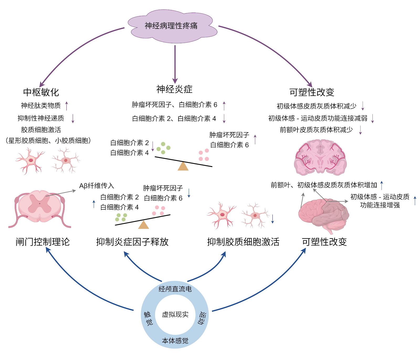
BACKGROUND: Virtual reality has demonstrated unique advantages as a novel non-pharmacological intervention in the treatment of neuropathic pain following spinal cord injury. OBJECTIVE: To systematically review the pathogenesis of neuropathic pain following spinal cord injury, summarize the specific application modes and mechanisms of virtual reality therapy for this condition, and explore key factors influencing treatment efficacy, thereby providing novel therapeutic options for patients with neuropathic pain following spinal cord injury. METHODS: A literature search was conducted in the CNKI, PubMed, Web of Science, and Cochrane Library databases up to June 2025. The Chinese and English search terms included “spinal cord injury, central sensitization, cerebral cortex, noninvasive therapy, neuropathic pain, virtual reality.” A total of 1 352 articles were retrieved, and 73 articles that met the inclusion criteria were ultimately included in the analysis. RESULTS AND CONCLUSION: The development of neuropathic pain following spinal cord injury involves complex mechanisms in both the spinal cord and brain, closely associated with central sensitization at the spinal level and abnormal plasticity changes in the brain. Virtual reality, as a novel intervention approach, can be used alone or in combination with other intervention methods, showing unique therapeutic value for the multidimensional pathogenesis of neuropathic pain after spinal cord injury. Standalone virtual reality applications primarily involve virtual walking interventions aimed at restoring abnormal brain plasticity changes, while combined virtual reality applications have shown certain effects in both inhibiting central sensitization and modulating brain plasticity. Although virtual reality intervention shows great potential in the treatment of neuropathic pain following spinal cord injury, current research and applications still have certain limitations. Future efforts should focus on addressing these issues to fully realize the therapeutic value of virtual reality in neuropathic pain following spinal cord injury.

Virtual reality therapy on neuropathic pain following spinal cord injury
Graphical Abstract
Original ResearchVol 1896, Issue 24 • pp. 100-112DOI: 10.12307/2026.21247Jan 15, 2026

The role of exercise-regulated mitophagy in cardiovascular diseases

Authors: JI Long, GONG Guopan, KONG Xiangkui, JIN Pan, CHEN Ziyang, PU Rui

BACKGROUND: Mitophagy plays a crucial regulatory role in the occurrence and development of cardiovascular diseases. Exercise intervention can mediate mitophagy to improve cardiovascular function, which provides new insights for the clinical prevention and treatment of cardiovascular diseases. OBJECTIVE: To summarize the regulatory role of mitophagy in cardiovascular diseases, the influence of exercise on mitophagy, and the mechanism by which exercise-mediated mitophagy improves cardiovascular diseases. METHODS: PubMed and CNKI databases were searched for relevant literature using the search terms of “mitophagy, mitochondrial function, cardiovascular disease, aerobic exercise, resistance training, combined aerobic resistance exercise, high-intensity interval training” in Chinese and English, respectively. Based on the inclusion and exclusion criteria, totally 88 documents were finally included for summary and analysis. RESULTS AND CONCLUSION: (1) Mitophagy plays a crucial role in the regulation of cardiovascular diseases such as heart failure, myocardial hypertrophy, atherosclerosis, and myocardial ischemia-reperfusion injury. Moreover, mitophagy imbalance or disorder can exacerbate the pathological process of cardiovascular diseases. (2) Various exercise modalities can activate mitophagy by regulating the expression of mitophagy-related factors. Among them, aerobic exercise can promote the formation of autophagosomes, thereby enhancing the regulation of mitophagy; resistance exercise can regulate mitochondrial biogenesis; combined aerobic and resistance exercise can further influence mitophagy by promoting lysosomal biogenesis; high-intensity interval training enhances mitophagy function by regulating the expression of mitochondrial dynamics-related proteins. (3) Exercise regulates mitophagy to alleviate myocardial fibrosis, inhibit cardiomyocyte apoptosis, regulate myocardial oxidative stress, and improve endothelial cell function, thereby playing a key role in the prevention and treatment of cardiovascular diseases, providing a new perspective for exercise promoting health and preventing cardiovascular diseases.

The role of exercise-regulated mitophagy in cardiovascular diseases
Graphical Abstract
Original ResearchVol 1896, Issue 24 • pp. 100-112DOI: 10.12307/2026.21242Jan 15, 2026

Forkhead box transcription factor O3 affects bone metabolism and participates in the pathological processes of various bone-related diseases

Authors: Han Jie, Hu Tianfa, Wu Yachao, Nong Bin, Yu Kailong

BACKGROUND: Bone metabolism disorders can cause the occurrence of bone-related diseases, and forkhead box transcription factor O3 (FoxO3a) can affect the processes of proliferation, differentiation and apoptosis of bone tissue cells by regulating oxidative stress and autophagy levels, and thereby regulate the bone metabolism. OBJECTIVE: To systematically analyze the relevant research literature on the regulation of bone metabolism by FoxO3a and its mechanism of action in bone diseases and to provide a reference for subsequent studies targeting FoxO3a in the treatment of bone diseases. METHODS: Literature searches were conducted using the following strategies: CNKI (China National Knowledge Infrastructure): SU=FoxO3a OR SU=Foxo3 OR SU=Forkhead box O3 OR SU=AND SU=Forkhead box transcription factor O3) AND SU=bone; WanFang Medical Database: Subject:("FoxO3a") OR Subject:("Foxo3") OR Subject:("Forkhead box O3") OR Subject:("Forkhead box transcription factor O3") AND Subject:("bone"); PubMed: ((FoxO3a) OR (Foxo3) OR (Forkhead box O3))AND ((bone) OR (Skeleton)). Outdated, repetitive, low-quality, and irrelevant studies were excluded, and 56 articles were finally included for review. RESULTS AND CONCLUSION: ①FoxO3a and bone marrow mesenchymal stem cells: FoxO3a can promote the formation of osteogenic lineage and promote early osteogenic differentiation by activating autophagy. Meanwhile, FoxO3a exhibits antioxidant properties in bone marrow mesenchymal stem cells, protecting cells from oxidative stress-induced senescence. ②FoxO3a and osteoblasts: FoxO3a can inhibit osteogenesis by interfering with the Wnt/β-catenin pathway in osteoblasts, while it can activate antioxidant enzymes to protect mature osteoblasts. FoxO3a can promote the proliferation of osteoprogenitor cells and promote osteogenic differentiation by activating autophagy. ③FoxO3a and osteoclasts: FoxO3a expression can resist oxidative stress and activate autophagy to inhibit osteoclastogenesis. ④FoxO3a and osteocytes: FoxO3a can protect osteocytes through antioxidant effects, and can also reduce bone loss by inhibiting p16 and p53 signaling pathways and inhibiting senescence-associated secretory phenotype. ⑤FoxO3a and chondrocytes: FoxO3a plays a protective role in chondrocytes in osteoarthritis, inhibiting chondrocyte breakdown or apoptosis, promoting chondrocyte extracellular matrix synthesis, and inhibiting chondrocyte hypertrophy; however, high co-expression of FoxO3a and Runt-related transcription factor 1 in chondrocytes promotes early chondrogenesis and terminal hypertrophy of chondroprogenitor cells. ⑥FoxO3a affects bone metabolism by participating in processes such as oxidative stress resistance and regulation of autophagy, and participates in the pathological processes of various bone-related diseases.

Forkhead box transcription factor O3 affects bone metabolism and participates in the pathological processes of various bone-related diseases
Graphical Abstract
Original ResearchVol 1896, Issue 24 • pp. 100-112DOI: 10.12307/2026.21248Jan 15, 2026

Transcription factor EB improves Alzheimer’s disease via the autophagy-lysosome pathway

Authors: Hu Yalin, Huang Fengqin, Yang Boyin, Luo Xingmei

BACKGROUND: Studies have confirmed that transcription factor EB and its dependent autophagy-lysosome pathway play a role in the development of various neurodegenerative diseases, including Alzheimer’s disease. OBJECTIVE: To summarize the role of transcription factor EB-mediated autophagy-lysosome pathway in Alzheimer’s disease. METHODS: Electronic databases including PubMed, Web of Science, Cochrane Library, CNKI, WanFang Medical Network, and VIP were searched. The search period was from database inception to January 2025. The search terms were “Alzheimer Disease, AD, Transcription Factor EB, TFEB, Autophagy-lysosome Pathway, Autophagy, Lysosomes, Amyloid beta, Aβ, Tau, Tau protein” in English as well as “Alzheimer’s disease, transcription factor EB, autophagic lysosomes, autophagy, lysosomes, β-amyloid, Tau” in Chinese. Additionally, the references of relevant reviews and grey literature were manually supplemented. A total of 100 articles were finally included for review. RESULTS AND CONCLUSION: Amyloid-β deposition and Tau protein phosphorylation are the key pathological features of Alzheimer’s disease. Abnormalities in the autophagy-lysosome pathway promote the formation of neurotoxic protein aggregates such as amyloid-β and Tau, leading to the clinical manifestations of Alzheimer’s disease characterized by cognitive dysfunction and behavioral abnormalities. Transcription factor EB is a key regulator of the autophagy-lysosome pathway, controlling the transcription of many autophagy-related genes and lysosomal biogenesis. After entering the nucleus, transcription factor EB upregulates the expression of autophagy-lysosome pathway-related genes, significantly increasing the clearance rate of amyloid-β and Tau and significantly reducing neuronal toxicity. Therefore, increasing attention focuses on targeting transcription factor EB to influence autophagy-lysosome biological activity, thereby improving Alzheimer’s disease pathology and behavioral deficits. For example, both pharmacological and non-pharmacological interventions can activate transcription factor EB, reducing amyloid-β deposition and Tau phosphorylation, and improving cognitive function in Alzheimer’s disease. However, sustained activation of transcription factor EB may pose risks such as lysosomal storage disorders, and current intracranial delivery systems have insufficient targeting efficiency. Future development of pathological microenvironment-responsive carriers and combination therapies is needed to achieve precise intervention.

Transcription factor EB improves Alzheimer’s disease via the autophagy-lysosome pathway
Graphical Abstract
Original ResearchVol 1896, Issue 24 • pp. 100-112DOI: 10.12307/2026.21235Jan 15, 2026

Biomarkers for diabetic foot ulcers: single-cell transcriptomics bioinformatics analysis and experimental validation

Authors: YANG Wenyan, WANG Huayu, YANG Like, PANG Xue, WANG Yutao

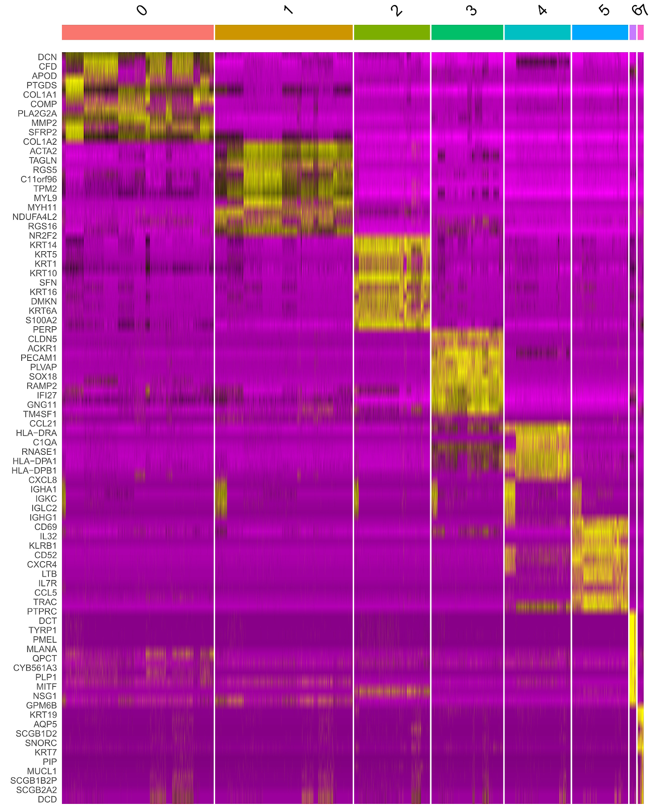
BACKGROUND: Factors such as infection, limb ischemia, and histiocyte activation are involved in diabetic foot ulcers, but the key cell subpopulations influencing diabetic foot ulcer healing remain unclear, and specific biomarkers for diabetic foot ulcers have yet to be identified. Gene Expression Omnibus (GEO) is a publicly accessible database managed by the National Center for Biotechnology Information that stores high-throughput gene expression data, allowing users to freely submit, share, query, and analyze data. Secondary analysis of published data can save research costs and uncover new research targets and ideas. OBJECTIVE: To screen biomarkers for diabetic foot ulcers using single-cell transcriptome and conventional transcriptome bioinformatics analysis, high-dimensional weighted gene co-expression network analysis (hdWGCNA), and weighted gene co-expression network analysis (WGCNA). METHODS: The single-cell transcriptome dataset GSE165816, containing non-healing ulcer tissue samples from diabetic foot ulcer patients and foot skin samples from diabetic patients, was downloaded from GEO. After data quality control, dimensionality reduction, differential analysis, cell type annotation, and pseudotime analysis, cell types spanning the entire course of diabetic foot ulcers were identified, and differentially expressed genes (DEGs) were obtained. hdWGCNA identified gene modules highly correlated with diabetic foot ulcers. Conventional transcriptome datasets GSE68183 and GSE80178, containing non-healing ulcer tissue samples from diabetic foot ulcer patients and foot skin samples from diabetic patients, were downloaded for differential analysis to screen DEGs, and WGCNA was used to identify diabetic foot ulcer-related gene modules. The DEGs from single-cell transcriptome, DEGs from conventional transcriptome samples, and module genes from WGCNA and hdWGCNA were integrated to screen biomarkers for diabetic foot ulcers. The GSE134431 dataset was downloaded as a validation conventional transcriptome dataset, and the expression levels of diabetic foot ulcer biomarkers were compared in single-cell transcriptome and validation conventional transcriptome datasets. Diabetic and diabetic foot ulcer rat models were replicated, wound tissue was collected, and immunohistochemistry and western blot were used to detect biomarker expression levels. RESULTS AND CONCLUSION: Single-cell transcriptome data analysis showed that epithelial cell differentiation spanned the entire pathological process of diabetic foot ulcers. A total of 146 DEGs were obtained from single-cell transcriptome between groups, including 59 upregulated and 87 downregulated DEGs. hdWGCNA identified 19 gene modules related to diabetic foot ulcers, containing 476 core genes. Conventional transcriptome data analysis yielded a total of 913 DEGs, including 343 upregulated and 570 downregulated DEGs. WGCNA obtained 19 diabetic foot ulcer-related gene modules, containing 887 genes. Two biomarkers for diabetic foot ulcers were screened: S100A14 and SFN. The expression levels of these two genes in diabetic foot ulcer samples were higher than those in diabetic foot skin samples in both single-cell transcriptome and validation conventional transcriptome datasets. Animal experiments showed that the expression levels of S100A14 and SFN in wound tissue of diabetic foot ulcer rats were higher than those in back skin tissue of diabetic rats. The results indicate that the pathological process of diabetic foot ulcers involves multiple cell types, among which epithelial cells are the key cell subpopulation. S100A14 and SFN are significantly upregulated in diabetic foot ulcer samples and are potential targets for the treatment of diabetic foot ulcers.

Biomarkers for diabetic foot ulcers: single-cell transcriptomics bioinformatics analysis and experimental validation
Graphical Abstract
Original ResearchVol 1896, Issue 24 • pp. 100-112DOI: 10.12307/2026.21250Jan 15, 2026

The relationship between inflammatory cytokines and frozen shoulder: a large-sample analysis of the European population based on the FinnGen GWAS database

Authors: Yan Wei, Kong Lingjun, He Tianxiang, Zhu Qingguang, Xi Xiaobing, Fang Min

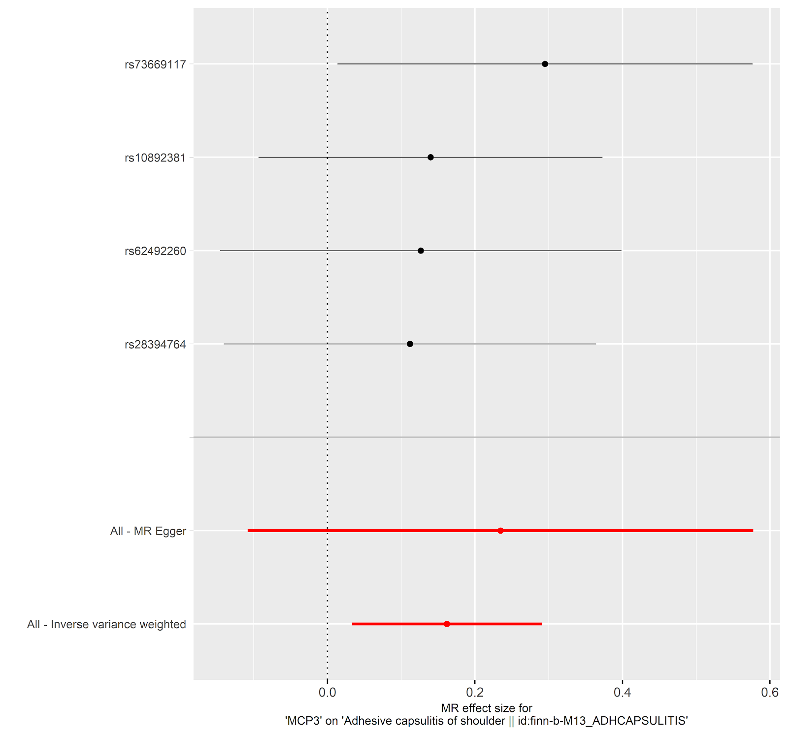
BACKGROUND: Frozen shoulder is a common disease in orthopedics, but there is no specific clinical indicator for diagnosis. There is a significant association between inflammatory cytokines and frozen shoulder, but the specific causal relationship is not yet clear. This study used summary statistical data from genome-wide association studies (GWAS) for Mendelian randomization analysis. GWAS data are based on large-sample genetic variation information, which can reduce environmental confounding factors and more reliably infer the causal relationship between inflammatory cytokines and frozen shoulder, making up for the limitation that traditional observational studies cannot determine causal associations. OBJECTIVE: To explore the causal relationship between inflammatory cytokines and the onset of frozen shoulder using bidirectional two-sample Mendelian randomization. METHODS: Using summary statistics from GWAS in the FinnGen database, we analyzed the causal relationship between 41 inflammatory cytokines and frozen shoulder. The FinnGen database, jointly initiated by the Finnish National Institute for Health and Welfare (THL), the University of Helsinki, and other Finnish research institutions, includes 2,942 cases and 167,641 European-ancestry controls, integrating genomic, clinical phenotype, and biochemical indicator data from hundreds of thousands to millions of individuals, supporting genetic association studies of diseases. This study is based on publicly available summary statistics databases and does not require ethical approval. Bidirectional Mendelian randomization analyses were performed using inverse variance weighting, weighted median, weighted model, simple model, MR-Egger regression, and sensitivity analyses (including MR-Egger, MR-PRESSO, Cochran's Q test). RESULTS AND CONCLUSION: Monocyte chemoattractant protein-3 (MCP-3) showed significant causal effects in both directions. In the forward analysis, MCP-3 was positively associated with frozen shoulder risk (OR=1.176, 95%CI: 1.034-1.338, P=0.014); in the reverse analysis, frozen shoulder was negatively associated with MCP-3 levels (OR=0.782, 95%CI: 0.625-0.979, P=0.032). Additionally, a significant association was found between tumor necrosis factor beta (TNF-β) and frozen shoulder risk (OR=1.126, 95%CI: 1.002-1.264, P=0.046); in the reverse analysis, stromal cell-derived factor 1 alpha (SDF-1α) was also significantly associated with frozen shoulder risk (OR=1.1, 95%CI: 1.011-1.196, P=0.028), indicating reliable correlations of TNF-β and SDF-1α with frozen shoulder. This bidirectional Mendelian randomization study reveals a complex interaction between MCP-3 and frozen shoulder, suggesting that MCP-3 may serve as a potential therapeutic target. Furthermore, the study indicates that TNF-β is associated with frozen shoulder risk and may be a potential risk factor; while frozen shoulder is also associated with elevated SDF-1α levels, and SDF-1α has the potential to become a diagnostic marker for frozen shoulder. However, further research is needed to elucidate the biological mechanisms underlying these causal relationships. Additionally, the analysis of international databases provides candidate molecules and causal inference paradigms for Chinese research, but it needs to be combined with local data for precise translation.

The relationship between inflammatory cytokines and frozen shoulder: a large-sample analysis of the European population based on the FinnGen GWAS database
Graphical Abstract
Original ResearchVol 1896, Issue 24 • pp. 100-112DOI: 10.12307/2026.21251Jan 15, 2026

Shared genetic basis and causal relationship between nutrition, nutritional status and inflammatory bowel disease

Authors: Liao Guibin, Wu Yixuan, Tang Jing, Huang Jinke, Wang Jun, Yan Ziqi, Liu Shujun, Zhang Haiyan

BACKGROUND: Inflammatory bowel disease, encompassing ulcerative colitis and Crohn’s disease, is a chronic condition linked to malnutrition, sarcopenia, and disease severity, with limited research on their genetic associations. OBJECTIVE: To systematically explore the common genetic basis and causal relationships between nutrition, nutritional status, and inflammatory bowel disease using advanced statistical genetics. METHODS: Single nucleotide polymorphism data for nutritional markers (minerals, vitamins, albumin, hemoglobin, fatty acids) and sarcopenia traits (appendicular lean mass and hand grip strength) were obtained from the GWAS Catalog, and inflammatory bowel disease and its subtypes from the FinnGen database R10. Advanced statistical genetics methods, including linkage disequilibrium score regression, cross-phenotype association analysis, and Mendelian randomization, were used to infer associations. RESULTS AND CONCLUSION: Significant genetic correlations were found: vitamin D with inflammatory bowel disease (rg=-0.080, P=0.029) and ulcerative colitis (rg=-0.087, P=0.027); appendicular lean mass with inflammatory bowel disease (rg=-0.100, P=0.0002), ulcerative colitis (rg=-0.100, P=0.0002), and small intestine Crohn’s disease (rg=-0.081, P=0.035); hand grip strength with small intestine Crohn’s disease (rg=-0.125, P=0.035). Mendelian randomization indicated a positive causal effect of magnesium levels on inflammatory bowel disease (OR=1.41, P=0.036) and small intestine Crohn’s disease (OR=1.78, P=0.035). Cross-phenotype analysis identified shared single nucleotide polymorphisms, particularly in the human leukocyte antigen region, affecting both nutritional status and inflammatory bowel disease. These findings further explain the genetic link between nutrition, sarcopenia, and inflammatory bowel disease, suggesting that targeted nutritional management may be key to slowing disease progression. This study provides new perspectives for personalized treatment and has potential implications for prevention strategies of inflammatory bowel disease.

Shared genetic basis and causal relationship between nutrition, nutritional status and inflammatory bowel disease
Graphical Abstract
Original ResearchVol 1896, Issue 24 • pp. 100-112DOI: 10.12307/2026.21246Jan 15, 2026

Role of fibrosis in tissue injury repair

Authors: LI Feihong, WANG Linrong, CHENG Leping

BACKGROUND: Fibrosis results from dysregulated tissue healing, characterized by abnormal proliferation of fibroblasts and excessive deposition of extracellular matrix, affecting nearly all organs including liver, kidney, heart, lung, and skin. OBJECTIVE: To summarize fibrosis-related diseases such as liver, kidney, cardiac, and pulmonary fibrosis, focusing on the major abnormal cells, signaling pathways, and therapeutic approaches. METHODS: PubMed and CNKI were searched using English terms "fibrosis, fibroblasts, fibrotic organs, extracellular matrix, tissue repair, inflammatory response" and Chinese equivalents. After screening according to inclusion and exclusion criteria, 200 articles were included for review. RESULTS AND CONCLUSION: Key abnormal cells in fibrosis include immune cells (macrophages, neutrophils, lymphocytes), fibroblasts, epithelial cells, and endothelial cells, with fibroblasts playing a central role. Major abnormal pathways include TGF-β, Wnt/β-catenin, Notch, TLR4/MyD88/NF-κB, and Hippo/YAP signaling, whose dysregulation drives fibrosis. Epigenetic modifications (DNA methylation, histone modification, non-coding RNA regulation) modulate fibrosis progression. Anti-fibrotic therapies include pharmacological, cellular, and gene-based approaches, targeting signaling pathways to inhibit persistent fibroblast activation or modulating extracellular matrix deposition to alleviate fibrosis and improve organ function.

Role of fibrosis in tissue injury repair
Graphical Abstract
Original ResearchVol 1896, Issue 24 • pp. 100-112DOI: 10.12307/2026.21244Jan 15, 2026

Strategies for the application of miRNA-targeted therapy in the treatment of osteoporosis

Authors: WU Lingjie, ZHENG Kaiyuan, WANG Guangrong, YIN Chong

BACKGROUND: Studies have shown that miRNAs, as important post-transcriptional regulators of genes, play a key role in the onset and progression of osteoporosis. Through in-depth research on the biology of miRNA regulation of osteoporosis, its potential healing mechanisms have been revealed, and this field has become a hot focus of current research. OBJECTIVE: To explore the regulatory role of miRNAs in the development of osteoporosis and their molecular mechanisms, and to provide an overview of the key difficulties encountered in the therapeutic strategies for osteoporosis targeting miRNAs and their solutions. METHODS: We searched PubMed, Web of Science and CNKI databases for relevant literature published up to March 2025. The search terms were “miRNA, osteoporosis, angiogenesis, osteogenesis, gene therapy, drug delivery” in English and “miRNA, osteoporosis, gene therapy, ribonucleic acid drugs, delivery carrier” in Chinese. After reading the titles and abstracts for preliminary screening, we excluded the literature with poor relevance, old information, or repetitive views and lack of authority, and finally included 138 papers for review. RESULTS AND CONCLUSION: (1) miRNAs are highly efficient non-coding RNAs with a wide range of applications that can precisely regulate cellular activities, and they show significant therapeutic potential in regulating osteoblast function and bone angiogenesis. (2) Although miRNA-based targeted drugs have entered preclinical research in other disease areas, clinical translation still faces challenges of insufficient nucleic acid stability in vivo and off-target effects. (3) To address these challenges, researchers have proposed various strategies, including precise targeting of miRNA target genes to reduce off-target effects; chemical modification to improve the stability of nucleic acid drugs in vivo; reducing nucleic acid production costs to advance research; and utilizing viral vectors, exosomes, and various biomaterials to optimize nucleic acid drug delivery routes. (4) Advances in technology continue to innovate in improving the performance of nucleic acid drug carriers, and in the future, precise and efficient drug delivery and targeted therapeutic effects will be achieved.

Strategies for the application of miRNA-targeted therapy in the treatment of osteoporosis
Graphical Abstract
Original ResearchVol 1896, Issue 24 • pp. 100-112DOI: 10.12307/2026.21249Jan 15, 2026

Visualization analysis of dynamic evolution of hot topics in the field of physical activity and neural plasticity

Authors: Deng Qing, Zhang Yeting, Mou Xiangqian, Wang Qingjun

BACKGROUND: In recent years, numerous studies have shown that physical activity significantly promotes neural plasticity, contributing to improvements in cognitive abilities and neurological health. However, the current state and dynamic evolution of the research field on "physical activity and neural plasticity" have not been systematically organized and analyzed. This lack of comprehensive understanding may hinder accurate predictions regarding the future development trends in this area. OBJECTIVE: Using bibliometric methods, we analyzed the dynamic evolution trajectory of research topics in the field of physical activity and neural plasticity, organized the development trends and the evolution of the knowledge framework in this area, and provided directional references for subsequent research. METHODS: Relevant literature was retrieved from the Web of Science Core Collection database (www.webofscience.com) using the following search strategy: TS=(("physical activity" OR exercise OR "motor activity" OR "physical exercise" OR "aerobic exercise" OR "physical training") AND ("neuroplasticity" OR "brain plasticity" OR "neural plasticity" OR "cognitive plasticity" OR "brain adaptability")). A total of 2,098 eligible articles were included. Co-word analysis and visualization were performed using SciMAT software to generate keyword topic overlay maps, strategic coordinate maps, and topic evolution path maps, revealing the dynamic evolution process of research topics in this field. RESULTS AND CONCLUSION: (1) Research in the field of physical activity and neural plasticity is flourishing, with increasingly in-depth studies and ample room for future development. (2) The field comprises five main evolution directions: "physical activity", "adult rats", "stem cells", "individual differences", and "randomized controlled trials", forming 15 sub-evolution paths. (3) Topics such as "older adults", "animal models", and "disease" may become future research focuses. (4) Therefore, it is recommended that future research employ randomized controlled trials and optimized animal model designs to investigate the long-term effects and mechanisms of physical activity on neural plasticity and cognitive function, and focus on clinical validation of multimodal interventions in special populations.

Visualization analysis of dynamic evolution of hot topics in the field of physical activity and neural plasticity
Graphical Abstract
Original ResearchVol 1896, Issue 24 • pp. 100-112DOI: 10.12307/2026.21245Jan 15, 2026

Postbiotic targeting muscle aging: mechanistic insights and application prospects of urolithin A

Authors: YANG Zijiang, GUO Chenggen, DENG Ziao, XUE Xinxuan

BACKGROUND: Urolithin A is a natural active compound produced by the metabolism of dietary polyphenols, which has multiple biological effects such as promoting mitochondrial function, antioxidation and anti-inflammation. In recent years, the development of urolithin A in delaying aging-related diseases has received extensive attention. However, the specific mechanism of its action in improving muscle aging remains unclear and further systematic research is still needed. OBJECTIVE: To systematically explore the mechanism of action of urolithin A in muscle aging, providing a theoretical basis for its potential application value in delaying muscle aging. METHODS: The Web of Science, PubMed, China National Knowledge Infrastructure (CNKI) and WanFang Database were retrieved from January 2000 to April 2025. The search terms were "urolithin A, muscle aging, mitochondrial function, mitophagy, inflammation, oxidative stress, muscle function, skeletal muscle" in English and Chinese. According to the inclusion and exclusion criteria, 80 literatures were finally selected for review. RESULTS AND CONCLUSION: (1) Urolithin A is a metabolite generated by the conversion of dietary polyphenolic compounds ellagic acid and ellagic acid under the action of intestinal microorganisms, and is widely present in pomegranates, berries and nuts. It has a unique α-benzocoumarin structure, with a small molecular weight, strong lipophilicity and easy absorption. The production of urolithin A depends on an individual’s intestinal microbiota and can be classified into different metabolic types. Moreover, this ability weakens with age, reflecting changes in intestinal function and physiological state. (2) Muscle aging is a process driven by mitochondrial dysfunction, chronic inflammation, and neuromuscular degeneration. With age, mitochondrial energy metabolism declines, reactive oxygen species accumulation aggravates cellular damage; chronic low-grade inflammation accelerates protein breakdown, inhibits synthesis, and impairs muscle repair; neuromuscular junction degeneration and signal transduction disorders lead to muscle denervation and atrophy. These factors interact, leading to a continuous decline in muscle mass and function. (3) Urolithin A improves muscle aging through multi-target mechanisms, mainly including: activating PTEN-induced kinase 1/Parkin signaling pathway to clear damaged mitochondria and enhance metabolic functions such as tricarboxylic acid cycle, fatty acid oxidation, and oxidative phosphorylation; inhibiting nuclear factor κB and protein kinase B/mitogen-activated protein kinase signaling pathways, upregulating anti-inflammatory factors such as interleukin-10 and transforming growth factor β1, downregulating pro-inflammatory factors such as interleukin-1β and tumor necrosis factor α, achieving inflammatory regulation; enhancing antioxidant enzyme activities such as catalase, superoxide dismutase, and glutathione peroxidase, inhibiting reactive oxygen species generation, and alleviating oxidative stress. (4) Urolithin A promotes the proliferation and differentiation of muscle stem cells, activates anabolic pathways, enhances protein synthesis, improves muscle strength, and regulates molecular mechanisms related to endurance and anti-fatigue, thereby comprehensively improving muscle performance. (5) Urolithin A shows broad prospects in delaying muscle aging and promoting muscle health, but existing studies are limited by small sample sizes, short durations, and individual differences. Future large-scale, long-term clinical studies are urgently needed to clarify dose-response relationships, explore individualized and combined intervention strategies, and focus on the potential of urolithin A as a sports nutrition supplement in enhancing physical fitness and healthy aging.

Original ResearchVol 1896, Issue 24 • pp. 100-112DOI: 10.12307/2026.21215Jan 15, 2026

Transverse tibial bone transfer accelerates healing of foot ulcers in a rabbit model of type 2 diabetes mellitus: involvement and regulation of circular RNA

Authors: Sun Zuyan, Huang Wenliang, Xu Lin, Li Haojie, Xie Tongliang, Yang Zhihang, Deng Jiang

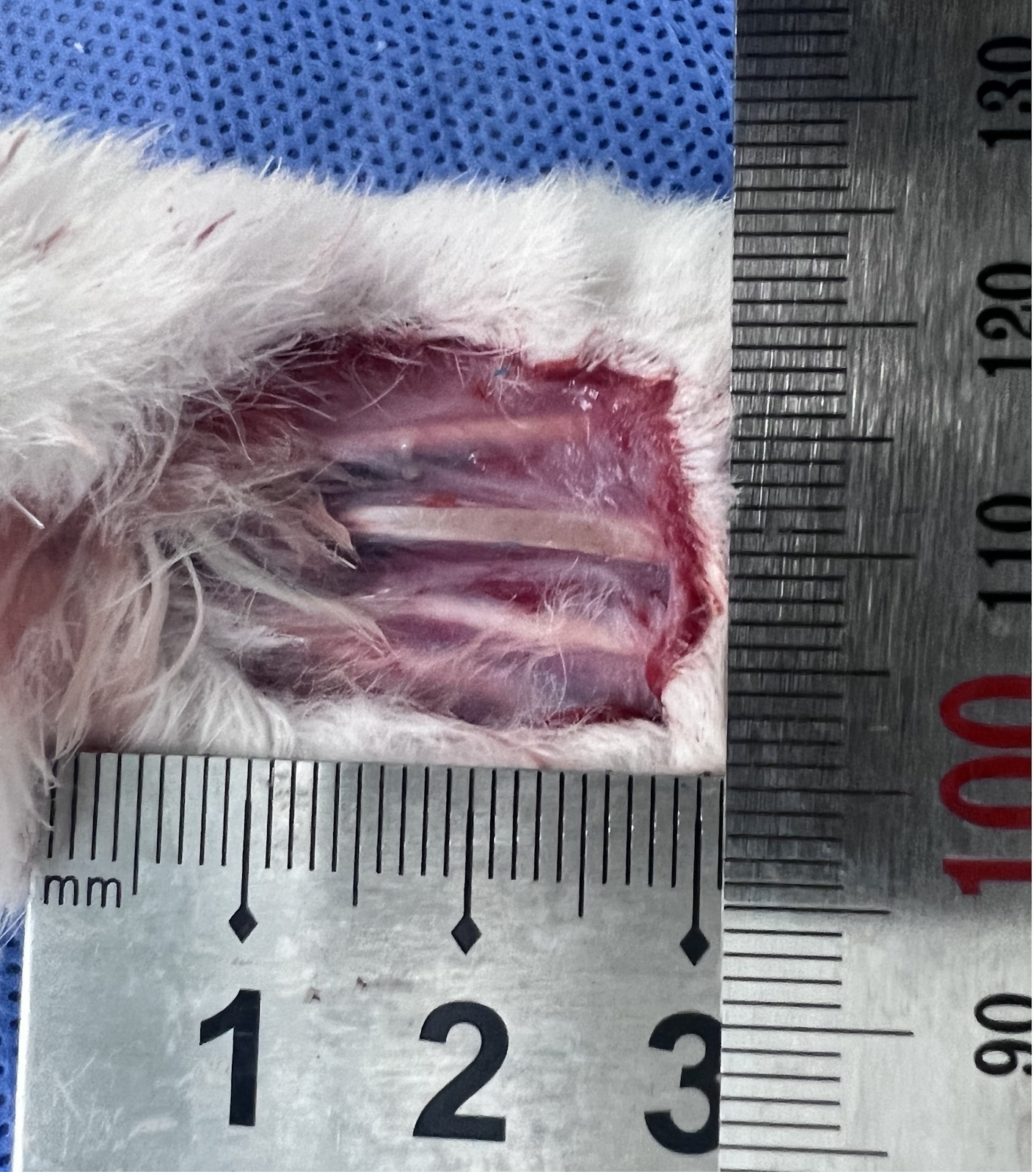
BACKGROUND: Transverse tibial bone transfer is an emerging surgical technique that enhances local blood circulation and promotes angiogenesis, thereby accelerating the healing of diabetic foot ulcers. Although this technique has demonstrated positive clinical outcomes, its specific molecular mechanisms remain unclear. Recently, the role of circular RNA in angiogenesis and wound healing has gained increasing recognition. Circular RNA may influence the healing process by regulating the expression of related genes; however, its involvement in the treatment of diabetic foot ulcers through transverse tibial bone transfer has yet to be explored. OBJECTIVE: To investigate the therapeutic effects of transverse tibial bone transfer on diabetic foot ulcers in a rabbit model and the mechanism of action. METHODS: Eighteen 3-month-old male New Zealand rabbits, weighing 2.8–3.6 kg, were included in this study. After being fed a high-sugar, high-fat diet for 1 month, type II diabetic rabbit models were induced by intravenous injection of alloxan monohydrate. After successful modeling, the right femoral artery at the mid-upper segment was ligated, and full-thickness skin on the ipsilateral foot dorsum was excised to simulate the pathological features of diabetic foot ulcers. Subsequently, the successfully modeled rabbits were randomly divided into 4 groups (4 rabbits per group): blank group (no additional treatment), dressing change group (routine iodophor disinfection after modeling), sham surgery group (installation of transverse tibial bone transfer scaffold without bone transfer), and surgery group (installation of scaffold and bone transfer). At 7 and 14 days post-surgery, the healing of foot ulcer wounds was observed. At 7, 14, and 21 days post-surgery, serum levels of vascular endothelial growth factor A (VEGF-A) and CD31 were measured by enzyme-linked immunosorbent assay. At 14 days post-surgery, ulcer tissue samples were collected for hematoxylin-eosin staining, CD31 immunofluorescence staining, and western blot analysis of VEGF-A and CD31 protein expression. At 7, 14, and 21 days post-surgery, venous blood from the surgery group was collected for whole-genome sequencing to analyze differential expression of circular RNAs. RESULTS AND CONCLUSION: At 7 and 14 days post-surgery, the surgery group showed significantly better recovery of diabetic foot ulcers compared to the other three groups, with superior promotion of epidermal repair, collagen fiber deposition, and angiogenesis. At 14 and 21 days post-surgery, serum levels of VEGF-A and CD31 in the surgery group were significantly higher than those in the other three groups (P < 0.01). Gene sequencing analysis revealed that the most significant changes in circular RNAs occurred at 21 days post-surgery, especially the expression of circular RNA PDS5B (circPDS5B) adhesion-related factor B, which gradually decreased over time, suggesting that circPDS5B may be closely related to angiogenesis and tissue repair. These results indicate that transverse tibial bone transfer can effectively promote the healing of diabetic foot ulcer wounds in rabbits. Gene sequencing results showed differential expression of circular RNAs, especially significant downregulation of circPDS5B, suggesting that transverse tibial bone transfer may promote wound repair and angiogenesis by activating related molecular pathways.

Transverse tibial bone transfer accelerates healing of foot ulcers in a rabbit model of type 2 diabetes mellitus: involvement and regulation of circular RNA
Graphical Abstract
Original ResearchVol 1896, Issue 24 • pp. 100-112DOI: 10.12307/2026.21228Jan 15, 2026

Potential mechanism by which iroquois homeobox 3 regulates the browning of perivascular adipose tissue in vascular injury

Authors: Hu Xiaoyong, Song Qianhua, Yang Zhaoying, Tang Rui, Li Hongjian

BACKGROUND: Vascular injury-related diseases have garnered significant attention in the medical field, and the browning of perivascular adipose tissue is closely linked to these diseases. However, the regulatory mechanisms of specific genes involved in this process remain unclear. OBJECTIVE: To investigate the potential mechanism by which iroquois homeobox 3 regulates the browning of perivascular adipose tissue in vascular injury. METHODS: The perivascular adipose tissue-related single-cell sequencing data matrix GSE275779 was analyzed to investigate the expression levels and functions of iroquois homeobox 3 in various cell subpopulations. In conjunction with adipocyte-related microarray and sequencing data GSE44059, GSE7032, GSE185518, and GSE168387, differentially expressed genes were identified, and the expression level of iroquois homeobox 3 during the differentiation of browning adipocytes was validated. The downstream target genes of iroquois homeobox 3 were screened using the msigdb database and the ChIP-seq database GTRD. By disrupting iroquois homeobox 3 and overexpressing retinol saturase in adipocyte precursor cells, the mRNA and protein expression levels of browning-related genes were detected by qPCR and western blot. RESULTS AND CONCLUSION: Bioinformatics analysis showed that adipocyte characteristic factors such as PR domain containing 16, cell death-inducing DFFA-like effector A, and uncoupling protein 1 were significantly downregulated in perivascular adipose tissue of diabetic patients, and these genes are involved in adipose browning. Combined with high-throughput sequencing data analysis, it was found that iroquois homeobox 3 is highly expressed in brown adipose tissue and participates in brown adipocyte differentiation. Further screening identified retinol saturase as a downstream target gene of iroquois homeobox 3, and its level was differentially expressed during brown adipocyte differentiation. In mature brown adipocytes, knockdown of iroquois homeobox 3 led to decreased expression of retinol saturase and browning-related markers (uncoupling protein 1, peroxisome proliferator-activated receptor gamma coactivator 1 alpha, PR domain containing 16). In the retinol saturase rescue experiment, overexpression of retinol saturase significantly upregulated the protein levels of browning-related markers but did not affect the expression of iroquois homeobox 3. This study preliminarily reveals the potential mechanism by which iroquois homeobox 3 regulates perivascular adipose tissue browning during vascular injury.

Potential mechanism by which iroquois homeobox 3 regulates the browning of perivascular adipose tissue in vascular injury
Graphical Abstract
Original ResearchVol 1896, Issue 24 • pp. 100-112DOI: 10.12307/2026.21241Jan 15, 2026

Effects of piRNA CFAPIR in doxorubicin-induced ferroptosis models of rat and human cardiomyocytes

Authors: Tao Xiangyu, Wang Shuang, Li Yuhan, Cao Jimin, Sun Teng

BACKGROUND: Ferroptosis plays a critical role in doxorubicin-induced cardiomyopathy; however, its specific regulatory mechanisms require further elucidation. Piwi-interacting RNA 413 (piRNA413) regulates ferroptosis in doxorubicin-induced cardiomyocytes, designated as cardiac ferroptosis-associated piRNA (CFAPIR). However, the specific regulatory mechanism needs to be further elucidated. OBJECTIVE: To investigate the role and regulatory mechanism of piRNA CFAPIR in doxorubicin-induced cardiomyocyte ferroptosis and cardiomyopathy. METHODS: (1) Intraperitoneal injection of doxorubicin was used to induce cardiomyopathy in mice. The myocardium was in situ injected with CFAPIR knocking down lentivirus. The body mass and survival rate of mice were monitored and recorded; cardiac function, heart volume and mass, inflammation, and cardiac fibrosis were assessed. (2) Doxorubicin was used to induce ferroptosis in AC16 cardiomyocytes, and CFAPIR inhibitor was transfected into cells. Cell damage, ferroptosis (expression levels of ferroptosis markers, iron ion, malondialdehyde, and reduced glutathione content), and mitochondrial dysfunction were detected. The effect of CFAPIR on ABCB8 expression was also examined. RESULTS AND CONCLUSION: (1) CFAPIR levels were significantly upregulated in both doxorubicin-induced cardiomyopathy animal models (P < 0.0001) and cardiomyocyte ferroptosis models (P < 0.01). (2) In vivo, knockdown of CFAPIR significantly alleviated doxorubicin-induced cardiotoxicity, including inhibition of body weight loss (P < 0.05), improved survival rate, improved cardiac function (P < 0.01), reduced cardiac atrophy (P < 0.05), inhibited lactate dehydrogenase activity increase (P < 0.05), and reduced cardiac fibrosis (P < 0.0001). (3) In vitro, knockdown of CFAPIR significantly ameliorated doxorubicin-induced cardiomyocyte ferroptosis, manifested by increased cell viability (P < 0.05), decreased lactate dehydrogenase activity (P < 0.01), upregulated expression of ferroptosis markers xCT (P < 0.01) and glutathione peroxidase 4 (P < 0.001), downregulated mRNA level of prostaglandin-endoperoxide synthase 2 (P < 0.05), reduced iron overload (P < 0.05), decreased malondialdehyde content (P < 0.05), increased reduced glutathione content (P < 0.01), reduced reactive oxygen species accumulation (P < 0.01), and increased mitochondrial membrane potential (P < 0.05). (4) Knockdown of CFAPIR significantly attenuated the doxorubicin-induced decrease in iron transporter ABCB8 expression (P < 0.05). (5) These results indicate that CFAPIR levels are significantly upregulated in both animal models of doxorubicin-induced cardiomyopathy and cellular ferroptosis models, and knockdown of CFAPIR significantly improves doxorubicin-induced cardiotoxicity and cardiomyocyte ferroptosis, possibly by targeting mitochondrial iron transporter ABCB8.

Effects of piRNA CFAPIR in doxorubicin-induced ferroptosis models of rat and human cardiomyocytes
Graphical Abstract
Original ResearchVol 1896, Issue 24 • pp. 100-112DOI: 10.12307/2026.21213Jan 15, 2026

U-shaped association between magnesium intake and all-cause and cancer mortality in patients with osteoarthritis

Authors: Zhou Haidong, Lu Yaohong, Fan Shaoyong

BACKGROUND: Clarifying the relationship between dietary magnesium intake and mortality risk in patients with osteoarthritis can provide theoretical basis for optimizing dietary interventions, reducing all-cause mortality and cardiovascular disease mortality, and provide reference value for nutritional management in patients with osteoarthritis. OBJECTIVE: To assess the association between dietary magnesium intake and all-cause and etiological mortality in adults with osteoarthritis in the United States. METHODS: We enrolled data of adults with osteoarthritis from the National Health and Nutrition Examination Survey (conducted by the National Center for Health Statistics under the U.S. Centers for Disease Control and Prevention, it aims to assess the health and nutritional status of the civilian population in the United States) between 2003 and 2020. The association between dietary magnesium intake and mortality in patients with osteoarthritis was evaluated by Cox proportional hazard model and two-stage Cox model. To assess whether the association between dietary magnesium intake and all-cause mortality was consistent across the population and to identify potential high-risk groups, subgroup analyses in terms of age, sex, body mass index, hypertension, diabetes mellitus, physical activity, smoking status, and drinking status were performed and tested for interactions. RESULTS AND CONCLUSION: (1) A total of 2 868 patients with osteoarthritis were included, and 699 all-cause deaths, 281 cardiovascular disease deaths, and 143 cancer deaths were recorded during follow-up. After adjusting for multiple variables, higher dietary magnesium intake was significantly associated with reduced risk of cardiovascular disease mortality; each 1-unit increase in dietary magnesium intake was associated with a 78% reduction in cardiovascular disease mortality (P=0.022 2), and quartile analysis of dietary magnesium intake was consistent with this. Dietary magnesium intake showed a U-shaped association with all-cause mortality and cancer mortality, with thresholds for lowest mortality risk at 0.38 g/d and 0.40 g/d, respectively. When magnesium intake was below the threshold, higher intake was significantly associated with lower all-cause mortality [HR=0.17, 95%CI(0.06, 0.50)] and cancer mortality [HR=0.16, 95%CI(0.01, 1.50)]; but above the threshold, magnesium intake was significantly associated with increased all-cause mortality [HR=2.94, 95%CI(0.55, 15.84)] and cancer mortality [HR=26.30, 95%CI(1.46, 474.73)]. Subgroup analyses further verified the robustness of the results. (2) The results indicate that insufficient dietary magnesium intake may play an adverse role in cardiovascular health, and there is a U-shaped relationship between dietary magnesium intake and all-cause mortality and cancer mortality, with both too low and too high magnesium intake potentially increasing mortality risk. This finding provides new insights into the potential impact of dietary magnesium intake on cardiovascular disease, cancer, and all-cause mortality, provides scientific basis for nutritional intervention in patients with osteoarthritis, and provides theoretical support for the prevention and management of cardiovascular disease and cancer in China, especially in the context of high cardiovascular disease incidence, reasonable magnesium intake helps reduce related mortality.

U-shaped association between magnesium intake and all-cause and cancer mortality in patients with osteoarthritis
Graphical Abstract
Original ResearchVol 1896, Issue 24 • pp. 100-112DOI: 10.12307/2026.21222Jan 15, 2026

Cobalt chloride-induced hypoxic environment accelerates knee cartilage degeneration in New Zealand rabbits

Authors: XU Peng, JIANG Wei, YU You, LEI Zhengliang, TIAN Yang, ZHANG Jie, LIU Luchang

BACKGROUND: Cobalt chloride solution is commonly used to induce osteoarthritis cell models in vitro. However, its ability to construct animal models of osteoarthritis by intra-articular injection remains unknown. OBJECTIVE: To investigate the effect of intra-articular injection of different concentrations of cobalt chloride solution on cartilage degeneration in the knee joint. METHODS: Thirty-six healthy adult male New Zealand rabbits were randomly divided into four groups: low, medium and high dose cobalt chloride groups and control group. The right hind knee was intra-articularly injected with 100, 200, and 300 μmol/(L·kg) of cobalt chloride, while the left hind knee served as the control knee and was injected with an equal amount of normal saline. At 4, 8 and 12 weeks after operation, four rabbits were killed respectively. The cartilage on the surface of the femur was exposed for gross morphological observation, and then the cartilage tissues were taken for hematoxylin-eosin staining, safranine O-fast green staining, the Osteoarthritis Research Society International scoring, and immunohistochemical staining of interleukin 1 and tumor necrosis factor α, to determine cartilage degeneration in various aspects. RESULTS AND CONCLUSION: (1) Gross observation: At the same postoperative time point, with the increase of cobalt chloride concentration, cartilage degeneration showed a progressive aggravation trend, and the high-dose cobalt chloride group even involved the deep layer of cartilage and subchondral bone; under the same concentration of cobalt chloride, with the prolongation of modeling time, cartilage degeneration progressed progressively. (2) Hematoxylin-eosin staining, safranine O-fast green staining, and Osteoarthritis Research Society International scoring showed that at the same postoperative time point, with the increase of cobalt chloride concentration, the cartilage surface gradually became rough, the superficial layer became thinner, and the destruction aggravated, and the Osteoarthritis Research Society International score gradually increased (P < 0.05); under the same concentration of cobalt chloride, with the prolongation of modeling time, the arrangement of chondrocytes tended to be disordered, polarity was lost, and the destruction of superficial cartilage and subchondral bone progressively aggravated, and the Osteoarthritis Research Society International score gradually increased (P < 0.05). (3) Immunohistochemistry showed that at the same postoperative time point, with the increase of cobalt chloride concentration, cartilage degeneration aggravated, intracellular brown particles increased, and the positive expression of interleukin 1 and tumor necrosis factor α increased (P < 0.01); under the same concentration of cobalt chloride, with the prolongation of modeling time, cartilage destruction and fissures aggravated, and the positive expression of interleukin 1 and tumor necrosis factor α increased (P < 0.01). This experiment successfully established an osteoarthritis model of New Zealand rabbits induced by intra-articular injection of cobalt chloride solution, preliminarily verified the stability and reliability of the animal model, and also proved that with the increase of modeling concentration and the prolongation of modeling time, cartilage degeneration progressed progressively.

Cobalt chloride-induced hypoxic environment accelerates knee cartilage degeneration in New Zealand rabbits
Graphical Abstract
Original ResearchVol 1896, Issue 24 • pp. 100-112DOI: 10.12307/2026.21239Jan 15, 2026

Effects of high-intensity interval training combined with estrogen on satellite cells, myonuclear domain and ribosome function in ovariectomized rats

Authors: Sun Yuan, Shu Jun, Ren Shuang, Wang Chenyu

BACKGROUND: Estrogen deficiency can lead to a decrease in skeletal muscle mass and muscle strength in postmenopausal women, thereby affecting their quality of life. Muscle mass is maintained by satellite cells, which are regulated by estrogen. Regular exercise, especially high impact exercise (such as resistance training and high-intensity interval training), can induce muscle hypertrophy, but the role and mechanism of estrogen are still unclear. OBJECTIVE: To explore the effects of high-intensity interval training combined with estrogen therapy on skeletal muscle hypertrophy in ovariectomized rats and reveal its possible mechanism. METHODS: Sixty 8-week-old female Sprague-Dawley rats were divided into five groups using a random number table method: sham operation, model sedentary group, model exercise group, model hormone group, or model combined group. Bilateral ovariectomy was used to establish an estrogen deficiency model. Twelve weeks after operation, the model exercise and model combined group performed high-intensity interval training for 8 weeks (3 times/week), and hormone treatment groups received abdominal subcutaneous injection of 17β-estradiol (once a day for 8 weeks). Seventy-two hours after the last training, the grip force of the forelimb was measured by an electronic grip force meter. The gastrocnemius muscle was separated, and muscle mass index was calculated as muscle mass/body mass ratio. Hematoxylin-eosin staining was used to obtain cell cross-sectional area. Immunofluorescence staining was used to classify muscle fiber types and obtain myonuclear number, myonuclear domain size, and activated satellite cell number. BCA method was used to determine total protein concentration. Trizol method was used to extract total RNA. Western blot was used to detect ribosomal protein S6 expression. Real-time quantitative PCR was used to detect ribosomal RNA expression. RESULTS AND CONCLUSION: Compared with the sham operation group, the model sedentary group showed increased body mass and myosin heavy chain type I fiber proportion (P < 0.05), while uterine mass index, gastrocnemius mass index, grip strength, cell cross-sectional area, myosin heavy chain type IIa proportion, satellite cell and myonuclear number, myonuclear domain size, total protein and RNA content, and ribosomal protein S6, 18S rRNA and 28S rRNA expression decreased (P < 0.05). Compared with the model sedentary group, the model exercise group showed decreased body mass and myosin heavy chain type IIb proportion (P < 0.05), while uterine mass index, gastrocnemius mass index, grip strength, cell cross-sectional area, myosin heavy chain type IIa proportion, satellite cell number, myonuclear domain size, total protein and RNA content, and ribosomal protein S6 and 28S rRNA expression increased (P < 0.05). Compared with the model exercise group and model hormone group, the model combined group showed higher gastrocnemius mass index, grip strength, cell cross-sectional area, satellite cell number, myonuclear domain size, total protein and RNA content, and ribosomal protein S6 and 28S rRNA expression (P < 0.05). These results indicate that estrogen can enhance the skeletal muscle hypertrophy response induced by high-intensity interval training in ovariectomized rats, and the mechanism may be related to satellite cell activation, increased myonuclear domain and ribosome biogenesis, and improved ribosome function.

Effects of high-intensity interval training combined with estrogen on satellite cells, myonuclear domain and ribosome function in ovariectomized rats
Graphical Abstract
Original ResearchVol 1896, Issue 24 • pp. 100-112DOI: 10.12307/2026.21233Jan 15, 2026

Gushukang Granule-containing drug serum improves dexamethasone-induced atrophy of C2C12 myotubes via regulating mitochondrial homeostasis

Authors: WEI Wei, LIU Hongfei, QI Xiaonan, LIU Yantong, WANG Deyu, YU Zhitong, QIAO Chunlin, WANG Shixuan, TENG Hai

BACKGROUND: Gushukang Granule is a Chinese herbal compound preparation, commonly used clinically for the treatment of osteoporosis and other bone-related diseases. However, its role in the regulation of muscle metabolism is not clear. OBJECTIVE: To investigate the inhibitory effect of Gushukang Granule-containing serum on dexamethasone-induced C2C12 muscle atrophy. METHODS: (1) In vivo experiment: Thirty-six 3-month-old female Sprague-Dawley rats were randomly divided into model group, blank group and Gushukang group, with 12 rats in each group. The rats in the model group were given 2.5 mg/kg dexamethasone by intragastric administration, once a day, for 1 week, followed by normal feeding for 3 weeks; the Gushukang group was given 0.48 g/kg Gushukang Granule suspension by gavage after modeling, once a day, for 3 weeks; the blank group was not modeled and given equal volume of distilled water by gavage, once a day, for 4 weeks. Two hours after the last administration, left femur and gastrocnemius muscle specimens were taken for hematoxylin-eosin staining, and RNA was extracted from right gastrocnemius and femur tissues; real-time fluorescence quantitative PCR was used to detect mRNA expression levels of mitochondrial function, autophagy and inflammation-related genes. (2) In vitro experiment: C2C12 cells were cultured and induced to differentiate into myotubes with 2% horse serum. Differentiated myotubes were divided into control group, model group and Gushukang-containing serum group. Muscle atrophy model was constructed by incubating with 4 µmol/L dexamethasone for 48 h, and the Gushukang group was treated with 10% Gushukang-containing serum for 24 h after dexamethasone treatment for 24 h. CCK-8 was used to detect cell viability, flow cytometry to detect reactive oxygen species levels, transmission electron microscopy to observe ultrastructural changes, and western blot to detect expression levels of mitochondrial function-related proteins, autophagy-related proteins and antioxidant-related proteins. RESULTS AND CONCLUSION: (1) Hematoxylin-eosin staining showed that compared with the model group, the muscle fiber structure of rats in the Gushukang group recovered better; real-time fluorescence quantitative PCR further verified that Gushukang Granule up-regulated mitochondrial function and autophagy-related genes, supporting its multi-target mechanism to alleviate muscle atrophy. (2) Flow cytometry and CCK-8 results showed that after dexamethasone treatment, reactive oxygen species levels in C2C12 cells were significantly increased (P < 0.05), and cell viability was significantly decreased (P < 0.05); transmission electron microscopy revealed that dexamethasone induced ultrastructural disorder, reduced number of organelles, atrophic and blurred mitochondria, accompanied by a large number of autophagosomes and cytoplasmic vacuoles; western blot results showed that after dexamethasone treatment, the expression of mitochondrial function-related proteins translocase of outer mitochondrial membrane 20 and heat shock protein 60 was significantly decreased, autophagy-related proteins LC3 and Beclin-1 were abnormally expressed (P < 0.05), and silent information regulator 1 expression was significantly decreased (P < 0.05), suggesting that dexamethasone disrupted mitochondrial homeostasis and inhibited autophagy. After treatment with Gushukang Granule-containing serum, reactive oxygen species levels were significantly decreased (P < 0.05), cell viability was significantly increased (P < 0.05); transmission electron microscopy showed that ultrastructure was improved and mitochondrial morphology was relatively restored; western blot results showed that the expression of translocase of outer mitochondrial membrane 20, heat shock protein 60, and silent information regulator 1 was significantly up-regulated (P < 0.05), and LC3 and Beclin-1 expression returned to normal levels (P < 0.05), indicating that Gushukang Granule-containing serum played a positive role in improving mitochondrial function, reducing oxidative stress, and regulating autophagy.

Gushukang Granule-containing drug serum improves dexamethasone-induced atrophy of C2C12 myotubes via regulating mitochondrial homeostasis
Graphical Abstract
Original ResearchVol 1896, Issue 24 • pp. 100-112DOI: 10.12307/2026.21238Jan 15, 2026

Establishing a diagnostic model for recurrent spontaneous abortion based on the levels of autophagy-related genes in the endometrium

Authors: Tang Cen, Hu Wanqin

BACKGROUND: The etiology of recurrent spontaneous abortion is complex. With the development of genetics and other fields, it has been found that the abnormal expression of autophagy-related genes may lead to the imbalance of cell homeostasis, thus triggers pathological processes, such as apoptosis, inflammatory response and immunosuppressive response, and affects the endometrial microenvironment, trophoblastic function and immune cell function, thereby leading to recurrent spontaneous abortion. By using the recurrent spontaneous abortion samples in the Gene Expression Omnibus database, the expression changes and regulatory mechanisms of autophagy-related genes were analyzed, which is helpful to reveal the mechanism of recurrent spontaneous abortion and develop new therapeutic strategies. However, the specific mechanism of autophagy-related genes in recurrent spontaneous abortion and their interaction with other biological processes still need to be further studied. OBJECTIVE: To establish a risk score prognostic model for patients with recurrent spontaneous abortion based on autophagy-related genes. METHODS: Endometrial gene expression matrix of patients with recurrent abortion was obtained from the Gene Expression Omnibus database, autophagy-related genes were obtained from the Human Autophagy Database (HADb), and 30 differentially co-expressed autophagy-related genes were identified. The biological functions of autophagy-related genes were analyzed by gene ontology (GO), Kyoto Encyclopedia of Genes and Genomes (KEGG) and DisGeNET enrichment. LASSO and logistic regression analysis identified 16 autophagy-related genes as potential biomarkers. A nomogram model was then constructed, and the predictive accuracy was evaluated using receiver operating characteristic curves. Six machine learning models (random forest, support vector machine, generalized linear model, etc.) were compared to select the optimal model. Nomogram, calibration curves, and decision curve analysis were used to validate the predictive efficacy. RESULTS AND CONCLUSION: GO analysis showed that autophagy-related genes in recurrent spontaneous abortion patients were mainly enriched in autophagy regulation, catabolic processes, and formation of mitochondrial or other organelle membranes. KEGG analysis showed enrichment in autophagy regulation, phosphatidylinositol 3-kinase/protein kinase B signaling pathway, human papillomavirus infection, and neurodegeneration pathways. GSEA analysis indicated involvement in copper detoxification, proton transmembrane transport, sarcoplasmic reticulum components, and regulation of nucleoside diphosphate phosphatase activity. A predictive risk model was constructed, identifying 16 specific autophagy-related genes as predictive targets. Based on the detection efficacy of machine models, the optimal neural network model was selected, and five most important autophagy-related gene variables (MAP2K7, CALCOCO2, SAR1A, TUSC1, and STK11) were identified. Using recurrent spontaneous abortion samples from the European population in the GEO database, the gene expression patterns, differentially expressed genes, and signaling pathways in the endometrium were analyzed from the genetic single nucleotide polymorphism level. The predictive model and machine learning model with good predictive efficacy can screen new therapeutic targets and potential biomarkers for recurrent spontaneous abortion, which has certain guiding significance for clinical work and mechanism research.

Establishing a diagnostic model for recurrent spontaneous abortion based on the levels of autophagy-related genes in the endometrium
Graphical Abstract
Original ResearchVol 1896, Issue 24 • pp. 100-112DOI: 10.12307/2026.21240Jan 15, 2026

Effects of protein kinase C on the expression of myocardial SarcKATP channels in model rats during exercise preconditioning

Authors: Wang Kai, Zhou Yuehui

BACKGROUND: Exercise preconditioning produces early and late myocardial protective effects, in which protein kinase C and myocardial ATP-sensitive potassium channels (SarcKATP) are mediators and effectors, respectively. Protein kinase C regulates the expression of myocardial SarcKATP channels. OBJECTIVE: To compare the effects of protein kinase C on the expression of myocardial SarcKATP channel subunits, inward recirculating potassium channel 6.2 (Kir6.2) and sulfonylurea receptor 2A (SUR2A), in exercise preconditioning. METHODS: Forty-eight Sprague-Dawley rats were randomly divided into five groups: control group (no intervention), early exercise preconditioning group, protein kinase C inhibitor (pre-exercise intraperitoneal injection) + early exercise preconditioning group, late exercise preconditioning group, and protein kinase C inhibitor + late exercise preconditioning group. After preconditioning, the distribution and expression changes of Kir6.2 and SUR2A mRNAs in the rat myocardium were observed and detected using real-time fluorescent quantitative PCR. The distribution and expression changes of Kir6.2 and SUR2A proteins were observed and detected using western blot. RESULTS AND CONCLUSION: (1) Compared with the control group, the mRNA expression of Kir6.2 and SUR2A showed no significant difference in the early and late exercise preconditioning group. (2) Compared with the early exercise preconditioning group, the protein kinase C inhibitor + early exercise preconditioning group showed decreased Kir6.2 mRNA expression but increased Kir6.2 protein expression; both SUR2A mRNA and protein expression decreased. (3) Compared with the late exercise preconditioning group, the protein kinase C inhibitor + late exercise preconditioning group showed decreased Kir6.2 mRNA and SUR2A mRNA expression, decreased Kir6.2 protein expression, but increased SUR2A protein expression. (4) These results indicate that for the same subunit (Kir6.2 or SUR2A), protein kinase C exerts coordinated and complementary regulatory effects in early and late exercise preconditioning; for different subunits (Kir6.2 and SUR2A), protein kinase C also exerts coordinated and complementary regulatory effects on their expression in early and late exercise preconditioning.

Effects of protein kinase C on the expression of myocardial SarcKATP channels in model rats during exercise preconditioning
Graphical Abstract
Original ResearchVol 1896, Issue 24 • pp. 100-112DOI: 10.12307/2026.21225Jan 15, 2026

Articular cartilage lesions at different stages of steroid-induced osteonecrosis of the femoral head: characteristics and mechanisms of crescent sign formation

Authors: Wan Ziyi, Jiang Mengyu, Zhou Yuehui, Xue Yuxuan, Wei Yangwenxiang, Zhou Chi

BACKGROUND: The crescent sign is a significant radiological feature in the progression of steroid-induced osteonecrosis of the femoral head (SIONFH), indicating the separation and defect of articular cartilage and subchondral bone. The appearance of the crescent sign is associated with the mid-to-late stages of the disease and poor prognosis. However, studies on the specific pathological characteristics and progression patterns of articular cartilage in SIONFH remain unclear. OBJECTIVE: To observe the pathological features of articular cartilage in specimens from different stages of SIONFH, explore the progression and pathological mechanisms, and elucidate the formation mechanism of the crescent sign, providing a theoretical basis for optimizing hip-preserving strategies. METHODS: Femoral head specimens were collected from patients with SIONFH who underwent total hip arthroplasty at the First Affiliated Hospital of Guangzhou University of Chinese Medicine from 2021 to 2024. According to the ARCO staging, they were divided into mild, moderate, and severe collapse groups, with fresh femoral neck fracture specimens as controls. All specimens were cut coronally, and the folded cartilage surface in the necrotic area was taken; control group took corresponding area. Hematoxylin-eosin staining and Safranin O-fast green staining were used for morphological observation, immunohistochemistry and western blot for biomarker expression, and apoptosis kit for apoptosis level. RESULTS AND CONCLUSION: (1) Gross observation: The control group showed smooth cartilage surface without folds or hyperplasia, no separation or defect between articular cartilage and subchondral bone, and tough texture. In SIONFH specimens, obvious folds were visible on the cartilage surface, with separation and defects between articular cartilage and subchondral bone, and a loose sensation on pressing. (2) Pathological observation: In the control group, chondrocytes in each layer were arranged neatly, cartilage matrix stained uniformly, tidemark was intact and continuous, calcified cartilage layer and subchondral bone connection was clear and complete, and bone trabeculae were arranged neatly. In SIONFH specimens, chondrocytes were disorganized, empty lacunae increased, matrix staining loss of varying degrees, tidemark duplication and loss, calcified cartilage layer showed numerous cavities and sclerosis, with granulation tissue invasion into cavities, separation and defects between calcified cartilage and subchondral bone, and abundant proliferative granulation tissue in subchondral bone trabecular spaces. (3) Immunohistochemistry: In SIONFH specimens, positive staining of Runt-related transcription factor 2, matrix metalloproteinase 13, matrix metalloproteinase 3, and collagen type I alpha 2 chain increased in calcified cartilage layer and deep cartilage; vascular endothelial growth factor A, hypoxia-inducible factor 1 alpha, interleukin-1 beta, and tumor necrosis factor alpha positive staining increased in subchondral bone trabecular spaces and deep cartilage granulation and scar tissue. (4) Western blot results showed decreased expression of collagen type II alpha 1 chain and SOX9, and increased expression of Runt-related transcription factor 2, matrix metalloproteinase 13, hypoxia-inducible factor 1 alpha, and vascular endothelial growth factor A in SIONFH specimens. (5) Caspase3/7 activity in SIONFH samples was significantly higher than that in the control group, positively correlated with the degree of collapse. (6) These results indicate that articular cartilage lesions in SIONFH mainly concentrate in the deep cartilage and calcified cartilage around the necrotic area. Necrosis of subchondral bone leads to changes in local microenvironment and elastic modulus, causing sclerosis of calcified cartilage. With continued weight-bearing, stress concentration at the necrosis-sclerosis junction leads to brittle fracture, which is the starting point of fracture. Bone and cartilage fracture leads to destruction of the subchondral cortical bone barrier, and invasion of granulation tissue from subchondral bone trabecular spaces directly stimulates calcified cartilage and deep cartilage, resulting in terminal differentiation, apoptosis, matrix degradation, and cavity formation of chondrocytes, leading to decreased repair capacity of articular cartilage. The diseased cartilage cannot properly interlock with subchondral bone, and with disease progression, extensive separation and defects eventually appear between bone and cartilage, manifesting as the crescent sign on imaging.

Articular cartilage lesions at different stages of steroid-induced osteonecrosis of the femoral head: characteristics and mechanisms of crescent sign formation
Graphical Abstract
Original ResearchVol 1896, Issue 24 • pp. 100-112DOI: 10.12307/2026.21237Jan 15, 2026

Prostaglandin E1 pretreatment inhibits ferroptosis in endothelial cells in a rat model of spinal cord ischemia-reperfusion injury

Authors: Huang Yushan, Wang Rongrong, Li Xiangmiao, Bai Jinzhu

BACKGROUND: Ferroptosis is an important pathological mechanism in spinal cord ischemia-reperfusion injury. Although studies have confirmed that prostaglandin E1 attenuates cerebral microvascular endothelial cell injury in the hippocampus induced by chronic cerebral hypoperfusion, its effect on ferroptosis of endothelial cells after spinal cord ischemia-reperfusion injury remains poorly studied. OBJECTIVE: To investigate whether prostaglandin E1 pretreatment attenuates spinal cord ischemia-reperfusion injury by inhibiting ferroptosis in endothelial cells and to elucidate possible mechanisms. METHODS: (1) Cell experiment: Rat spinal cord microvascular endothelial cells were divided into four groups. Control group was cultured under normoxia (20% O2) with complete medium. Model group was subjected to oxygen-glucose deprivation (OGD) for 3 hours (hypoxia chamber with 95% N2 and 5% CO2, glucose-free serum-free medium) followed by reoxygenation for 12 hours (normoxia, complete medium) to simulate spinal cord ischemia-reperfusion injury. Pretreatment group received prostaglandin E1 for 2 hours after OGD and before reoxygenation. Inhibitor group received ML385 (Nrf2 inhibitor) for 2 hours after OGD, then prostaglandin E1 for 2 hours, followed by reoxygenation for 12 hours. After treatment, intracellular malondialdehyde, glutathione, and Fe2+ levels were measured; cell viability was assessed by CCK-8; immunofluorescence staining and western blot were used to detect ACSL4 and GPX4 expression; flow cytometry measured reactive oxygen species; western blot detected Nrf2 and HO-1 protein expression. (2) Animal experiment: 45 rats were randomly divided into three groups: sham group (n=15) underwent laparotomy without aortic occlusion; model group (n=15) underwent occlusion of abdominal aorta for 30 minutes followed by tail vein injection of saline, then reperfusion; pretreatment group (n=15) underwent occlusion for 30 minutes followed by tail vein injection of prostaglandin E1, then reperfusion. At 24 hours after reperfusion, motor function and neuronal injury were assessed by BBB score, inclined plane test, and Nissl staining; blood-spinal cord barrier integrity and microvascular density were evaluated by spinal cord water content, immunofluorescence staining of ZO-1, and CD34 immunohistochemistry; ferroptosis in spinal cord tissue was assessed by immunofluorescence, Prussian blue staining, western blot, and biochemical assays. RESULTS AND CONCLUSION: (1) Cell experiment: OGD/reoxygenation reduced cell viability, induced ferroptosis, and downregulated Nrf2 and HO-1 protein expression in rat spinal cord microvascular endothelial cells. Prostaglandin E1 pretreatment inhibited these effects; ML385 partially reversed the protective effect of prostaglandin E1. (2) Animal experiment: Prostaglandin E1 pretreatment alleviated motor dysfunction, neuronal injury, and blood-spinal cord barrier damage, improved microvascular density, and inhibited ferroptosis in spinal cord tissue after spinal cord ischemia-reperfusion injury. (3) These results indicate that prostaglandin E1 pretreatment protects against spinal cord ischemia-reperfusion injury by activating the Nrf2/HO-1 signaling pathway to inhibit ferroptosis in endothelial cells.

Prostaglandin E1 pretreatment inhibits ferroptosis in endothelial cells in a rat model of spinal cord ischemia-reperfusion injury
Graphical Abstract
Original ResearchVol 1896, Issue 24 • pp. 100-112DOI: 10.12307/2026.21236Jan 15, 2026

Mechanism by which the paraventricular nucleus of the hypothalamus is involved in chronic pain and anxiety in mice with lumbar disc herniation

Authors: Shi Gaolong, Ge Caijun, Chen Jianpeng, Wang Yuanbin, Fan Zelin, Yan Jun, Wang Qianliang

BACKGROUND: Patients with lumbar disc herniation (LDH) often experience comorbid anxiety due to chronic pain and functional limitations, significantly affecting their quality of life. However, the mechanisms underlying the pain-anxiety comorbidity remain unclear. OBJECTIVE: To investigate the neural regulatory mechanisms of the paraventricular nucleus in the hypothalamus in a mouse model of lumbar disc herniation with chronic pain-anxiety comorbidity. METHODS: A total of 100 C57BL/6 mice were randomly divided into a normal group (24 mice) and a model group (76 mice). The lumbar disc herniation model was established in the model group using a needle puncture method. Seventy-two successfully modeled mice were randomly divided into the model group, oxytocin group, and oxytocin+Vasotocin group, with 24 mice in each group. Mice in the oxytocin group received a 200 nL injection of oxytocin (0.5 μg/μL) into the paraventricular nucleus of the hypothalamus. Mice in the oxytocin+Vasotocin group received a 200 nL injection of oxytocin into the paraventricular nucleus and a 20 μL intraperitoneal injection of Vasotocin (an oxytocin antagonist, 0.15 μg/μL). Anxiety-like behavioral changes were evaluated via the elevated plus maze and open field tests on day 20 after modeling. Mechanical paw withdrawal threshold and thermal paw withdrawal latency experiments were conducted for all groups before modeling and 21 days after modeling. On day 21 post-modeling, immunofluorescence staining was used to observe c-FOS expression in the paraventricular nucleus of the hypothalamus; qPCR was used to detect mRNA expression of inflammatory factors prostaglandin E2, tumor necrosis factor α, and interleukin-1β in dorsal root ganglion tissue; Western blot was used to detect oxytocin receptor and p-ERK1/2 protein expression in the paraventricular nucleus. RESULTS AND CONCLUSION: Compared with the normal group, the model group showed significantly decreased mechanical and thermal pain thresholds (P < 0.05), significantly reduced time and entries in the open arms of the elevated plus maze (P < 0.05), significantly reduced time and entries in the open field (P < 0.05), significantly increased mRNA expression of prostaglandin E2, tumor necrosis factor α, and interleukin-1β in dorsal root ganglion tissue (P < 0.05), significantly increased c-FOS expression in the paraventricular nucleus (P < 0.05), significantly decreased oxytocin receptor protein expression, and significantly increased p-ERK1/2 protein expression (P < 0.05). Compared with the model group, the oxytocin group showed significantly increased mechanical and thermal pain thresholds (P < 0.05), significantly increased time and entries in the open arms of the elevated plus maze (P < 0.05), significantly increased time and entries in the open field (P < 0.05), significantly decreased mRNA expression of prostaglandin E2, tumor necrosis factor α, and interleukin-1β in dorsal root ganglion tissue (P < 0.05), significantly decreased c-FOS expression in the paraventricular nucleus (P < 0.05), significantly increased oxytocin receptor protein expression, and significantly decreased p-ERK1/2 protein expression (P < 0.05). Compared with the oxytocin group, the use of Vasotocin reversed the beneficial effects of oxytocin on pain and anxiety, increased inflammatory factor expression, significantly decreased oxytocin receptor protein expression, and significantly increased p-ERK1/2 protein expression. These results indicate that oxytocin can significantly improve chronic pain and anxiety-like behavior in mice with lumbar disc herniation, inhibit dorsal root ganglion inflammation, and the mechanism may be related to activation of the ERK signaling pathway in the paraventricular nucleus and downregulation of inflammatory factor expression.

Mechanism by which the paraventricular nucleus of the hypothalamus is involved in chronic pain and anxiety in mice with lumbar disc herniation
Graphical Abstract