🧬 SinoBioData Academic Portal
🏛️ Indexed Academic JournalOriginal: 生物化学与生物物理学报

Acta Biochimica et Biophysica Sinica

Premier Chinese Biomedical Journal indexed in SinoBioData: Acta Biochimica et Biophysica Sinica (生物化学与生物物理学报).

Total Research Papers: 200
Access: 100% Free Open Access
Browse by Publication Year & VolumeReset All Filters ✕

Published Research PapersFiltered: Year 2026 • 58 • 2

Showing 13 of 200 peer-reviewed papers with full Graphical Abstracts.

Original ResearchVol. 58, Issue 2 • pp. 421-436DOI: 10.3724/abbs.2026018

Cardiac PTN-SIRT1 axis alleviates oxidative stress and promotes mitochondrial energy reprogramming to mitigate doxorubicin-induced cardiotoxicity through AMPK/PGC1α signaling

Authors: Yuxiao Sun, Tianwen Wei, Hongping Xu, Hongda Li, Chang Zhou, Xianliang Liu, Yafei Li, Shangwei Huang, Qi Zhang, Xia Duan

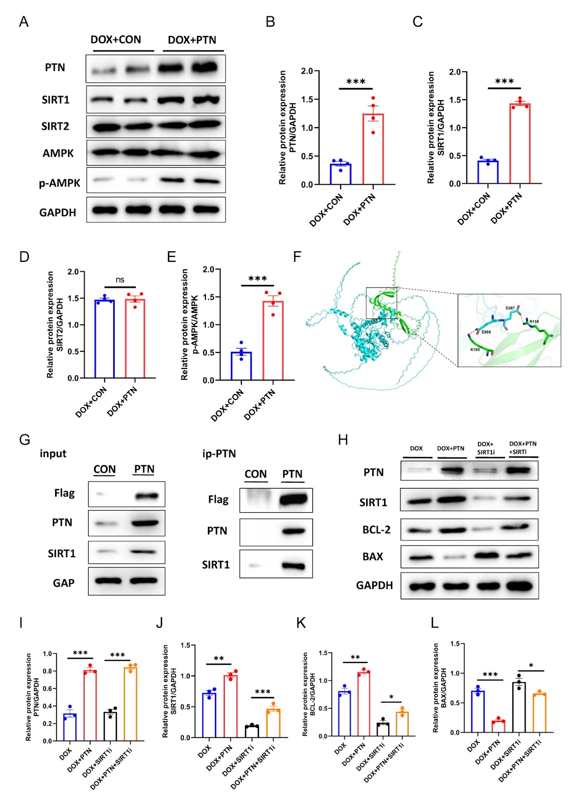
Doxorubicin (DOX) remains a cornerstone chemotherapeutic agent for malignancies, yet its clinical utility is severely limited by dose-dependent cardiotoxicity, which can lead to progressive left ventricular dysfunction and heart failure. Pleiotrophin (PTN), a heparin-binding growth factor with diverse physiological functions, regulates glucose and lipid metabolism and promotes oxidative energy pathways. However, whether PTN exerts protective effects against DOX-induced cardiotoxicity (DIC) remains unclear. In this study, we establish cellular and animal models of DIC. DOX administration induces pronounced myocardial injury in both models, characterized by impaired ventricular contractility, increased fibrotic remodeling, and reduced cell viability. Concurrently, PTN protein expression is significantly downregulated in cardiomyocytes under DOX treatment. Overexpression of PTN substantially alleviates these pathological changes. In vitro, PTN reduces mitochondrial oxidative stress and apoptosis while restoring energy production and cell viability. In vivo, PTN improves mitochondrial ultrastructure, decreases cardiomyocyte apoptosis, and enhances cardiac function. Mechanistically, PTN directly binds to SIRT1 and activates AMPK phosphorylation at Thr172, triggering a downstream cascade through the AMPK-PGC1α axis that reprograms mitochondrial energy metabolism and attenuates cardiotoxicity. In conclusion, the PTN-SIRT1 axis protects against DIC by reducing oxidative stress and promoting mitochondrial energy homeostasis via the AMPK/PGC1α pathway, highlighting its potential as a novel therapeutic target for preventing chemotherapy-related cardiac injury.
Cardiac PTN-SIRT1 axis alleviates oxidative stress and promotes mitochondrial energy reprogramming to mitigate doxorubicin-induced cardiotoxicity through AMPK/PGC1α signaling
Graphical Abstract
Original ResearchVol. 58, Issue 2 • pp. 216-230DOI: 10.3724/abbs.2025123

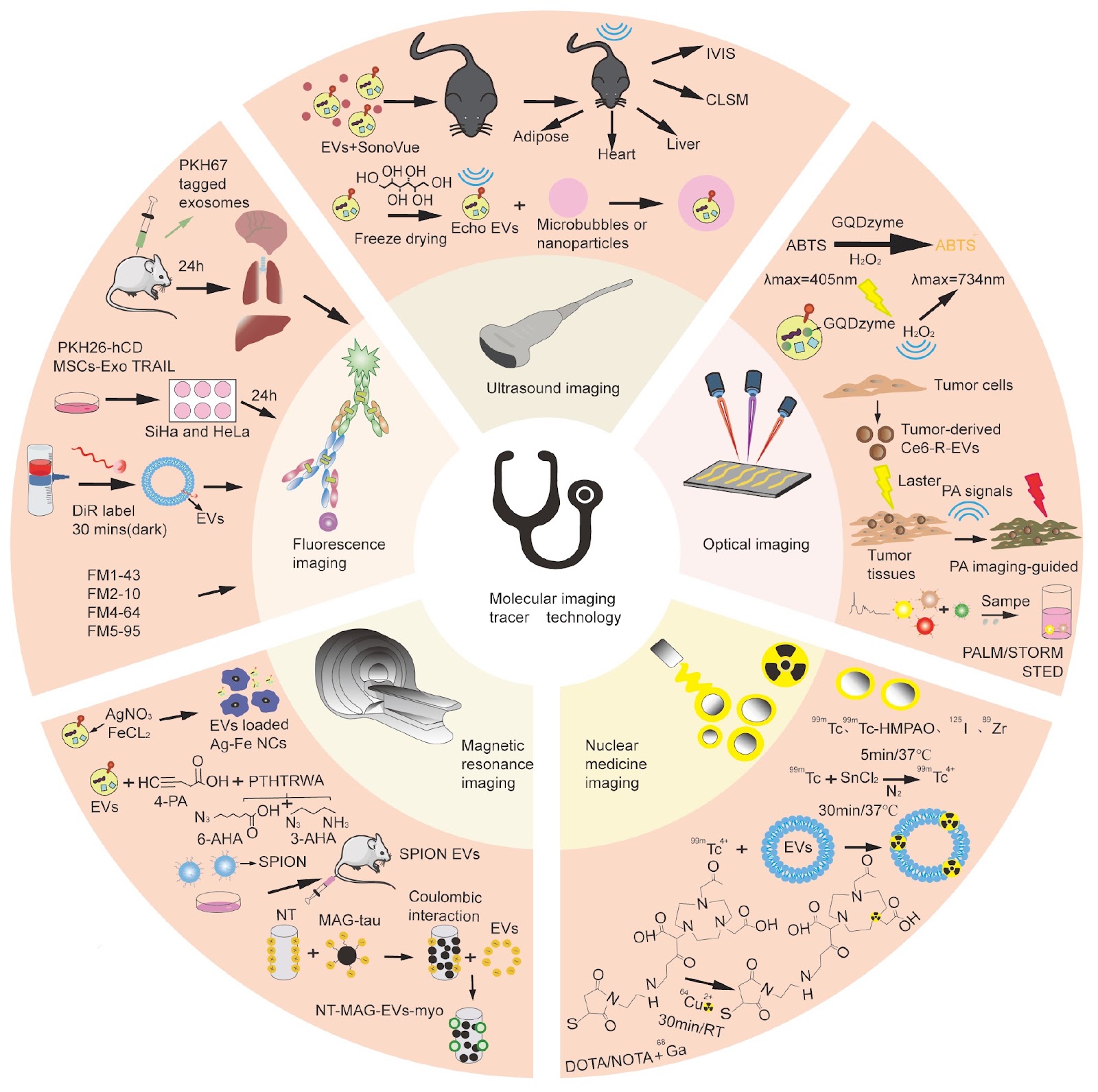
Unveiling the multifaceted roles of extracellular vesicles in cancer: insights from molecular imaging and engineering strategies

Authors: Yuqin Teng, Gang Huang, Hao Yang

Extracellular vesicles (EVs), a class of nanoscale, membrane-bound vesicles secreted by various cell types, have emerged as rapidly advancing fields of research in recent years. This heterogeneous vesicle is a versatile carrier system for a variety of biomolecules, including proteins, nucleic acids, and metabolites. EVs play pivotal roles in intercellular communication, immune regulation, and disease pathogenesis, with particular implications for cancer biology. On the one hand, EVs promote tumor progression and metastasis by facilitating communication between cancer cells and their microenvironment. On the other hand, EVs carry noncoding RNAs, such as miRNAs and other regulatory RNAs, which directly modulate immune cell function or exert antitumor effects by influencing cancer cell proliferation and apoptosis. In addition to their biological roles, EVs show great potential as drug delivery systems because of their ability to be effectively taken up by target cells and stably deliver therapeutic payloads. In the context of cancer therapy, natural EVs demonstrate inherent therapeutic potential, particularly in targeting highly metabolically active organs. Furthermore, engineered EVs, which serve as both therapeutic vehicles and molecular imaging probes, have demonstrated significant potential for cancer theranostics. This review focuses on elucidating the dynamic changes and biological functions of EVs in vivo, with the aim of exploring the translational potential of EV-based molecular imaging and tracing technologies in cancer treatment. This work seeks to provide critical insights that may enhance the precision and efficacy of tumor therapies, offering a foundation for future clinical applications.

Unveiling the multifaceted roles of extracellular vesicles in cancer: insights from molecular imaging and engineering strategies
Graphical Abstract
Original ResearchVol. 58, Issue 2 • pp. 353-368DOI: 10.3724/abbs.2025108

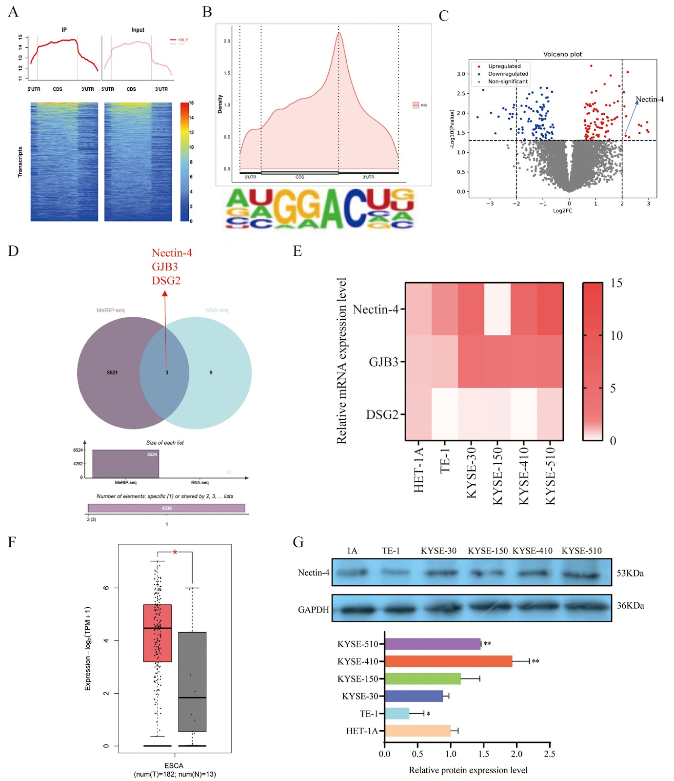
METTL3-mediated m6A modification facilitates Nectin-4-induced VNN1 upregulation and promotion of ESCC progression

Authors: Yuanfeng Long, Hang Yang, Ruolan Zhang, Quanneng Zhao, Mi Yang, Guiqin Song, Kang Liu

Esophageal squamous cell carcinoma (ESCC) is a highly aggressive malignancy with poor prognosis and limited therapeutic options. N6-methyladenosine (m6A) RNA modification plays a role in tumorigenesis, but its contributions to ESCC and the regulation of cell adhesion molecules such as Nectin-4 are not fully elucidated. In this study, we investigate the role and the regulatory mechanisms of Nectin-4 in ESCC, particularly regarding the influence of m6A modification and its downstream metabolic effects. Our study demonstrates that methyltransferase-like protein 3 (METTL3) enhances Nectin-4 mRNA stability and expression through m6A methylation in ESCC, as validated by actinomycin D assay, MeRIP-qPCR, and dual-luciferase reporter assay. Both METTL3 and Nectin-4 are highly expressed in ESCC tissues and promote malignant phenotypes such as proliferation, migration, and invasion. Further analysis identifies pantothenate esterase 1 (VNN1) as a downstream target of Nectin-4, mediating the oncogenic effects of the METTL3/Nectin-4 axis and promoting the biosynthesis of pantothenic acid and coenzyme A, thus driving ESCC progression. By integrating transcriptomic data, this study elucidates a key pathogenic mechanism in which the METTL3/Nectin-4/VNN1 axis regulates metabolic reprogramming to promote ESCC development. These findings provide new insights into the molecular pathology of ESCC and offer potential biomarkers and therapeutic targets for early screening, prognosis, and precision treatment for ESSC.

METTL3-mediated m6A modification facilitates Nectin-4-induced VNN1 upregulation and promotion of ESCC progression
Graphical Abstract
Original ResearchVol. 58, Issue 2 • pp. 437-452DOI: 10.3724/abbs.2025197

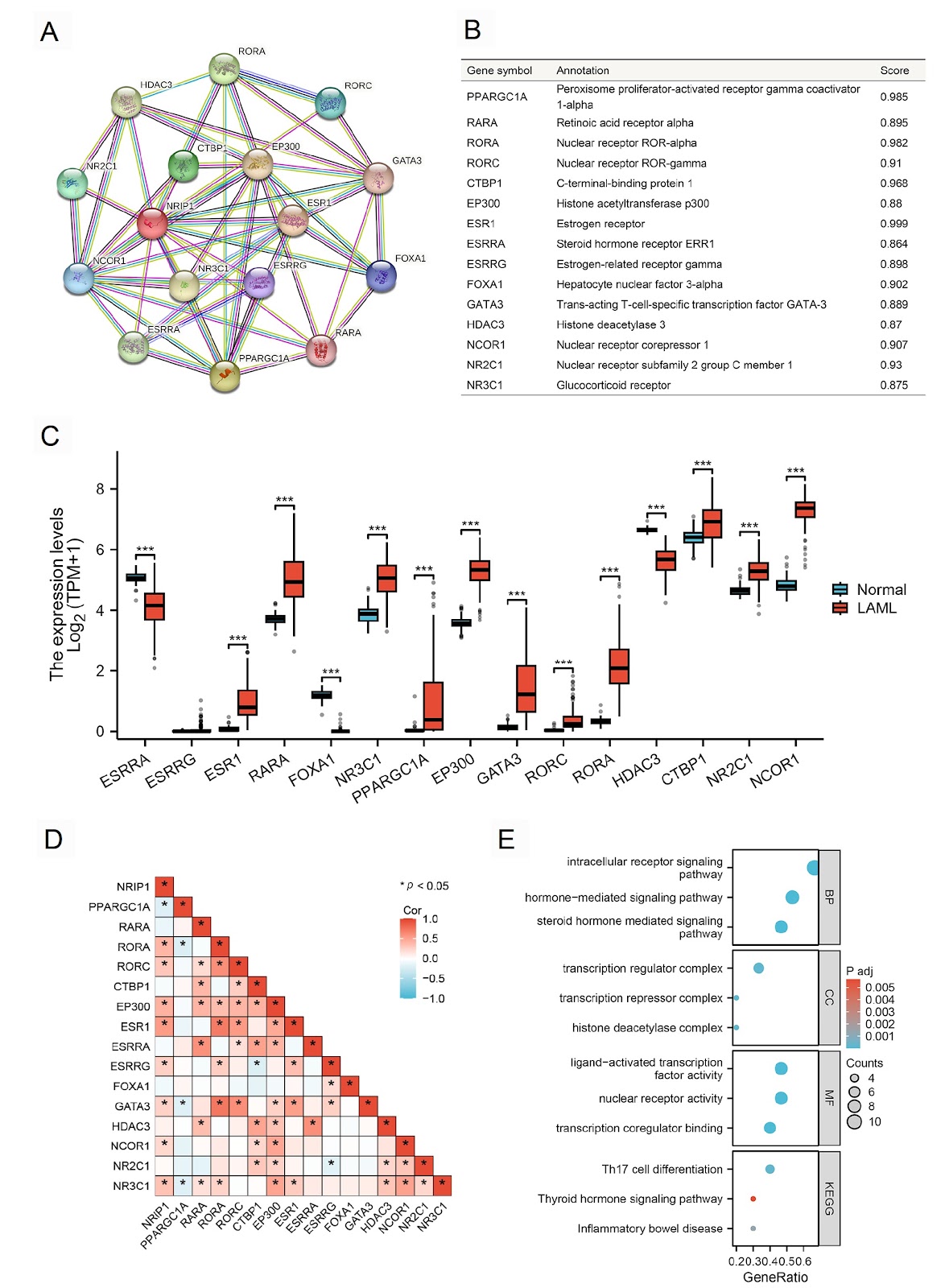
The prognostic marker NRIP1 is associated with tumor progression and immune infiltration in acute myeloid leukemia

Authors: Xunxun Zhu, Mingyan Zhang, Jingjing Zhang, Yanling Tao, Hao Zhang

Acute myeloid leukemia (AML) is a clinically aggressive hematologic malignancy characterized by high relapse rates and treatment resistance, highlighting the need for novel biomarkers to improve clinical outcomes. In this study, we explore the roles of nuclear receptor-interacting protein 1 (NRIP1) in AML, focusing on its associations with tumor progression and immune infiltration. Analysis of public AML gene expression datasets reveals that NRIP1 expression is significantly increased in AML patients. Those with high NRIP1 expression have markedly shorter overall survival than those with low expression. Furthermore, NRIP1 expression is significantly associated with the infiltration of diverse immune cells, including B cells, dendritic cells, T cells, mast cells, eosinophils, and T helper cells, suggesting that NRIP1 may be a regulator of immune cell infiltration. Functional enrichment analysis indicates that NRIP1 and its interacting partners are involved in tumorigenesis, immune microenvironment remodeling, and metabolic reprogramming. Survival analysis confirms the prognostic value of NRIP1. Importantly, functional validation in AML cell lines confirms that NRIP1 knockdown suppresses proliferation and induces apoptosis. Our study identifies NRIP1 as a multifaceted regulator that promotes AML by driving tumor progression, regulating immune cell infiltration, and modulating ferroptosis, highlighting its role as a novel prognostic biomarker.

The prognostic marker NRIP1 is associated with tumor progression and immune infiltration in acute myeloid leukemia
Graphical Abstract
Original ResearchVol. 58, Issue 2 • pp. 258-274DOI: 10.3724/abbs.2025075

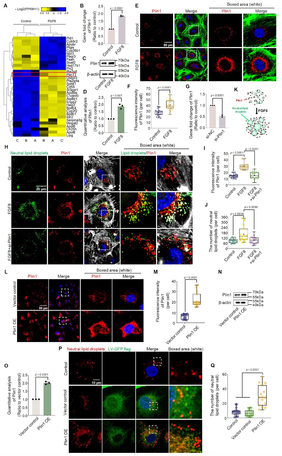
FGF8 promotes lipid droplet accumulation via the FGFR1/p-p38 axis in chondrocytes

Authors: Minglei Huang, Haoran Chen, Jieya Wei, Caixia Pi, Mengmeng Duan, Xiaohua Pu, Zhixing Niu, Siqun Xu, Shasha Tu, Sijun Liu, Jiazhou Li, Li Zhang, Yang Liu, Hao Chen, Chunming Xu, Jing Xie

Chondrocytes store lipids in the form of lipid droplets (LDs) and maintain cartilage lipid metabolic homeostasis by consuming or regenerating LDs. This modulation is largely mediated by a series of biochemical factors. Fibroblast growth factor 8 (FGF8) is one of the most important factors involved in the proliferation, differentiation, and migration of chondrocytes and has attracted increasing attention in the physiology and pathology of cartilage. However, the effect of FGF8 on LD accumulation in chondrocytes remains unclear. This study aims to elucidate the role of FGF8 in LDs and explore the underlying biomechanism involved. The results reveal that FGF8 promotes LD accumulation in chondrocytes by upregulating perilipin1 (Plin1) expression. FGF8 activates the cytoplasmic p-p38 signaling pathway via fibroblast growth factor receptor 1 (FGFR1) to increase LD accumulation in chondrocytes. Subsequent experiments with siRNAs and specific inhibitors further confirm the importance of the FGFR1/p38 axis for LD accumulation in chondrocytes exposed to FGF8. The results increase our understanding of the role of FGF8 in the lipid metabolic homeostasis of chondrocytes and provide insights into the physiology and pathology of cartilage.

FGF8 promotes lipid droplet accumulation via the FGFR1/p-p38 axis in chondrocytes
Graphical Abstract
Original ResearchVol. 58, Issue 2 • pp. 396-405DOI: 10.3724/abbs.2025159

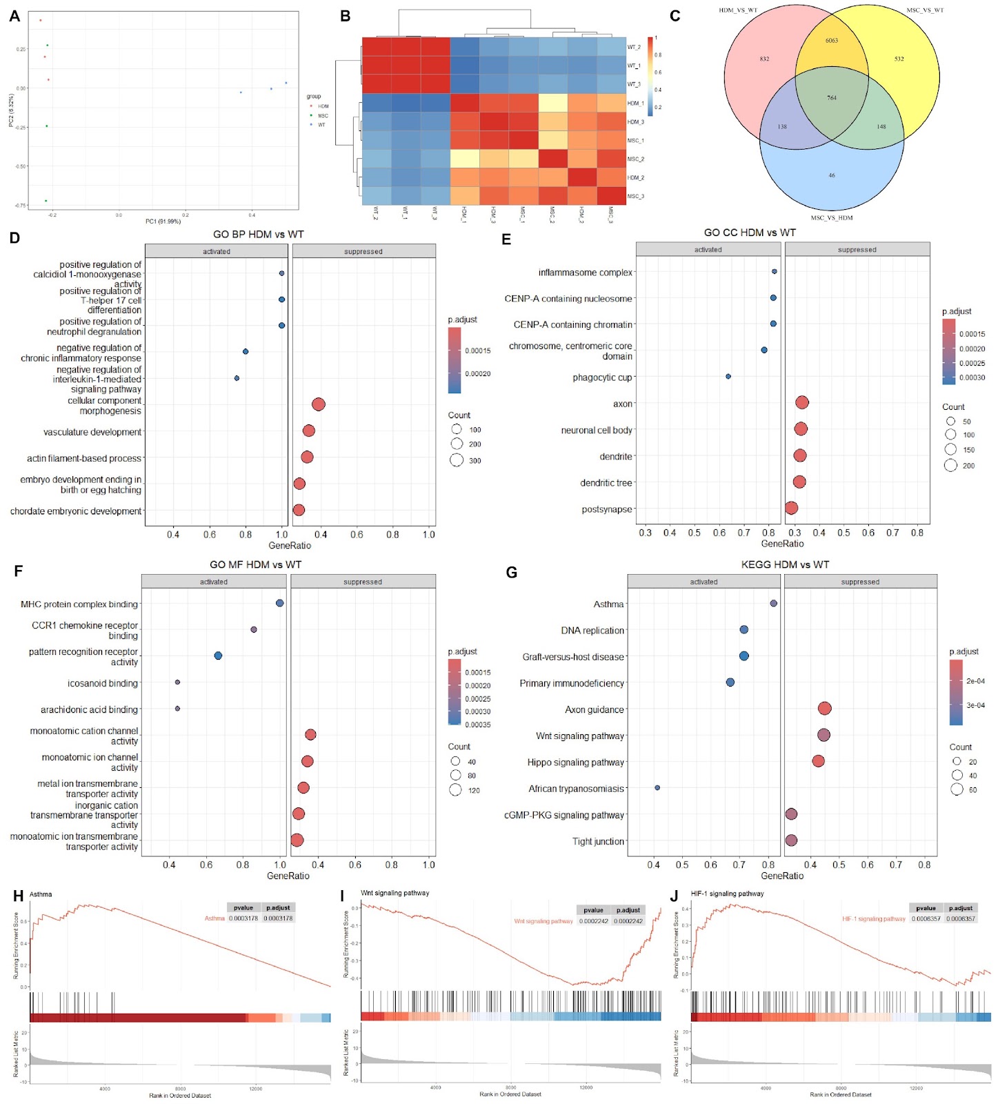
MSCs attenuate airway remodeling in HDM-induced asthma by inhibiting the Timp1-Wnt2b axis

Authors: Kai Yu, Xinyu Feng, Rong Zhang, Jian Fan, Jiaying Yuan, Yan Shang, Jiayi Zhao

MSCs have demonstrated their unique therapeutic potential in early clinical trials for a variety of respiratory diseases in recent years, but their use in the treatment of asthma has rarely been reported. In this study, a chronic murine asthma model that is more similar to clinical asthma is constructed via sustained HDM induction for 70 days, followed by treatment via tail vein injection of MSCs after modeling. The mechanism by which MSCs alleviate airway remodeling is investigated via RNA-seq. The airways on the day following treatment are used to screen for transcriptomic changes resulting from the MSC treatment under study, filtering for differentially expressed genes (DEGs), identifying their enrichment pathways, and finally confirming the DEGs gained via western blot analysis. After HDM treatment, airway remodeling is reversed, asthma and the HIF-1 signaling pathway are inhibited, and the expression levels of Timp1 and Wnt2b in the fibrosis pathway are also significantly decreased. STRING analysis reveals a reciprocal interaction in their expression, which is also confirmed by western blot analysis. To verify whether MSCs alleviate airway remodeling by inhibiting Timp1, we construct MSCs overexpressing Timp1 and evaluate their effects in vitro and in vivo. The ability of MSCs to alleviate airway remodeling is reversed after Timp1 is overexpressed. These findings demonstrate that MSCs alleviate asthma-induced airway remodeling by inhibiting the Timp1-Wnt2b axis.

MSCs attenuate airway remodeling in HDM-induced asthma by inhibiting the Timp1-Wnt2b axis
Graphical Abstract
Original ResearchVol. 58, Issue 2 • pp. 231-244DOI: 10.3724/abbs.2025175

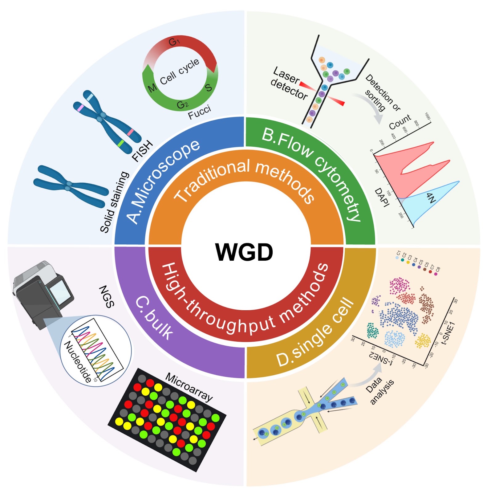
The dual role of whole-genome duplication: biological mechanisms, functional consequences, and detection advances

Authors: Yawei Song, Jiajie Yang, Shuheng Wu, Wei Wu

Whole-genome duplication (WGD) represents an evolutionarily conserved process occurring in prokaryotes, eukaryotes, and somatic mammalian tissues. While developmentally programmed WGD supports normal tissue regeneration, unscheduled WGD drives chromosomal instability and oncogenic progression in cancer. Recent studies have clarified dual roles of WGD across physiological homeostasis and disease pathogenesis. Here, we review the prevalence of WGD, the molecular mechanisms driving its major causes and its biological consequences. In addition, we highlight recent advancements in WGD detection, including both conventional cytogenetic techniques and newly developed high-throughput sequencing approaches. The integration of multi-omics and machine learning further improves ploidy analysis, particularly in cancer research. Together, these insights establish WGD as a critical regulator of development, regeneration, and disease and underscore the importance of emerging computational and sequencing tools for its precise characterization.

The dual role of whole-genome duplication: biological mechanisms, functional consequences, and detection advances
Graphical Abstract
Original ResearchVol. 58, Issue 2 • pp. 245-257DOI: 10.3724/abbs.2025086

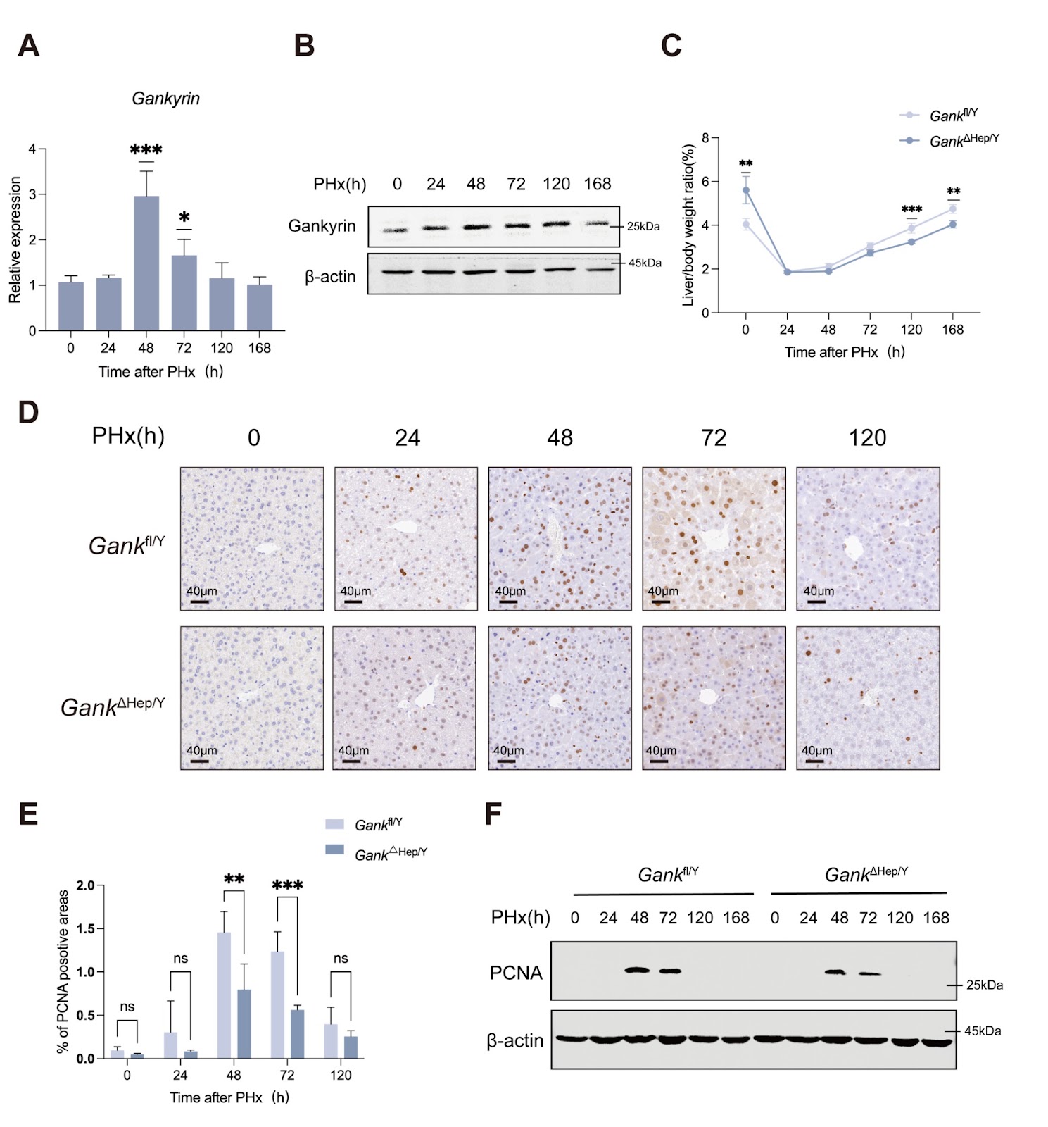
Gankyrin-deficiency reprograms intrahepatic glucose and lipid metabolism to delay liver regeneration

Authors: Yitian Liu, Yiwei Sun, Lv Jin, Ying Xu, Bibo Wang, Ting Yu, Xiaofei Wei, Jing Xu, Yating Wei, Shuai Yang, Min Yu, Hongyang Wang, Yao Chen

Liver regeneration is a critical adaptive response to hepatic injury, requiring precise metabolic reprogramming to meet the energetic and biosynthetic demands of proliferating hepatocytes. While the oncoprotein Gankyrin is well-established as a promoter of liver fibrosis and hepatocarcinogenesis, its role in metabolic adaptations underlying liver regeneration remains unclear. In this study, we demonstrate that Gankyrin deficiency in the liver (Gank△Hep/Y) induces hepatic hypertrophy and aberrant glycogen accumulation. Gankyrin expression is significantly upregulated after partial hepatectomy (PHx), whereas Gank△Hep/Y -PHx mice exhibit impaired liver regeneration. This impairment is marked by a delayed restoration of the liver-to-body weight ratio, blunted glycogenolysis, and reduced fatty acid uptake. Mechanistically, Gankyrin activates Pygl and Cd36, key regulators of glycogenolysis and lipid uptake, respectively. Pharmacological inhibition of PYGL activity retards liver regeneration. Furthermore, we identify a novel interaction between Gankyrin and FOXO1, wherein Gankyrin promotes FOXO1 ubiquitination and subsequent proteasomal degradation. This Gankyrin-dependent suppression of FOXO1 leads to the transcriptional upregulation of Pygl and Cd36, thereby fueling hepatocyte proliferation. Collectively, our findings reveal Gankyrin as a master regulator of liver regeneration, integrating metabolic reprogramming with proliferative signaling through the FOXO1-PYGL/CD36 axis. These insights not only elucidate the mechanistic underpinnings of liver regeneration but also unveil the therapeutic potential of targeting the Gankyrin/FOXO1 pathway to mitigate hepatic insufficiency and enhance regenerative capacity in clinical settings.

Gankyrin-deficiency reprograms intrahepatic glucose and lipid metabolism to delay liver regeneration
Graphical Abstract
Original ResearchVol. 58, Issue 2 • pp. 369-382DOI: 10.3724/abbs.2025103

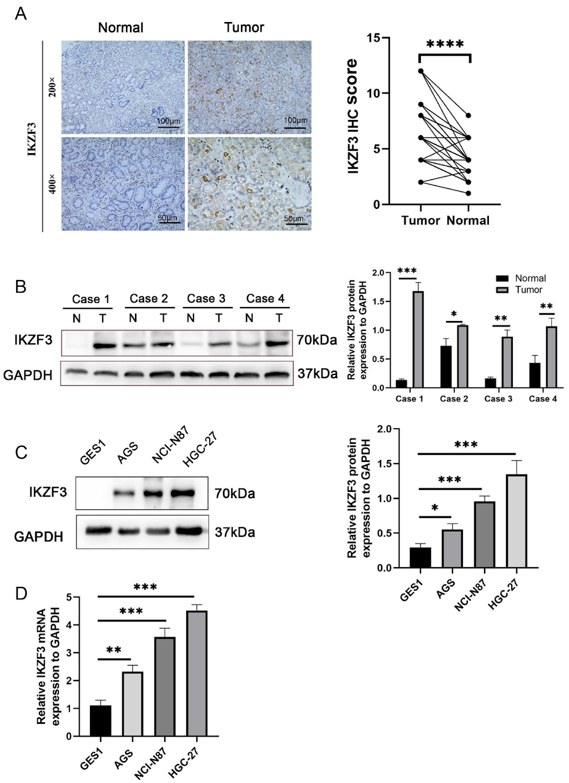
IKZF3 promotes gastric cancer progression via Hedgehog signaling activation and is targetable by SANT-1

Authors: Muhammad Ali, Shantanu Baral, Jun Ren, Liuhua Wang, Bin Liu, Sen Wang, Daorong Wang

Elevated expression of Aiolos family zinc finger 3 (IKZF3), a transcription factor crucial for lymphocyte maturation, is observed in hematological cancers. However, its role in gastric cancer (GC) remains unclear. We detect the increased IKZF3 levels in GC tissues using immunohistochemical, qRT-PCR and western blot analysis. The function of IKZF3 in GC cells is further studied through CCK-8, Transwell, colony formation, scratch wound healing, and flow cytometry assays. IKZF3 overexpression significantly promotes GC cell invasion, migration, and proliferation, whereas IKZF3 knockdown induces cell cycle arrest at the G1/S phase. Flow cytometry confirms these alterations in cell cycle dynamics. Using the JASPAR database, we determine that IKZF3 binds to the SMO promoter region, thereby activating SMO expression. Notably, the SMO inhibitor SANT-1 effectively reverses IKZF3-mediated effects. Furthermore, IKZF3 promotes GC tumor growth in xenograft models. Our findings highlight the pivotal role of IKZF3 in GC progression by modulating SMO expression and activating the Hedgehog signaling pathway. Therapeutically, targeting IKZF3 with SANT-1 is promising for mitigating GC proliferation and invasion. This study provides insights into potential therapeutic approaches targeting IKZF3 for GC treatment.

IKZF3 promotes gastric cancer progression via Hedgehog signaling activation and is targetable by SANT-1
Graphical Abstract
Original ResearchVol. 58, Issue 2 • pp. 458-462DOI: 10.3724/abbs.2025118

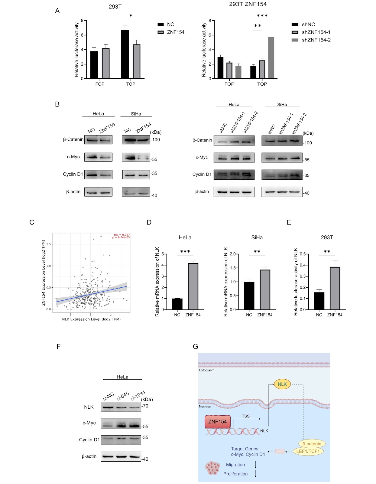
Zinc finger protein 154 inhibits the growth and metastasis of cervical cancer cells through inhibiting Wnt/β-catenin signaling by upregulating NLK

Authors: Chulan Yang, Hongyu Zhao, Jing Tuo, Wei Zhao, Zhiwei Zhang, Zemin Pan, Lianghai Wang, Haixuan Zhao, Songhua Zhao, Hongtao Li

Cervical cancer represents a significant global health concern affecting women. The global cancer burden data published by the World Health Organization’s International Agency for Research on Cancer (IARC) indicated that the incidence and mortality of cervical cancer were the fourth most common malignancy in females worldwide in 2022 [1]. DNA methylation is recognized as a pivotal epigenetic mechanism for gene silencing, which may accumulate with disease severity [2]. Hypermethylation has been discovered in several tumor suppressor gene (TSG) promoters in human cancers, and further understanding of gene silencing mechanisms has led more studies to consider epigenetic disruption as an important mechanism leading to the silencing of tumor suppressor genes in tumor development [3]. Recent studies have reported that methylation of the zinc finger protein 154 (ZNF154) gene plays an oncogenic role in the development of several cancers [4]. ZNF154 has been shown to inhibit tumor cell proliferation in nasopharyngeal carcinoma by altering the expression of E-cadherin through the Wnt/β-catenin pathway, thereby inhibiting epithelial-to-mesenchymal transition (EMT) [5]. He et al. [6] demonstrated that ZNF154 could transcriptionally regulate the expressions of tumor suppressor genes involved in the cell cycle, the p53 signaling pathway, and the Wnt/β-catenin signaling pathway in esophageal squamous cell carcinoma. Thus, ZNF154 can be considered a novel cancer biomarker of clinical significance. However, the role of ZNF154 in cervical cancer remains unclear. In the present study, we analyzed ZNF154 expression and its potential biological functions and molecular mechanisms in cervical cancer. ZNF154 was found to be downregulated by promoter methylation in cervical cancer tissue. Its overexpression in cervical cancer cells inhibited cell proliferation and migration. Mechanistically, ZNF154 inhibits the Wnt/β-catenin signaling pathway by directly targeting and positively modulating Nemo-like kinase (NLK) activity. Collectively, our findings indicate the crucial role of ZNF154 in the proliferation and migration of cervical cancer cells, indicating that ZNF154 may serve as a promising target for future therapeutic development.

Zinc finger protein 154 inhibits the growth and metastasis of cervical cancer cells through inhibiting Wnt/β-catenin signaling by upregulating NLK
Graphical Abstract
Original ResearchVol. 58, Issue 2 • pp. 463-465DOI: 10.3724/abbs.2025140

ADD domain added new binding partners for the nuclear hub protein ATRX

Authors: Yan Chen, Yang Luo, Jielin Sun, Shouhua Wang, Bingbing Wan

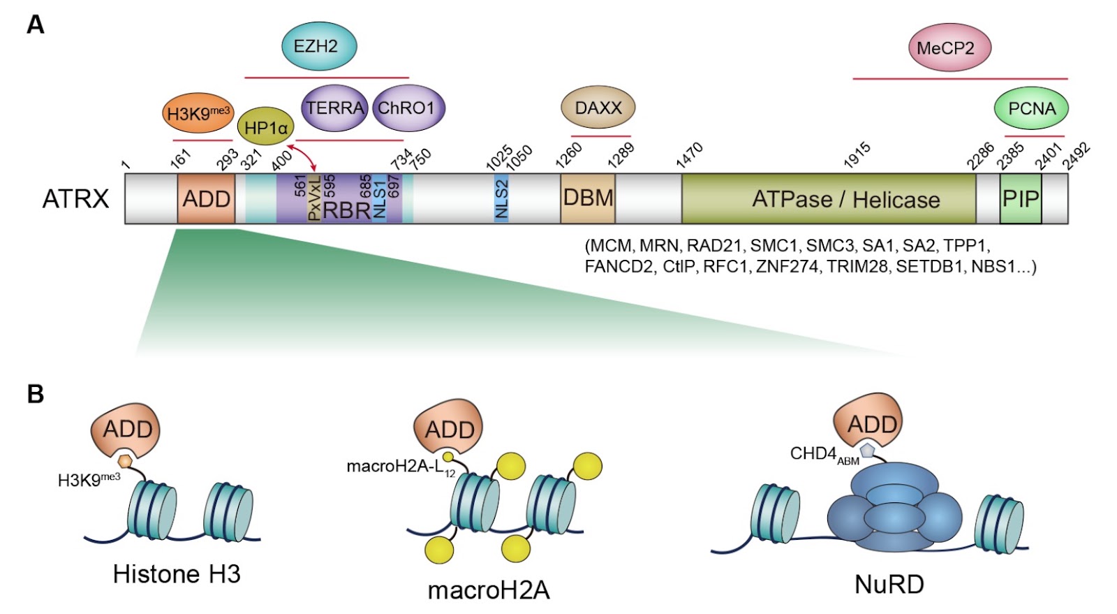
ATRX is a large, multi-domain nuclear protein that functions as a crucial ATP-dependent chromatin remodeler, transcriptional regulator, and guardian of telomeric and genomic integrity. As a member of the SWI/SNF family of chromatin remodeling proteins, a primary and well-defined function of ATRX is to facilitate the replication-independent deposition of the histone variant H3.3 at specific genomic loci, predominantly repetitive sequences such as telomeres, pericentromeric heterochromatin, and ribosomal DNA (rDNA). Mutations in the ATRX gene are associated with a severe X-linked neurodevelopmental disorder and alpha-thalassemia. Moreover, ATRX dysfunction can lead to genomic instability, contributing to the development and progression of various cancers, including gliomas and pancreatic neuroendocrine tumors (PanNETs). ATRX orchestrates chromatin dynamics through its modular domains. Its N-terminal ADD domain and a PxVxL-like motif recognize histone H3K9me3 and interact with the heterochromatin-binding protein HP1α, respectively. Collaborating with the histone chaperone DAXX, ATRX then utilizes its C-terminal ATPase/Helicase domain to provide the energy needed to remodel chromatin and deposit the histone variant H3.3 into repetitive DNA regions. The Chen lab and other two groups previously elucidated the minimal elements for DAXX interaction, demonstrating that a short 1260‒1289 residues motif (DAXX-binding motif, DBM) of ATRX is solely responsible for ATRX-DAXX heterodimer formation. Beyond its role in chromatin remodeling, ATRX employs its RBR (RNA-binding region) to engage the telomeric long non-coding RNA (lncRNA) TERRA, thereby regulating TERRA-mediated R-loops and telomeric G-quadruplex (G4) structures. Interestingly, this same RBR also binds the muscle-specific lncRNA ChRO1 to coordinate constitutive heterochromatin reorganization and regulate cell differentiation. Notably, nearly half of the disease-causing mutations in ATRX, leading to a severe neurodevelopmental disorder, are clustered within the ADD domain, highlighting this relatively small domain’s critical functional importance and warranting more intense investigation. This Research Highlight discusses recent findings by Yan et al. that the histone variant macroH2A binds the ATRX ADD domain, expanding the known binding partners of this domain and providing structural insights into the interaction.

ADD domain added new binding partners for the nuclear hub protein ATRX
Graphical Abstract
Original ResearchVol. 58, Issue 2 • pp. 201-215DOI: 10.3724/abbs.2025106

Energy stress and adaptation strategy of tumor cells in different microenvironments: from primary tumors to distant metastases

Authors: Mingzhe Xu, Junjie Fei, Zhi-Xiong Xiao, Yong Yi

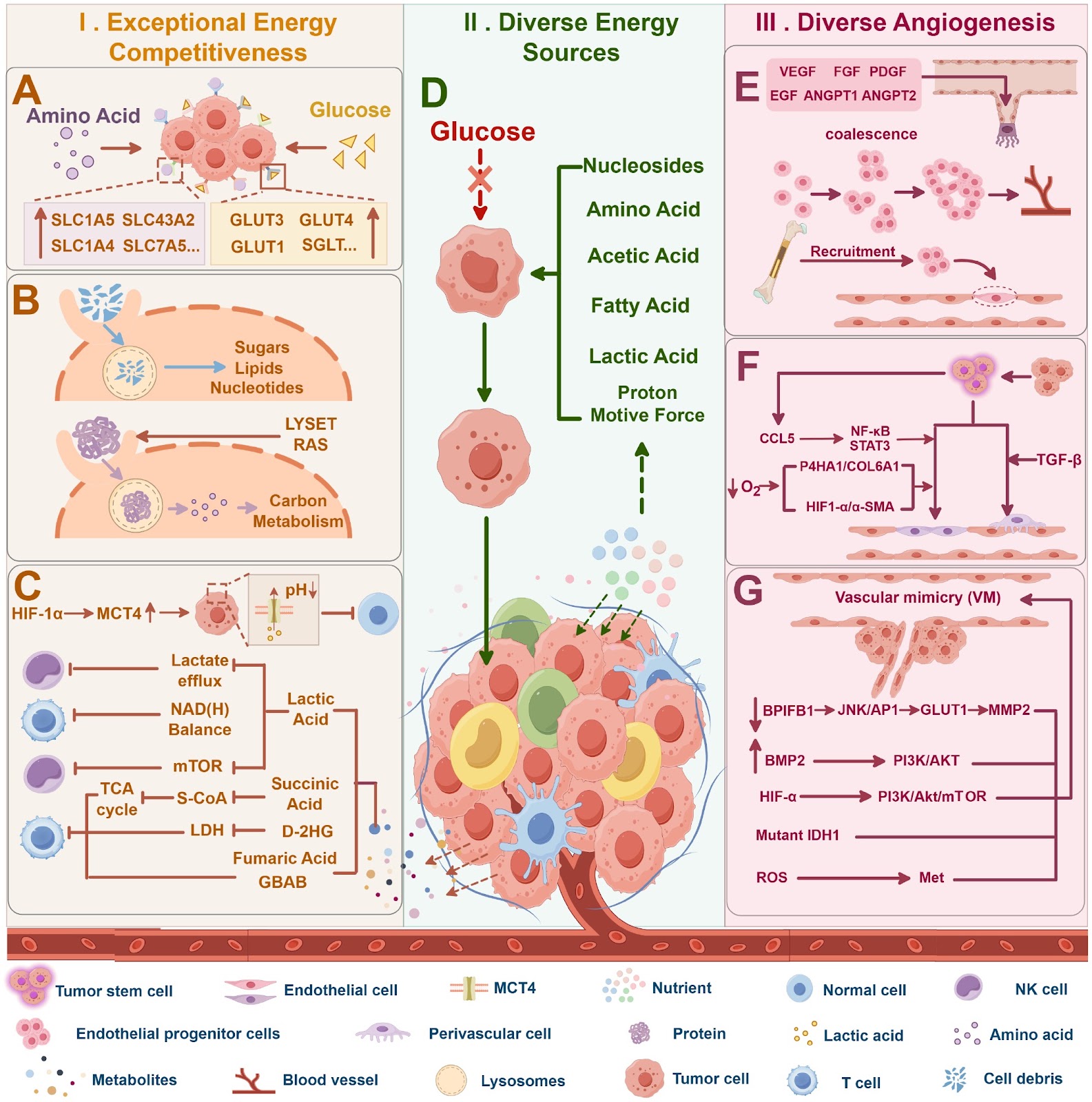
Since the Warburg effect was first described in the 1920s, tumor energy metabolism has been a central focus of cancer research, emerging as a potential therapeutic target. The tumor microenvironment—including blood vessels, immune cells, stromal components, and other cell types—profoundly influences tumor cell metabolism. Variations in energy supply, oxygen availability, nutrient composition, and the accumulation of metabolic waste across different microenvironments challenge tumor cell survival and progression. In response, tumor cells adapt through flexible regulation and reprogramming of metabolic pathways. Although recent studies have explored metabolic adaptation mechanisms in various tumor microenvironments, the full spectrum from primary tumors to distant metastases remains unexplored. This review summarizes energy stress and adaptation maneuvers in tumor cells across different stages of tumor progression and offers a new perspective for comprehensive research to explore therapeutic strategies targeting tumor metabolism.

Energy stress and adaptation strategy of tumor cells in different microenvironments: from primary tumors to distant metastases
Graphical Abstract
Original ResearchVol. 58, Issue 2 • pp. 303-310DOI: 10.3724/abbs.2025120

Structural insight into Vibrio cholerae EIIC sugar transporter dimer captured in a substrate-free inward-facing state

Authors: Hanhan Guo, Qiaoshuo Zhang, Zhao Wang, Kuo Zhang, Yang Fu

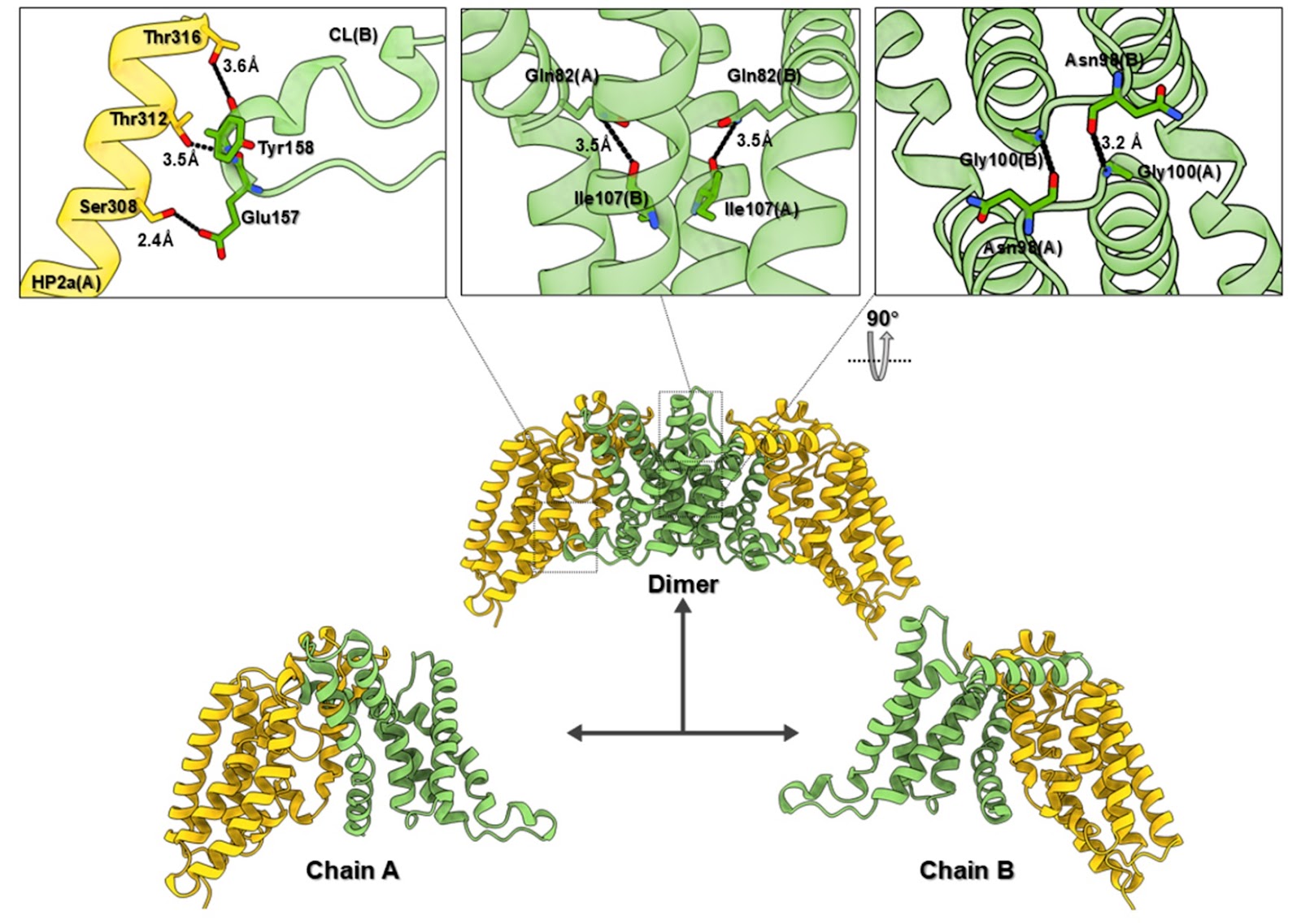
The phosphoenolpyruvate-dependent sugar phosphotransferase system (PTS) is a central pathway for carbohydrate transport in bacteria and plays a critical role in nutrient acquisition, metabolism, and virulence. In Vibrio cholerae, the glucose-specific EIIC transporter is a key component of the PTS system, mediating the transport of sugars into the bacterial cell, coupled with phosphorylation during translocation. Here, we present the 3.68 Å cryo-electron microscopy (cryo-EM) structure of the dimeric EIIC transporter from Vibrio cholerae in its inward-facing, substrate-free conformation. The structure reveals a detailed arrangement of the scaffold and transport domains, stabilized by extensive inter- and intraprotomer interactions. Comparative analysis with substrate-bound inward-facing structures of EIIC from E. coli highlights conformational changes, providing insights into substrate release and the structural transitions required for alternating access. Notably, the observed substrate-free inward-facing conformation features a larger substrate-binding pocket, which is consistent with a state poised for glucose release into the cytoplasm. The formation of a unique intraprotomer disulfide bond between residues C240 and C254 stabilizes the interface between the scaffold and transport domains, potentially regulating transporter dynamics. These findings elucidate the structural basis for substrate release in the PTS system and underscore the dynamic nature of EIIC-mediated sugar transport. Our study enhances the understanding of PTS system function in Vibrio cholerae and highlights the EIIC transporter as a promising target for antimicrobial drug development. Disruption of sugar transport in this essential pathway could impair bacterial growth and virulence, suggesting a novel therapeutic strategy against cholera. These results provide a foundation for future investigations into the structural and functional dynamics of bacterial sugar transporters.

Structural insight into Vibrio cholerae EIIC sugar transporter dimer captured in a substrate-free inward-facing state
Graphical Abstract