🧬 SinoBioData Academic Portal
🏛️ Indexed Academic JournalOriginal: 干细胞研究与转化

Stem Cell Research & Therapy

Premier Chinese Biomedical Journal indexed in SinoBioData: Stem Cell Research & Therapy (干细胞研究与转化).

Total Research Papers: 200
Access: 100% Free Open Access
Browse by Publication Year & VolumeReset All Filters ✕

Published Research PapersFiltered: Year 2026 • 17 • None

Showing 26 of 200 peer-reviewed papers with full Graphical Abstracts.

Original ResearchVol. 17, None • pp. 191DOI: 10.1186/s13287-026-04989-4

Isorhamnetin-preconditioned MSC-derived exosomes restore ovarian function by inhibiting ferroptosis in chemotherapy-induced POF

Authors: Qiang Zhang, Jinyu Yu, Yan Zheng, Jinlan Jiang, Lianwen Zheng

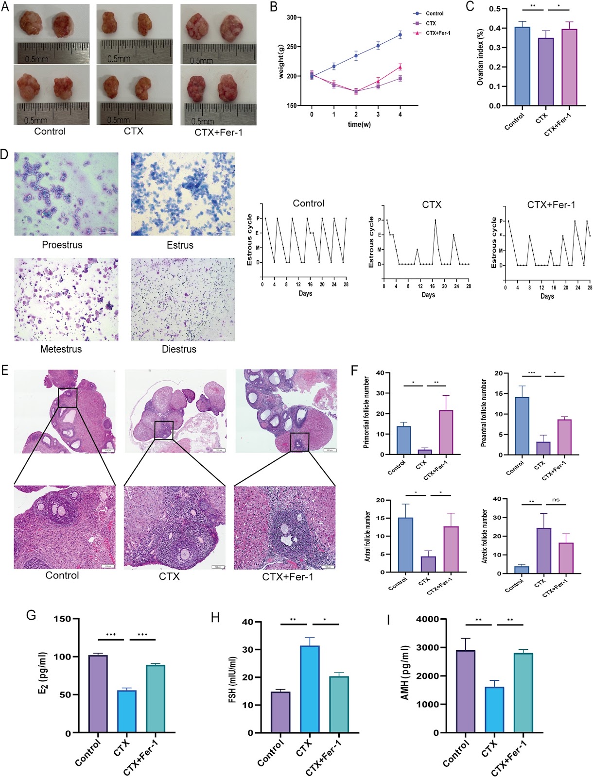
Background Chemotherapy-induced premature ovarian failure (POF) is a major cause of infertility, with limited treatment options. Mesenchymal stem cell-derived exosomes (MSC-Exos) have therapeutic potential. This study investigated whether preconditioning MSCs with the antioxidant flavonoid isorhamnetin (ISO) enhances the efficacy of their exosomes (ISO-MSC-Exos) against POF. Methods A cyclophosphamide-induced POF rat model was established, and the role of the ferroptosis inhibitor ferrostatin-1 was evaluated. MSC-Exos and ISO-MSC-Exos were isolated by ultracentrifugation and administered via tail vein injection. Ovarian recovery was assessed by monitoring the oestrous cycle, serum hormone levels, and histological findings. Lipid peroxidation and iron metabolism were evaluated by quantifying malondialdehyde, glutathione, iron deposition, and mitochondrial ultrastructure. Immunohistochemistry was used to assess the expression levels of GPX4, ACSL4, and FTH1. Proteomic analyses were performed to explore the underlying mechanisms. Results Ferroptosis plays a pivotal role in the cyclophosphamide-induced POF rat model. Both exosome therapies improved ovarian function and suppressed ferroptosis, with ISO-MSC-Exos showing superior efficacy. ISO-MSC-Exos significantly restored hormone levels, ameliorated oestrous cycle disorders, reduced follicular atresia, and enhanced fertility. Furthermore, ISO-MSC-Exos more effectively elevated glutathione levels, reduced malondialdehyde and Fe2⁺ levels, and reversed the abnormal expression of ferroptosis-related proteins GPX4, ACSL4, and FTH1. Proteomic analysis suggested that ISO-MSC-Exos effectively inhibit ferroptosis by downregulating Alox15 and Tf, thereby reducing lipid peroxidation substrates and cellular iron uptake. This finding represents a potential molecular mechanism underlying their superior efficacy compared with that of MSC-Exos. Conclusions ISO-MSC-Exos showed superior efficacy compared with MSC-Exos in restoring ovarian function and inhibiting ferroptosis, suggesting that ISO pretreatment enhances the therapeutic effect of MSC-Exos in the POF
Isorhamnetin-preconditioned MSC-derived exosomes restore ovarian function by inhibiting ferroptosis in chemotherapy-induced POF
Graphical Abstract
Original ResearchVol. 17, None • pp. 177DOI: 10.1186/s13287-026-04995-6

Peptide-based Wnt signal activation enables scalable production of clinical-grade patient-derived intestinal organoids for regenerative cell therapy

Authors: Hady Yuki Sugihara, Sayaka Nagata, Sho Kawasaki, Junichi Takahashi, Yui Hiraguri, Masayoshi Fukuda, Kohei Suzuki, Tatsuro Murano, Satoru Fujii, Toshimitsu Fujii, Hiromichi Shimizu, Kazuo Ohtsuka, Mamoru Watanabe, Ryuichi Okamoto, Tomohiro Mizutani

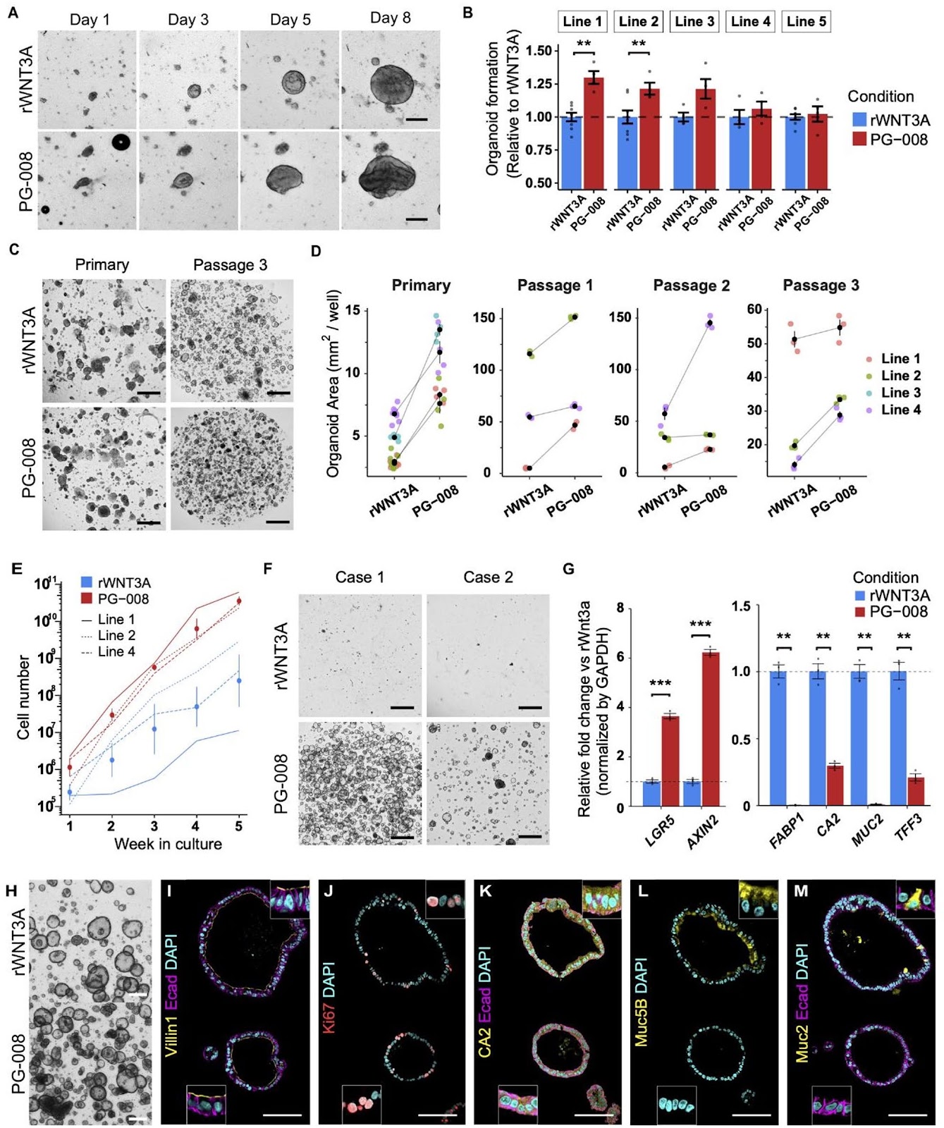
Background Gastrointestinal diseases often involve cellular damage, degeneration or dysfunction in the tract, frequently requiring surgical interventions risking complications and lowered quality of life. Regenerative medicine holds great promise in improving patient care and providing novel treatment options for previously irreparable and untreatable tissues. Despite the clinical potential of intestinal organoids as a resource for regenerative cell therapy and bioengineering, the lack of clinical-grade cultures has hampered further development. Moreover, strategies to efficiently and reliably expand clinical-grade cultures at the scale required for application is limited. Methods A GMP-compliant protocol was developed to generate patient-derived colonic organoids from endoscopic biopsies. Clinical-grade colonic organoids cultured and expanded in Type-I collagen were compared to conventional Matrigel cultured organoids. To improve the culture-, cost-, and time-efficiency of culture expansion, several strategies were developed including organoid area-based passaging, one well plate culture, and the incorporation of Wnt activating peptide, PG-008. Conventional recombinant WNT3A culture was compared to the peptide PG-008 culture using single cell RNA sequencing. Results Clinical-grade collagen cultured organoids exhibited similar culture efficiency to Matrigel. Organoid establishment rate from 60 patients using the GMP-compliant protocol was 82%. The incorporation of PG-008 significantly enhanced organoid growth and stabilized patient-patient variability through intestinal stem cell (ISC) enrichment. Single cell RNA sequencing revealed that PG-008 resulted in remarkably pure culture consisting of ISCs

Peptide-based Wnt signal activation enables scalable production of clinical-grade patient-derived intestinal organoids for regenerative cell therapy
Graphical Abstract
Original ResearchVol. 17, NoneDOI: 10.1186/s13287-026-04965-y

Targeting p75NTR activity alleviates the neurotoxic effect of high glucose on iPSC-derived dopaminergic neurons

Authors: Konstantina Chanoumidou, Ioanna Zota, Maria Anna Papadopoulou, Chrystalla Konstantinou, Alexandros Tsimpolis, Electra Tsagliotis, Maria Tziortziou, Katerina Ntarntani, Anne Grünewald, Matthieu David Lavigne, Achille Gravanis, Ioannis Charalampopoulos

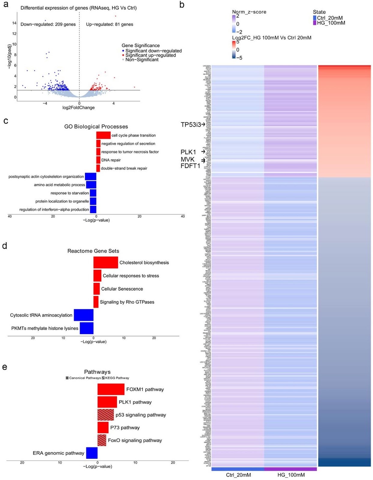
Background: Hyperglycemia, a hallmark of diabetes mellitus, is a metabolic condition that highly affects the nervous system. While evidence from epidemiological and animal studies links diabetes to dopaminergic dysfunction and an increased risk of Parkinson’s disease, the underlying mechanisms remain unclear. Here, we examined the effects of high glucose on human iPSC-derived dopaminergic neurons and glial cells to better understand the pathogenic alterations that lead to neurotoxicity. Previous implication of neurotrophins in the neurological manifestations of diabetes prompted us to focus on the role of p75NTR neurotrophin receptor (p75NTR) in dopaminergic neurodegeneration under hyperglycemic conditions. Methods: iPSC-derived dopaminergic neurons, astrocytes and microglia were treated with high glucose (50mM, 100mM) for 48 h to simulate hyperglycemia. Cytotoxicity assays, RNA sequencing and DNA damage assessments were employed to investigate the pathological alterations induced by high glucose exposure in neurons. Pharmacological targeting of p75NTR activity allowed investigation of its involvement in glucose neurotoxicity. Glial-mediated neurotoxicity was evaluated using conditioned media and inflammatory marker analysis. Results: High glucose treatment led to DNA damage, activation of JNK signaling and cell death in neurons. Importantly, we observed upregulation of p75NTR and its pro-apoptotic ligand pro-NGF, suggesting activation of the pro-NGF/p75NTR axis in high glucose-treated neurons. Inhibition of p75NTR activity rescued neuronal cell death, identifying p75NTR as a central mediator of glucose neurotoxicity. Furthermore, glucose overload sensitized neurons to 6-hydroxydopamine (6-OHDA), increasing their vulnerability to neurotoxic insults—an effect reversed by p75NTR blockade. Treatment with BNN27, a synthetic NGF mimetic, prevented neuronal loss through p75NTR and TrkA receptors, suggesting neurotrophin signaling as a potential therapeutic target for combating high glucose-induced neuronal damage. Finally, we demonstrated the contribution of glial cells to neurodegeneration since high glucose

Targeting p75NTR activity alleviates the neurotoxic effect of high glucose on iPSC-derived dopaminergic neurons
Graphical Abstract
Original ResearchVol. 17, None • pp. 237DOI: 10.1186/s13287-026-05051-z

Awakening endogenous repair: salidroside boosts mitophagy in NPMSCs via SIRT1/FOXO3 to combat intervertebral disc degeneration

Authors: Zhengguang Li, Yiming Wu, Benkui Hua, Hua Sun, Huofeng Wu, Shuangjia Zai, Chen Liu, Yongbo Zhang, Zhaoyu Li, Xuan You, Yufeng Huang, Xuhua Lu, Guoyong Yin, Liang Zhang

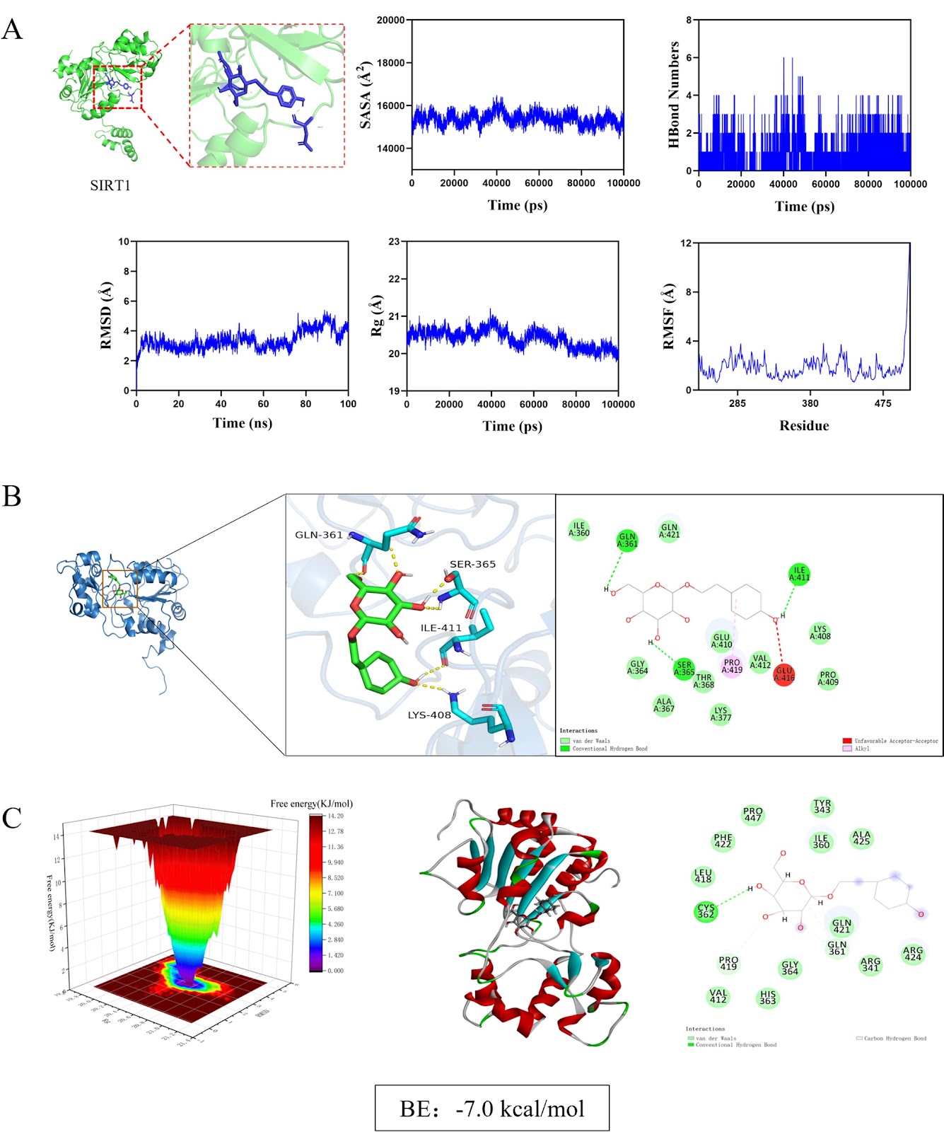
Background Intervertebral disc degeneration (IVDD) is a major cause of low back pain, characterized by mitochondrial dysfunction and impaired mitophagy in nucleus pulposus-derived mesenchymal stem cells (NPMSCs). Sirtuin 1 (SIRT1), a key regulator of mitochondrial quality control, is downregulated in degenerated discs. Salidroside (Sal), a natural compound from Rhodiola rosea, has shown potential in enhancing mitophagy, but its mechanism in IVDD remains unclear. Methods Using network pharmacology, molecular docking, and dynamics simulations, we identified SIRT1 as a key target of Sal. Human and rat NPMSCs were isolated and treated with tert-butyl hydroperoxide (TBHP) to induce degeneration. In vitro assays included CCK-8, EdU, SA-β-Gal, JC-1, Western blot, immunofluorescence, and transmission electron microscope (TEM). An in vivo rat IVDD model was established via needle puncture and treated with Sal and/or the autophagy inhibitor 3-MA. Histological, immunohistochemical, and imaging analyses were performed to evaluate IVDD. Results Sal bound stably to SIRT1 and activated SIRT1/FOXO3 pathway, promoting mitophagic flux, reducing reactive oxygen species accumulation, and suppressing apoptosis in NPMSCs. SIRT1 knockdown or 3-MA treatment abolished Sal’s protective effects. In vivo, Sal treatment preserved disc height, reduced apoptosis, and enhanced mitophagy, while 3-MA exacerbated degeneration.

Awakening endogenous repair: salidroside boosts mitophagy in NPMSCs via SIRT1/FOXO3 to combat intervertebral disc degeneration
Graphical Abstract
Original ResearchVol. 17, None • pp. 153DOI: 10.1186/s13287-026-04954-1

Tanshinone IIA-pretreated mesenchymal stem cells alleviate neuroinflammation in 3×Tg-AD mice via the TREM2/PI3K/Akt pathway

Authors: Jingjing Wu, Ying Ge, Li Zhang, Juan Huang, Nanqu Huang, Yong Luo

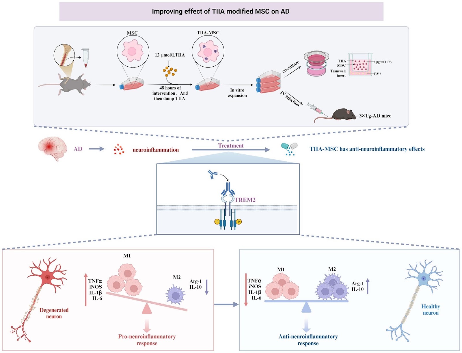
Neuroinflammation is a key pathogenic factor for neurodegenerative diseases. Mesenchymal stem cell (MSC) transplantation, as a potential strategy for regulating neuroinflammation, has received extensive attention. Our previous research revealed that compared with ordinary MSC, MSC pretreated with tanshinone IIA (TIIA), referred to as TIIA-MSC, exhibited superior anti-neuroinflammatory activity, but the mechanism of action remains unclear. To clarify the underlying mechanism, this study integrated in vitro and in vivo experiments and evaluated the therapeutic effect of TIIA-MSC in a triple-transgenic Alzheimer’s disease mouse model (3×Tg-AD mice) and explored its mechanism of action in a lipopolysaccharide (LPS)-induced BV2 microglial cell inflammation model. The results showed that TIIA-MSC could significantly improve the cognitive function of 3×Tg-AD mice, increase brain glucose metabolism levels, promote the recovery of synaptic and mitochondrial structures, and effectively alleviate neuroinflammatory responses. In vitro experiments further verified the superior inhibitory effect of TIIA-MSC on microglial cell activation and proinflammatory factor release. Mechanistic studies have indicated that the triggering receptor expressed on myeloid cells 2 (TREM2) is the key molecule that mediates this process. The knockdown of TREM2 expression significantly weakened the anti-inflammatory effect of TIIA-MSC, suggesting that TREM2 plays a central role in this process. Further analysis revealed that by activating the phosphoinositide 3-kinase (PI3K)/protein kinase B (Akt) signaling pathway downstream of TREM2, TIIA-MSC may promote the transformation of the functional state of microglia from mainly proinflammatory to having neuroprotective and repair properties. This study systematically revealed the molecular mechanism by which TIIA-MSC regulate microglial cell phenotypic transformation through the TREM2/PI3K/Akt pathway and exert anti-neuroinflammatory effects, providing new ideas and an experimental basis for expanding the application of MSC in the treatment of neurodegenerative diseases.

Tanshinone IIA-pretreated mesenchymal stem cells alleviate neuroinflammation in 3×Tg-AD mice via the TREM2/PI3K/Akt pathway
Graphical Abstract
Original ResearchVol. 17, None • pp. 75DOI: 10.1186/s13287-026-04896-8

Deficiency of extracellular vesicles miR-32 from bone marrow mesenchymal stem cells alleviates vascular calcification in type 2 diabetes by inhibiting endothelial ferroptosis

Authors: Zhengjie Lin, Anqi Li, Jie Zheng, Kun Luo, Fei Liang, Shiyan Liu, Zhengfeng Liang, Wei Liu, Jian Tang, Xiaolin Zhong, Jianghua Liu

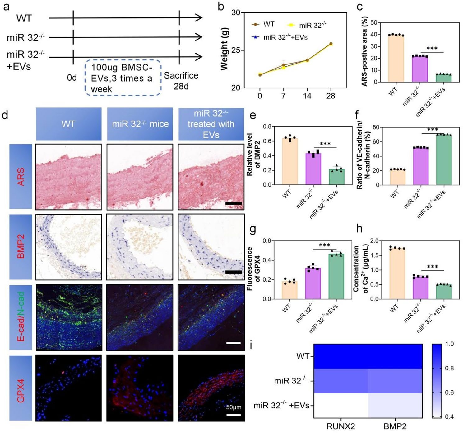
Background The development of vascular calcification (VC) in diabetes is closely related to the endothelial-to-mesenchymal transition (EndMT). We found that microRNA-32-5p (miR-32) was elevated in the plasma of calcification patients. However, it is unclear whether miR-32 mediates the function of bone marrow mesenchymal stem cell-derived extracellular vesicles (BMSC-EVs) in type 2 diabetes (T2D) VC. Methods BMSC-EVs were characterized by TEM, NTA, Western blotting, and confocal microscopy. Alizarin Red and ALP staining assessed the severity of VC. qRT-PCR and Western blotting evaluated the expression of BMP2, RUNX2, GPX4, SLC7A11, VE-cadherin, and N-cadherin, while immunofluorescence was used for detecting VE-cadherin and N-cadherin. In vivo validation was performed using miR-32–/– and ApoE–/– mice. RNA sequencing (RNA-seq) and bioinformatics analysis was conducted to explore underlying mechanisms. Results We demonstrated that BMSC-EVs attenuate VC in endothelial cells (ECs) and inhibit EndMT. In vivo, histological analysis showed that treatment with BMSC-EVs significantly reduced the severity of VC associated with T2D. Notably, knockout of miR-32 further enhanced the inhibitory effect of BMSC-EVs on VC. Mechanistically, transcriptomic and functional analyses suggest that the protective effect of BMSC-EVs on VC is associated with regulation of the MAPK/FoxO signaling pathway, potentially mediated by modulation of ferroptosis. Conclusion These findings demonstrate that BMSC-EVs attenuate T2D-associated VC, partially through miR-32-mediated suppression of EC ferroptosis.

Deficiency of extracellular vesicles miR-32 from bone marrow mesenchymal stem cells alleviates vascular calcification in type 2 diabetes by inhibiting endothelial ferroptosis
Graphical Abstract
Original ResearchVol. 17, None • pp. 78DOI: 10.1186/s13287-026-04895-9

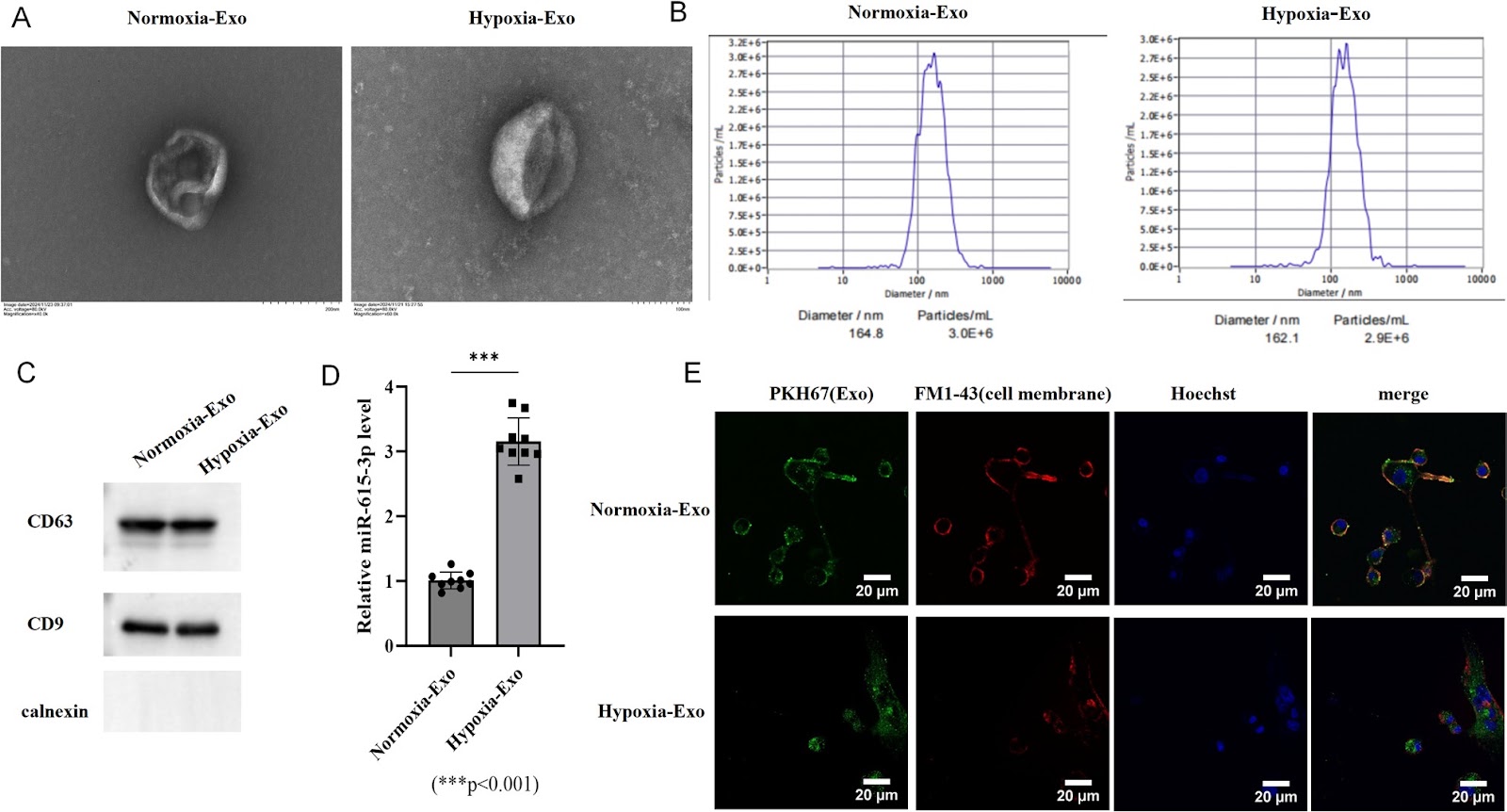
Hypoxia-conditioned BMSC exosomes improve short-term spinal cord injury outcomes via the miR-615-3p/PDE4C-mediated cAMP/PKA pathway

Authors: Wei Bian, Xiangyu Zeng, Ziwen Liu, Mingyan Guan, Tegeleqi Bu, Haoze Li, Zewei Gao, Jianyu Liu

Spinal cord injury (SCI) remains a significant global health challenge with limited effective therapeutic options. Exosomes derived from mesenchymal stem cells (MSCs) have emerged as promising neuroprotective agents due to their biocompatibility and immunomodulatory properties. This study investigated the therapeutic potential of hypoxia-conditioned bone marrow MSC (BMSC)-derived exosomes in both in vitro and in vivo SCI models. Hypoxic preconditioning significantly enriched miR-615-3p in bone marrow mesenchymal stem cell (BMSC)-derived exosomes. In spinal neuron injury models, hypoxic exosomes enhanced cell viability, reduced apoptosis, and ameliorated dysfunction of the mitochondria-associated endoplasmic reticulum membranes (MAMs). Mechanistically, miR-615-3p directly targeted and suppressed phosphodiesterase 4 C (PDE4C), activating the cyclic adenosine monophosphate (cAMP)/protein kinase A (PKA) pathway. This in turn modulated calcium signaling, attenuated mitochondrial calcium overload, and reduced endoplasmic reticulum stress (ERS). In a mouse model of SCI, short-term treatment with hypoxic exosomes promoted functional recovery within a 14-day post-injury period, as evidenced by improved locomotor performance, reduced lesion volume, attenuated tissue edema, and decreased inflammatory cell infiltration. Furthermore, in vivo administration of hypoxic exosomes upregulated miR-615-3p and downregulated PDE4C expression in injured spinal cord tissues. These results demonstrate that hypoxia-conditioned BMSC-derived exosomes exert neuroprotective effects via the miR-615-3p/PDE4C axis, highlighting their potential as a novel therapeutic strategy for SCI by targeting calcium homeostasis and mitochondrial-ER dysfunction. These findings demonstrate the short-term therapeutic potential of hypoxia-conditioned exosomes in SCI. However, further preclinical studies, including long-term follow-up to assess the durability of recovery and potential late-onset effects, alongside clinical validation, are warranted before clinical translation.

Hypoxia-conditioned BMSC exosomes improve short-term spinal cord injury outcomes via the miR-615-3p/PDE4C-mediated cAMP/PKA pathway
Graphical Abstract
Original ResearchVol. 17, None • pp. 209DOI: 10.1186/s13287-026-05020-6

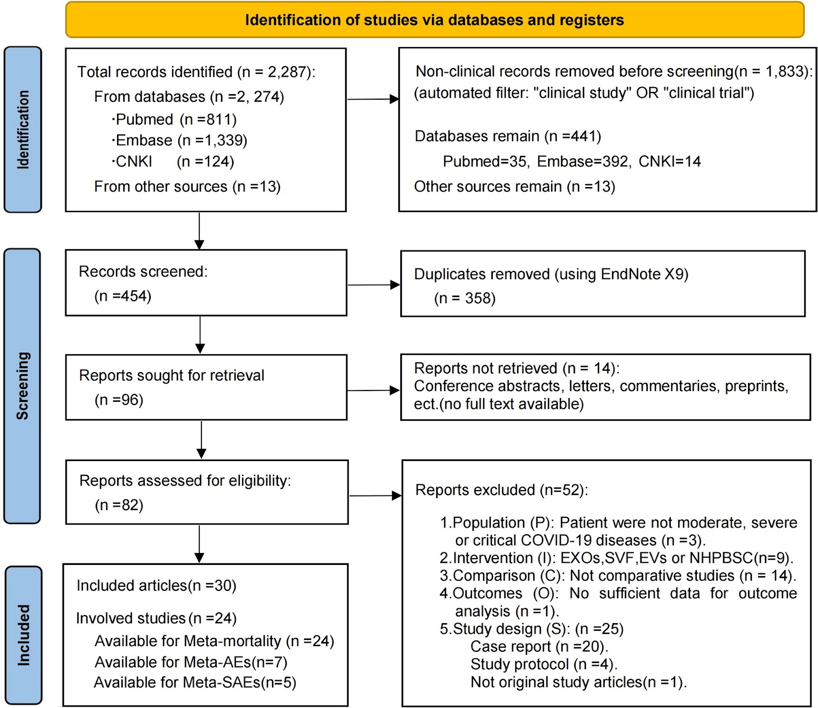
Therapeutic potential of mesenchymal stromal cells in COVID-19: a meta-analysis of clinical trials conducted since the pandemic onset

Authors: Meng-Qi Yuan, Yue-Fei Pan, Zi-Ying Zhang, Ya-Xin Wu, Kai-Di Zhu, Ze-Rui Wang, Ze-Yi Zhang, Jia-Qi Xiong, Zhe Xu, Lei Huang, Fu-Sheng Wang, Lei Shi

Background Severe acute respiratory syndrome coronavirus 2 (SARS-CoV-2) infection can induce immune dysregulation and multi-organ injury; mesenchymal stromal cell (MSC) therapy has shown promise in clinical trials for COVID-19 and may have broader applicability to pneumonia induced by respiratory viruses (e.g., the influenza virus). This meta-analysis synthesized the available comparative clinical evidence on the safety and efficacy of MSCs in patients with moderate to critical COVID-19 and examined the reported outcomes relevant to Long-COVID. Methods We searched the PubMed, Embase, and CNKI databases for original, comparative studies in moderate, severe, or critical COVID-19 published up to September 2, 2024. Twenty-four eligible studies (13 RCTs and 11 non-randomized controlled trials; n=1080) were included in the mortality meta-analysis. Patients were assigned to either the intervention group (MSC therapy plus standard care) or the control group (standard care with or without placebo). The primary efficacy outcome was all-cause mortality, while the primary safety outcomes were adverse events (AEs) and serious adverse events (SAEs). Secondary outcomes included clinical recovery, hospitalization metrics, chest imaging, and inflammatory biomarkers. We performed a pooled meta-analysis on mortality with subgroup analyses (by disease severity, administration route, dosing frequency, and study design), assessment of publication bias (using funnel plots and Egger’s test), and evaluation of the quality of evidence via the GRADE approach. AEs/SAEs were analyzed using meta-analysis and descriptive statistics, while other secondary outcomes were summarized descriptively. Results MSC therapy significantly reduced all-cause mortality (MSC: 26.4% vs control: 31.9%; fixed-effect OR=0.74, 95% CI 0.55–0.99), with low heterogeneity (I2=2.8%, P=0.422[Q-test]) and no publication bias. The quality of evidence

Therapeutic potential of mesenchymal stromal cells in COVID-19: a meta-analysis of clinical trials conducted since the pandemic onset
Graphical Abstract
Original ResearchVol. 17, None • pp. 107DOI: 10.1186/s13287-026-04921-w

Inhalation of mesenchymal stromal cell-derived extracellular vesicles activates macrophage polarization through the miR-22-3p/NLRP3/IL-1β pathway, ameliorating lung ischemia-reperfusion injury

Authors: Tao Wang, Guodong Wu, Peigen Gao, Fenghui Zhuang, Zeyu Wang, Ziheng Zhou, Chongwu Li, Junqi Wu, Deping Zhao

Background Lung ischemia–reperfusion injury (IRI) is a major contributor to primary graft dysfunction (PGD) after lung transplantation. Mesenchymal stem cell-derived extracellular vesicles (MSC-EVs) have emerged as promising therapeutic agents in inflammatory diseases by ameliorating tissue damage and promoting repair. However, the anti-inflammatory efficacy of these approaches and the underlying mechanisms in lung ischemia–reperfusion injury remain incompletely understood. Methods The protective effects of mesenchymal stromal cell-derived extracellular vesicles (MSC-EVs) against lung ischemia–reperfusion injury were evaluated using two delivery approaches, inhalation and intravenous injection. Both in vivo and in vitro models were employed to assess the biological activity of MSC-EVs and to elucidate the underlying molecular mechanisms. In addition, a rat orthotopic lung transplantation (OLT) model was established to further examine the translational relevance of MSC-EVs. Results MSC–EVs treatment significantly ameliorated lung IRI, with inhalation showing superior efficacy over intravenous delivery. Mechanistically, miR-22-3p within MSC-EVs targeted macrophage NLRP3, suppressing activation of the NLRP3/Caspase-1/IL-1β pathway and promoting M2 polarization. The protective efficacy was confirmed in a clinically relevant rat OLT model, underscoring their translational potential

Inhalation of mesenchymal stromal cell-derived extracellular vesicles activates macrophage polarization through the miR-22-3p/NLRP3/IL-1β pathway, ameliorating lung ischemia-reperfusion injury
Graphical Abstract
Original ResearchVol. 17, None • pp. 100DOI: 10.1186/s13287-026-04903-y

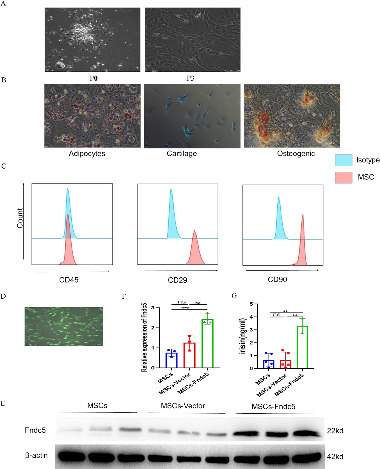
Fndc5 modification optimizes the therapeutic effect of rat MSCs on sepsis-induced ALI/ARDS via activating the PI3K/AKT signaling pathway

Authors: Yuling Luo, Minhua Li, Shan Lin, Zheng Gong, Sumei Wang, Ziqing Zhou, Shiyue Li

Background Acute lung injury/Acute respiratory distress syndrome (ALI/ARDS) is a life-threatening inflammatory lung disorder characterized by high mortality rates and a lack of effective treatment options. Although mesenchymal stem cell (MSC)-based therapies have emerged as a promising approach for ARDS management, optimizing their therapeutic efficacy remains a significant challenge. Recent advances in gene modification techniques have opened new avenues for enhancing MSC functionality. Among these, Fibronectin type III domain-containing protein 5 (Fndc5)/irisin has attracted considerable attention due to its ability to improve endothelial function. This study aims to evaluate the therapeutic potential of Fndc5-modified MSCs in sepsis-induced ALI/ARDS and to elucidate the underlying molecular mechanisms driving their protective effects. Methods To comprehensively evaluate the therapeutic potential of Fndc5-modified MSCs (MSCs-Fndc5) in ARDS, we employed both in vivo and in vitro experimental models. In vivo, a mouse model of sepsis-induced ALI was established through intraperitoneal injection of lipopolysaccharide (LPS), and the protective effects of MSCs-Fndc5 were systematically assessed by analyzing lung histopathology, inflammatory cytokine levels, vascular endothelial integrity, lung wet-to-dry weight ratio, and MSC retention in lung tissue. In parallel, in vitro studies were conducted to investigate the role of MSCs-Fndc5 in mitigating LPS-induced endothelial cell (EC) injury, with a focus on EC proliferation, angiogenesis, barrier permeability, apoptosis, and the regulation of key signaling pathways. Results Fndc5 modification significantly increased the retention rate of MSCs in sepsis-induced ALI murine model while augmenting their in vitro proliferation and migration potential. In vivo, treatment with Fndc5-modified MSCs markedly attenuated lung inflammation, as evidenced by reduced levels of pro-inflammatory cytokines, decreased neutrophil infiltration, and improved lung histopathology. Additionally, MSCs-Fndc5 alleviated pulmonary edema, reduced fibrosis, lowered the lung wet-to-dry weight ratio, and preserved vascular endothelial integrity. In vitro, Fndc5 modification optimizes the therapeutic effect of rat MSCs on sepsis-induced ALI/ARDS via activating the PI3K/AKT signaling pathway.

Fndc5 modification optimizes the therapeutic effect of rat MSCs on sepsis-induced ALI/ARDS via activating the PI3K/AKT signaling pathway
Graphical Abstract
Original ResearchVol. 17, NoneDOI: 10.1186/s13287-026-04991-w

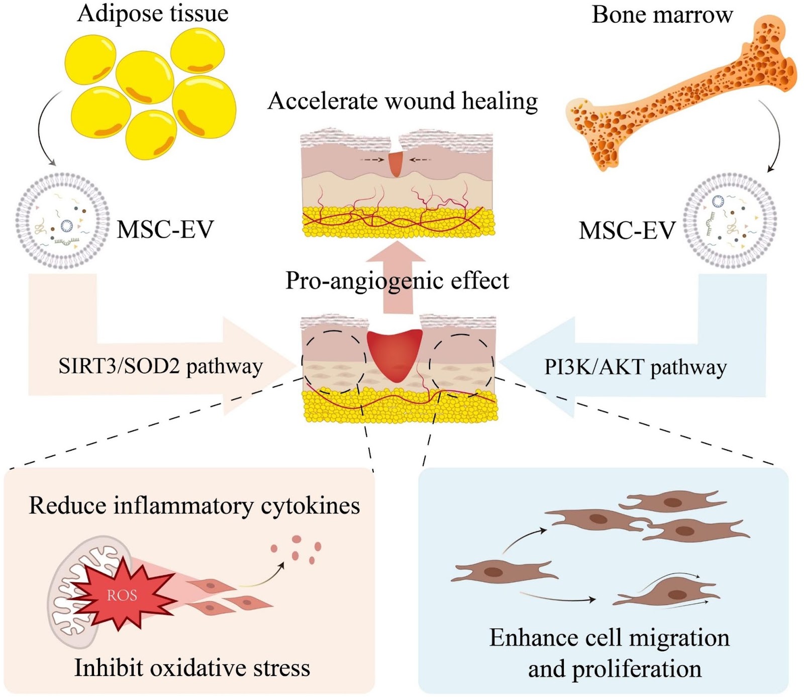
Mesenchymal stem cell-derived extracellular vesicles in the treatment of type 2 diabetes and its complications: current progress and future directions

Authors: Sha Zhang, Zong-Yu Zhang, Ruo-Nan Tang, Kai Zhang, Yu Fu, Hua Tian, Jing Ma, Yan Jin, Chen-Xi Zheng, Bing-Dong Sui

Type 2 diabetes (T2D) and its complications represent a complex disorder involving multiple pathophysiological processes. Although conventional therapeutic approaches partially regulate blood glucose, they fail to fundamentally reverse disease progression or effectively prevent complications. This review summarizes the current research advance and challenges of using different forms of mesenchymal stem cell-derived extracellular vesicles (MSC-EVs) in treating T2D and complications. It begins with an introduction to the characteristics of MSC-EVs. Subsequently, the mechanisms and therapeutic prospects of natural MSC-EVs are analyzed, with a focus on their roles in inflammatory modulation, tissue regeneration, and improving insulin resistance. Engineering MSC-EVs, covering strategies including optimizing MSC culture conditions, modifying EV contents, and establishing MSC-EV delivery systems based on bioactive materials are then discussed, which boost EV yield and quality while enhancing therapeutic efficacy. Current challenges, including the limited yield and high heterogeneity of natural MSC-EVs, as well as issues related to long-term safety, immunocompatibility, and large-scale production of engineered MSC-EVs are finally overviewed, with emphasizing artificial intelligence in guiding future research directions. These summaries are crucial for clinical translation of MSC-EVs and will ultimately provide T2D patients with an effective and safe treatment option.

Mesenchymal stem cell-derived extracellular vesicles in the treatment of type 2 diabetes and its complications: current progress and future directions
Graphical Abstract
Original ResearchVol. 17, None • pp. 204DOI: 10.1186/s13287-026-05026-0

DMOG pretreatment restores osteogenic–adipogenic balance and mitochondrial function in ONFH BMSCs through the HIF-1α/Homer3 pathway

Authors: Qiheng Chen, Lingxian Yi, Penghui Nie, Jie Wang, Jing Zhu, Jiang Peng, Tujun Weng

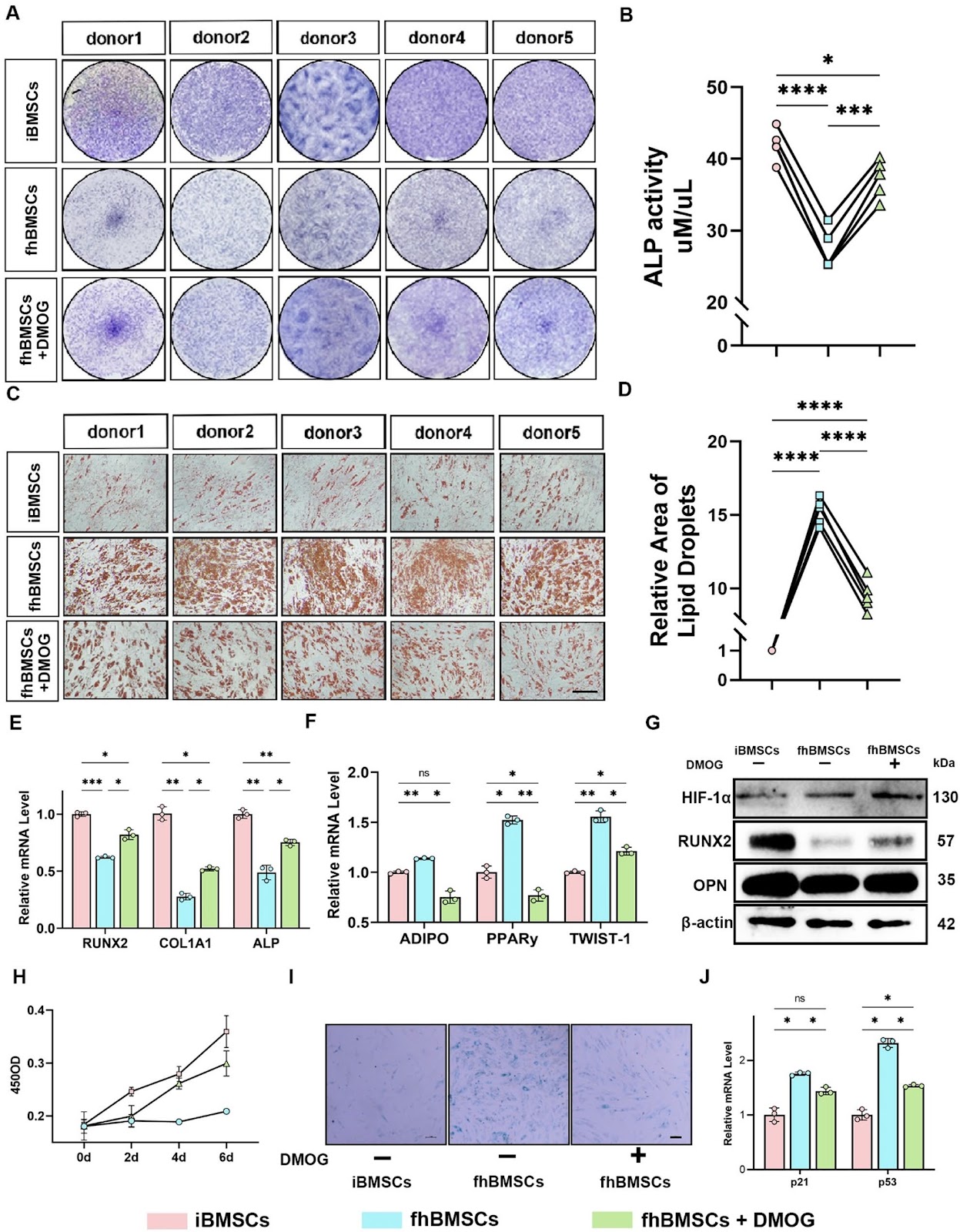
Background  Osteonecrosis of the femoral head (ONFH) is a progressive orthopedic disorder that often culminates in femoral head collapse and joint failure. Dysfunction of bone marrow mesenchymal stem cells (BMSCs), including impaired osteogenesis, enhanced adipogenesis, and mitochondrial dysfunction, has been increasingly recognized as a central driver of ONFH pathogenesis. However, the molecular mechanisms linking metabolic stress to lineage imbalance remain poorly defined. Methods  Paired BMSCs were isolated from necrotic femoral head regions (fhBMSCs) and the iliac crest (iBMSCs) of ONFH patients. Functional assays, RNA sequencing, and molecular analyses were performed to evaluate the effects of the hypoxia mimetic dimethyloxalylglycine (DMOG) on osteogenic–adipogenic balance, mitochondrial function, and senescence. Loss-of-function experiments targeting hypoxia-inducible factor-1α (HIF-1α) and Homer3 were conducted to elucidate mechanistic pathways. Results  Compared with iBMSCs, fhBMSCs exhibited impaired osteogenesis, enhanced adipogenesis, mitochondrial dysfunction, and increased senescence. DMOG pretreatment restored osteogenic differentiation, suppressed adipogenesis, improved mitochondrial dynamics, reduced oxidative stress, and enhanced bioenergetic metabolism. These protective effects were dependent on HIF-1α stabilization. Transcriptomic profiling identified Homer3 as a downstream negative regulator of HIF-1α. Homer3 was aberrantly upregulated in fhBMSCs but suppressed by DMOG, and its knockdown mimicked the effects of DMOG by promoting osteogenesis, inhibiting adipogenesis, enhancing mitophagy, and restoring mitochondrial function. Conversely, silencing HIF-1α abolished DMOG-mediated benefits and reinstated Homer3 expression.

DMOG pretreatment restores osteogenic–adipogenic balance and mitochondrial function in ONFH BMSCs through the HIF-1α/Homer3 pathway
Graphical Abstract
Original ResearchVol. 17, None • pp. 91DOI: 10.1186/s13287-026-04913-w

Extracorporeal cardiac shock wave stimulation enhances the therapeutic efficacy of intravenously delivered endothelial colony-forming cells via PI3K/AKT signaling in a rat myocardial infarction model

Authors: Mingqiang Wang, Dan Yang, Yiming Ma, Yunke Shi, Jinping Lun, Chaoyue Zhang, Xinbin Li, Yuchen Shi, Hongyan Cai

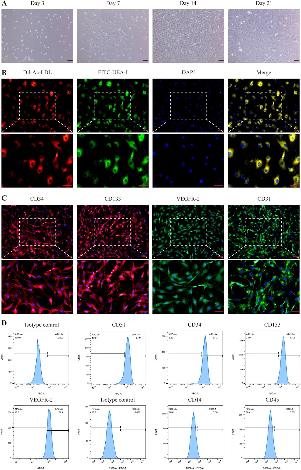
Background Extracorporeal cardiac shock wave (ECSW) therapy enhances the function of endothelial colony-forming cells (ECFCs), but whether it can serve as a preconditioning strategy to enhance myocardial infarction (MI) therapy remains unclear. This study investigated the efficacy and mechanism of intravenously delivered ECSW-preconditioned ECFCs (SW-ECFCs) in a rat MI model. Methods ECFCs were isolated from the bone marrow of ApoE-/- rats and fully characterized. RNA sequencing of control ECFCs versus SW-ECFCs revealed significant enrichment of the PI3K/AKT pathway. We therefore performed a series of in vitro functional assays on these cells, including Transwell migration, Matrigel tube formation, CCK-8 proliferation, flow cytometric apoptosis analysis, and VEGF-A ELISA. The role of the PI3K/AKT pathway was interrogated using the inhibitor LY294002. Subsequently, an acute MI model was established in ApoE-/- rats via left anterior descending coronary artery ligation. Rats were randomized into four groups: MI+PBS, MI+ECFCs, MI+SW-ECFCs, and MI+LY294002-pretreated SW-ECFCs (LY-SW-ECFCs), with sham-operated rats as controls. Comprehensive evaluations included echocardiography, serum injury biomarkers, TTC, and histopathological (H&E, Masson) staining, immunohistochemical detection of cardiomyocyte apoptosis and p-eNOS, immunofluorescence assessment of ECFC homing and vascular markers (CD31, α-SMA, VEGF-A), tissue/plasma nitric oxide measurement, and Western blot analysis of PI3K/AKT signaling proteins. Results Transcriptomic analysis revealed significant enrichment of the PI3K/AKT pathway in SW-ECFCs. Functionally, ECSW enhanced ECFCs migration, tube formation, proliferation, and VEGF-A secretion, while reducing apoptosis;

Extracorporeal cardiac shock wave stimulation enhances the therapeutic efficacy of intravenously delivered endothelial colony-forming cells via PI3K/AKT signaling in a rat myocardial infarction model
Graphical Abstract
Original ResearchVol. 17, None • pp. 146DOI: 10.1186/s13287-026-05022-4

An open phase I/IIa study evaluating safety, patient-reported outcomes and voice function after surgery, local administration of mesenchymal stromal cells and voice training in patients with vocal fold scarring and dysphonia

Authors: Erika Bergström Börlin, Ulrika Nygren, Maria Södersten, Svante Granqvist, Nadir Kadri, Ida Rasmusson Duprez, Katarina Le Blanc, Stellan Hertegård

Background Damage to the vocal folds can result in scarring, leading to chronic, severe voice impairments for which lasting and effective treatments are currently lacking. The aim of this clinical trial was to evaluate the safety and effectiveness of autologous bone marrow-derived Mesenchymal Stromal Cell (MSC) therapy for patients with vocal fold scarring and severe dysphonia. Additionally, the study sought to propose a post-operative voice training protocol and explore its potential role in facilitating voice improvement. Methods Eight patients with vocal fold scarring and chronic dysphonia underwent surgical scar resection and autologous MSC injection, followed by voice training. Safety was continuously monitored for up to 36 months postoperatively. Data to evaluate therapeutic efficacy was collected pre-treatment, 3 and 12 months post-treatment. Assessments included analysis of vocal fold vibrations, Phonation Threshold Pressure, and Maximum Phonation Time. Patient-reported measures were collected using the Voice Handicap Index, the Vocal Fatigue Index, and ratings of major symptoms and their impact on daily life. Treatment effectiveness was analyzed at both group and individual levels, with clinically relevant changes predefined.

An open phase I/IIa study evaluating safety, patient-reported outcomes and voice function after surgery, local administration of mesenchymal stromal cells and voice training in patients with vocal fold scarring and dysphonia
Graphical Abstract
Original ResearchVol. 17, None • pp. 189DOI: 10.1186/s13287-026-05000-w

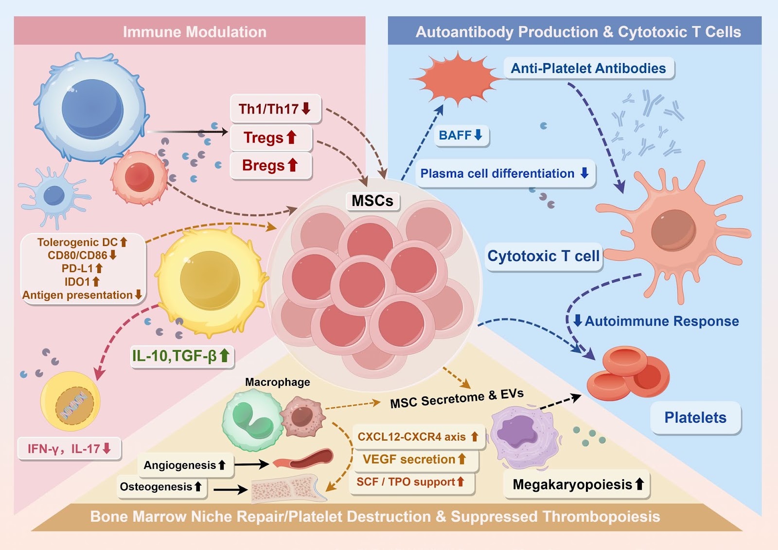
Innovative strategies for immune thrombocytopenia treatment: immunomodulatory mechanisms and clinical potential of mesenchymal stem cells

Authors: Xin Zhou, Ningning Shan

Immune thrombocytopenia (ITP) is a heterogeneous autoimmune disorder characterized by increased platelet destruction and impaired megakaryopoiesis within a dysregulated bone marrow niche. Conventional therapies often achieve only transient platelet recovery, failing to restore immune tolerance, thereby underscoring the need for mechanism-based therapeutic strategies. Mesenchymal stem cells (MSCs) have emerged as promising candidates due to their ability to modulate immune responses and repair the hematopoietic microenvironment. This review synthesizes current evidence regarding the biological properties, immunomodulatory mechanisms, and therapeutic applications of MSCs in ITP, emphasizing intrinsic abnormalities of patient-derived MSCs and the corrective potential of exogenous MSCs from distinct tissue sources. It further integrates emerging insights into MSC functional heterogeneity, optimization of culture conditions, priming strategies, and cellular engineering approaches that may enhance therapeutic efficacy and safety. By highlighting the interplay between immune tolerance restoration and bone marrow niche remodeling, this review provides a translational framework that links mechanistic understanding to the future clinical development of MSC-based therapies for ITP.

Innovative strategies for immune thrombocytopenia treatment: immunomodulatory mechanisms and clinical potential of mesenchymal stem cells
Graphical Abstract
Original ResearchVol. 17, None • pp. 82DOI: 10.1186/s13287-026-04902-z

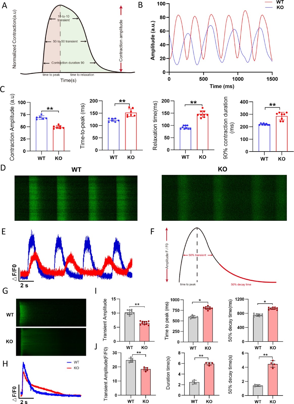
FHOD3 deficiency disrupts sarcomere organization and activates CaMKII signaling in human stem cell-derived cardiomyocytes

Authors: Mingyu Wei, Xiaojie Hou, Siyao Zhang, Xianjing Hu, Xi Chen, Zhen Gao, Shuwan Xu, Zhan Shi, Min Zhu, Feng Lan, Ming Cui

Background Inherited cardiomyopathy (ICM) is a genetic disorder characterized by abnormal myocardial structure and function, often progressing to heart failure. FHOD3, a member of the Formin gene family, plays a crucial role in cardiomyocyte cytoskeletal organization. Mutations in FHOD3 have been associated with various cardiomyopathies, including hypertrophic cardiomyopathy (HCM), dilated cardiomyopathy (DCM) and left ventricular noncompaction (LVNC). However, the molecular mechanisms underlying FHOD3 deficiency-induced cardiomyopathy remain elusive. Methods A FHOD3 knockout (FHOD3-/-) human embryonic stem cell (hESC) line was generated using the CRISPR/Cas9 system and subsequently differentiated into cardiomyocytes (hESC-CMs). Sarcomere structure, calcium handling, mitochondrial function, and contractility were evaluated via immunofluorescence, electron microscopy, Seahorse metabolic analysis, and high-definition video analysis, respectively. Transcriptomic sequencing was performed to identify differentially expressed genes and enriched pathways. Results FHOD3-deficient hESC-CMs exhibited marked sarcomere disorganization and degradation, impaired calcium handling and compromised mitochondrial function, ultimately leading to reduced contractility. Transcriptomic analysis revealed significant downregulation of sarcomere-related genes and calcium-handling genes, with enrichment in pathways associated with cardiomyopathy and calcium signaling. Furthermore, FHOD3 deficiency triggered the phosphorylation of CaMKII (Thr286), a key regulator of cardiac hypertrophy and remodeling, contributing to the progression of heart failure. Treatment with the myosin activator Omecamtiv mecarbil (OM) partially restored contractility without affecting calcium handling, highlighting its potential as a therapeutic strategy.

FHOD3 deficiency disrupts sarcomere organization and activates CaMKII signaling in human stem cell-derived cardiomyocytes
Graphical Abstract
Original ResearchVol. 17, NoneDOI: 10.1186/s13287-026-04979-6

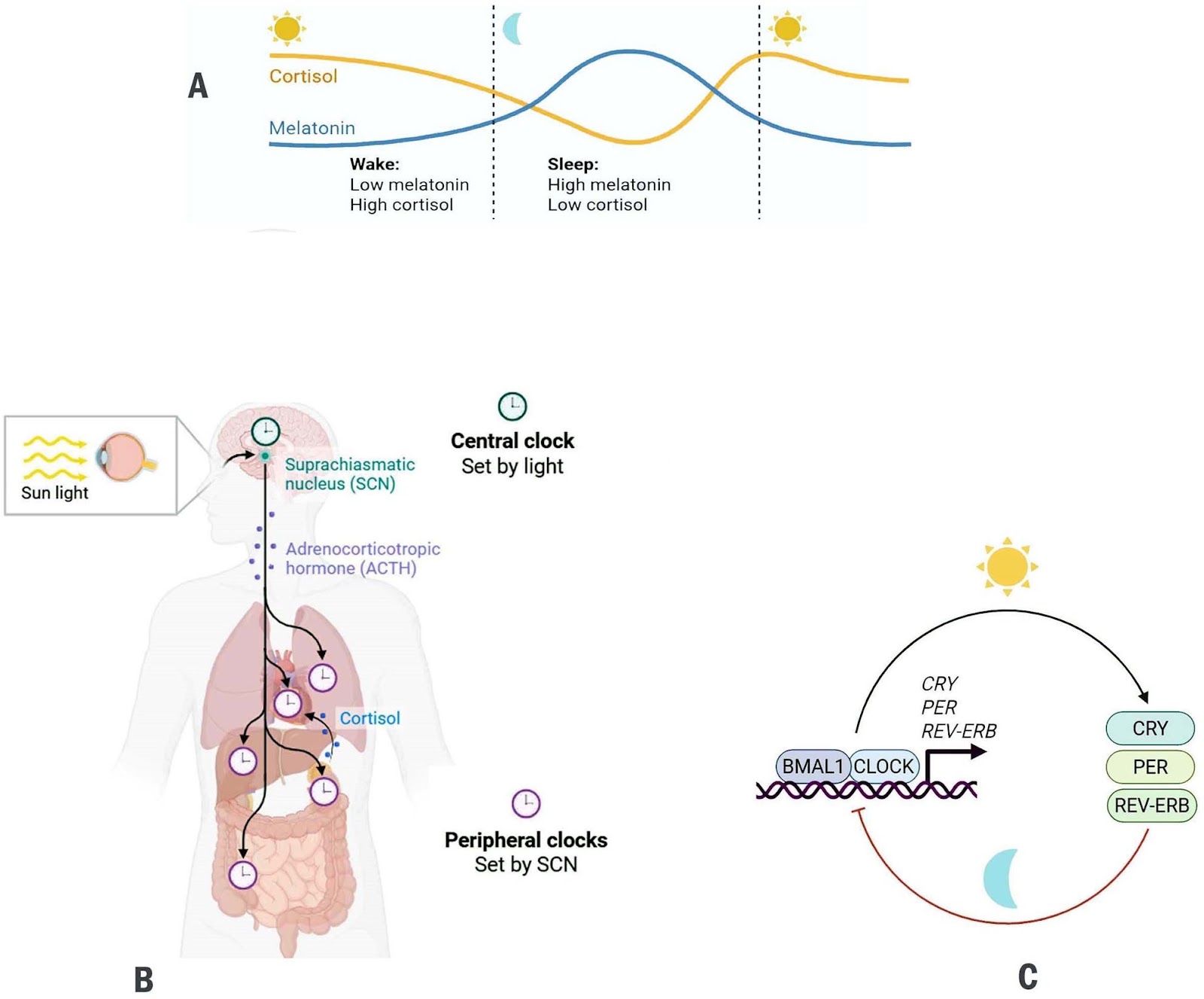
A review of the circadian regulation of stem cells: harnessing the internal body clock for enhanced regenerative therapies

Authors: Sulaiman Mohammed Alnasser

Background Circadian rhythms are endogenous, transcription-translation feedback loops that align cellular activities with the 24-h light–dark cycle. Stem-cell populations across tissues exhibit circadian oscillations that influence their self-renewal, proliferation, and differentiation. Key developmental pathways (Wnt/β-catenin, Notch, and Hedgehog) are increasingly recognized as both regulators and targets of circadian machinery. Objectives This review synthesizes current knowledge on the bidirectional crosstalk between circadian clock components and major stem-cell regulatory pathways, and evaluates how this interplay shapes tissue homeostasis, regenerative capacity, and therapeutic potential. Methods Literature examining molecular interfaces between circadian clock genes and Wnt, Notch, and Hedgehog signaling was surveyed, with emphasis on transcriptional regulation, chromatin dynamics, post-translational control, and functional outcomes for stem-cell behavior and regeneration. Results Evidence indicates that core clock components modulate stem-cell pathways through direct transcriptional control, shared enhancer architecture, altered chromatin accessibility, and rhythmic protein modification. In turn, Wnt, Notch, and Hedgehog signals feed back onto clock genes, influencing circadian amplitude and phase within stem-cell niches. Perturbation of this reciprocal regulation disrupts tissue maintenance, diminishes regenerative responses, alters metabolic equilibrium, and may promote tumorigenesis. Conclusions Circadian oscillators act as temporal gatekeepers of stem-cell function. Mapping the molecular interfaces between clock genes and developmental signaling pathways reveals new opportunities to refine regenerative therapies. Chronotherapeutic strategies, i.e. timing interventions to intrinsic circadian phases may enhance the efficacy, precision, and safety of stem-cell–based treatments.

A review of the circadian regulation of stem cells: harnessing the internal body clock for enhanced regenerative therapies
Graphical Abstract
Original ResearchVol. 17, None • pp. 176DOI: 10.1186/s13287-026-04964-z

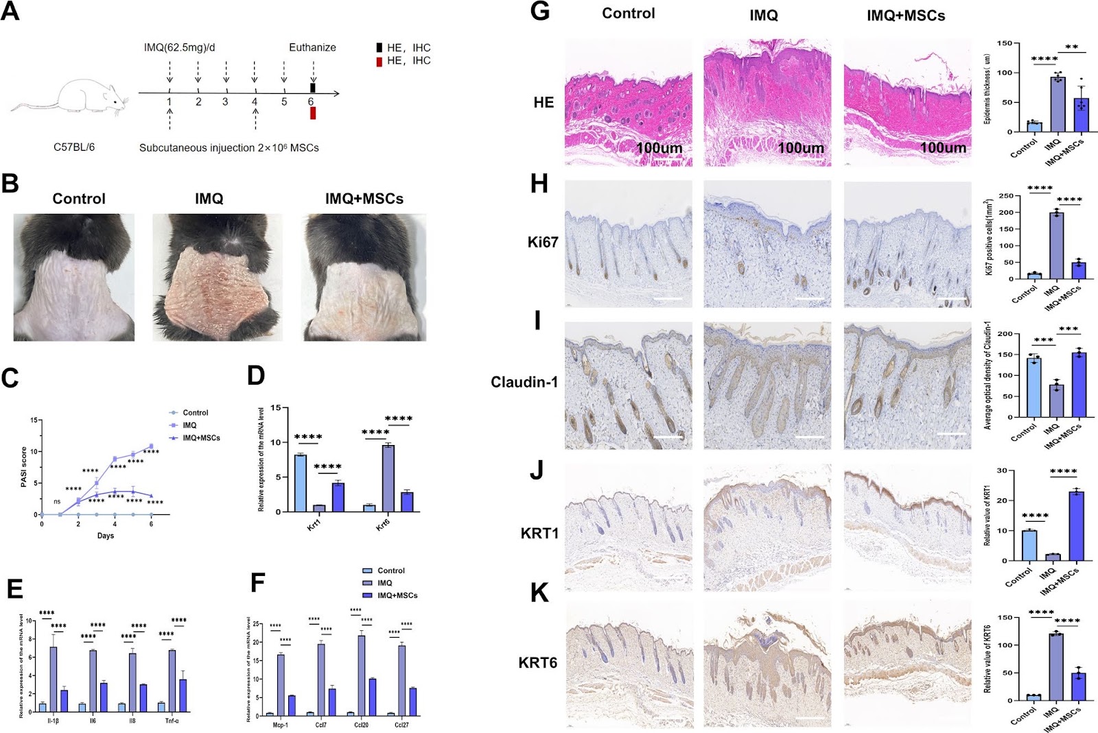
Research based on serine metabolism indicates mesenchymal stem cells alleviate psoriasis by regulating the PSPH-PINK1-Parkin-NLRP3 pathway in HaCaT

Authors: Qing Lin, Yunfei Ji, Bin Yang, Rongjia Zhu, Ping Song, Robert chunhua Zhao

Background Psoriasis is a refractory immune-related disease. In recent years, it has been discovered that mesenchymal stem cells (MSCs) can be used as a new therapeutic approach for psoriasis, but their potential therapeutic mechanism remains unclear. This study aims to explore the role of MSCs in the treatment of psoriasis. Methods We employed a mouse psoriasis model induced by imiquimod (IMQ) in vivo and a co-culture system of MSCs and HaCaT keratinocytes (KCs) cell line in vitro. These approaches allowed us to investigate the effect of MSCs on the levels of inflammatory factors and the activation of inflammasomes in both contexts. Mouse-targeted amino acid sequencing, transmission electron microscopy for in vitro observation, immunofluorescence for both in vivo and in vitro analyses, and siRNA transfection in vitro were employed in this study. Results Our results showed that MSCs significantly improved the skin lesion of mice with psoriasis, and reduced the levels of inflammatory factors and chemokines including IL-1β, IL-6, IL-8, TNF-α, MCP-1, CCL7, CCL20 and CCL27 in the mouse skin lesion areas and M5- induced psoriatic KCs models in vitro. Likewise, MSCs repaired the skin barrier by enhancing claudin-1 expression in vivo. In addition, MSCs increased KRT1 and decreased KRT6 levels in vivo and in vitro. Amino acid metabolism analysis showed that MSCs could improve the serine metabolism level in the mouse skins and upregulated the key enzyme phosphoserine phosphatase (PSPH) in serine metabolism. In vitro experiments demonstrated that knockdown of PSPH could reverse the therapeutic effects of MSCs on psoriasis. Furthermore, studies in vitro and in vivo revealed that MSCs can activate the PINK1-Parkin pathway. It was specifically manifested by elevated levels of PINK1, Parkin, p-Parkin, Beclin-1, and LC3B-II/I, coupled with a reduction in P62 protein. Subsequently, the activation of PINK1-Parkin led to decreased expressions of IL-1β, IL-6, IL-8, TNF-α, CCL7, CCL20, CCL27, and MCP-1. In vitro and in vivo experiments indicated that MSCs can reduce the levels of IL-1β, IL-6, IL-8, TNF-α, CCL7, CCL20, CCL27, and MCP-1 by inhibiting the activation of NLRP3 inflammasomes. Meanwhile, PSPH knockdown in vitro can reverse the activating effects of MSCs on the PINK1-Parkin, as shown by decreased levels of PINK, Parkin, p-Parkin, Beclin-1, and LC3B-II/I, concurrently with an elevation in P62. Conclusions The results of this study indicated that MSCs can alleviate IMQ-induced psoriasiform dermatitis in mice by upregulating serine metabolism. The key serine metabolism enzyme PSPH may enhance PINK1/Parkin-mediated mitochondrial autophagy in psoriatic HaCaT and inhibit NLRP3 inflammasome activation in HaCaT cells, thereby alleviating skin inflammatory responses and suppressing skin proliferation in psoriatic mice.

Research based on serine metabolism indicates mesenchymal stem cells alleviate psoriasis by regulating the PSPH-PINK1-Parkin-NLRP3 pathway in HaCaT
Graphical Abstract
Original ResearchVol. 17, NoneDOI: 10.1186/s13287-026-05059-5

Autologous bone marrow mesenchymal stem cell mitochondrial transplantation in recurrent assisted reproductive technology failure: a randomized controlled trial

Authors: Xiaoping Liu, Dandan Wang, Lei Jia, Weixi Chen, Rui Huang, Cong Fang, Cijie Du, Liang Yang, Xingguo Liu, Xiaoyan Liang

Background Mitochondrial dysfunction contributes to poor embryo quality and recurrent assisted reproductive technology (ART) failure. Mitochondrial transplantation (MIT), which involves supplementing oocytes with exogenous mitochondria, has been proposed as a novel strategy to improve ART outcomes. However, both its clinical efficacy and safety remain unclear. Methods In this single-center trial, 151 patients with a history of ≥ 2 failed ART cycles provided 1178 metaphase II (MII) oocytes. Sibling oocytes were randomized 1:1 to receive autologous bone marrow mesenchymal stem cells (BMSCs) mitochondria co-injection during intracytoplasmic sperm injection (ICSI) or standard ICSI. The primary outcome was the rate of day-3 good-quality embryos. Results MIT significantly accelerated early embryonic cleavage at the 3-cell stage and 5-cell stage, but this morphokinetic alteration did not translate into improvements in good-quality embryo rate, clinical pregnancy rate, or live birth rate. Long-term follow-up of 23 live births revealed no adverse effects, with all offspring exhibiting normal growth and development. Exploratory analysis revealed that oocytes yielding ≥ 70% transferable embryos after MIT harbored an elevated higher burden of medium frequency (0.05–0.5) mtDNA point mutations. Conclusions While autologous BMSCs-MIT transiently alters early cleavage kinetics, it does not demonstrate a clinical advantage in unselected patients with recurrent ART failure. Nevertheless, its observed safety profile and the identification of mtDNA mutation burden as a potential predictive biomarker provide a foundation for shifting future MIT research from a universal approach toward precision application in molecularly stratified populations.

Autologous bone marrow mesenchymal stem cell mitochondrial transplantation in recurrent assisted reproductive technology failure: a randomized controlled trial
Graphical Abstract
Original ResearchVol. 17, None • pp. 192DOI: 10.1186/s13287-026-05008-2

ATG5 overexpression enhances the therapeutic efficacy of mesenchymal stem cells in a mouse colitis model by augmenting anti-inflammatory and antioxidative mechanisms

Authors: Yang Sun, Huiyan Sun, Tengfei Zhao, Yanyun Zhao, Fengjun Xiao, Lisheng Wang

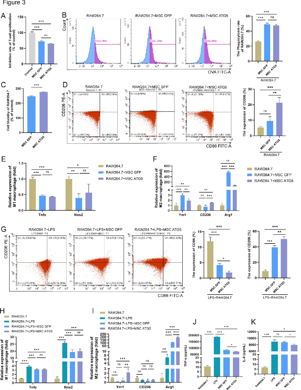
Background The therapeutic efficacy of mesenchymal stem cells (MSCs) can be improved by enhancing their adaptation to the inflammatory microenvironment. Autophagy maintains MSCs functionality, and autophagy-related gene 5 (ATG5) mediates autophagy and regulates the biological functions and therapeutic efficacy of these cells. The aim of this study was to investigate the role of ATG5 in the antioxidant capacity and evaluate the therapeutic effect of ATG5-engineered MSCs for colitis treatment. Methods Cell viability was assessed using a Cell Counting Kit-8. The mRNA expression of autophagy-, antioxidant-, and polarization-related genes was determined through real-time quantitative polymerase chain reaction, and protein expression was analyzed via western blotting. Macrophage polarization markers were analyzed using flow cytometry. Multiomics approaches, including RNA transcriptome sequencing, untargeted metabolomics, and 16S ribosomal RNA microbiota analysis, were also used. Mice with dextran sulfate sodium-induced colitis were used to evaluate the therapeutic efficacy of MSCs. Results Preconditioning MSCs with hypoxia (1% O₂) and serum deprivation significantly enhanced autophagy and upregulated ATG5 expression. Adenovirus-mediated ATG5 overexpression in MSCs (MSCs-ATG5) enhanced their autophagic activity and antioxidant capacity, upregulated HMOX-1, SOD2, and CAT expression, and increased glutathione peroxidase and catalase enzymatic activity, while enhancing cell proliferation, without altering surface marker expression. Further, MSCs-ATG5 significantly promoted M2 macrophage polarization and regulated oxidative stress-related signaling pathways. Additionally, MSCs-ATG5-based therapy markedly ameliorated colitis disease signs in mice. Transcriptome analysis revealed that MSCs-ATG5 suppressed the IL-17/NF-κB inflammatory signaling pathway. This treatment also regulated levels of the anti-inflammatory metabolite prostaglandin D2 (PGD2) in colon tissues.

ATG5 overexpression enhances the therapeutic efficacy of mesenchymal stem cells in a mouse colitis model by augmenting anti-inflammatory and antioxidative mechanisms
Graphical Abstract
Original ResearchVol. 17, None • pp. 116DOI: 10.1186/s13287-026-04938-1

Research progress on the effects of M1/M2 macrophages on the differentiation and maturation of stem cell-derived cardiomyocytes: a review

Authors: Xi Wu, Fan Zhou, Junsheng Mu

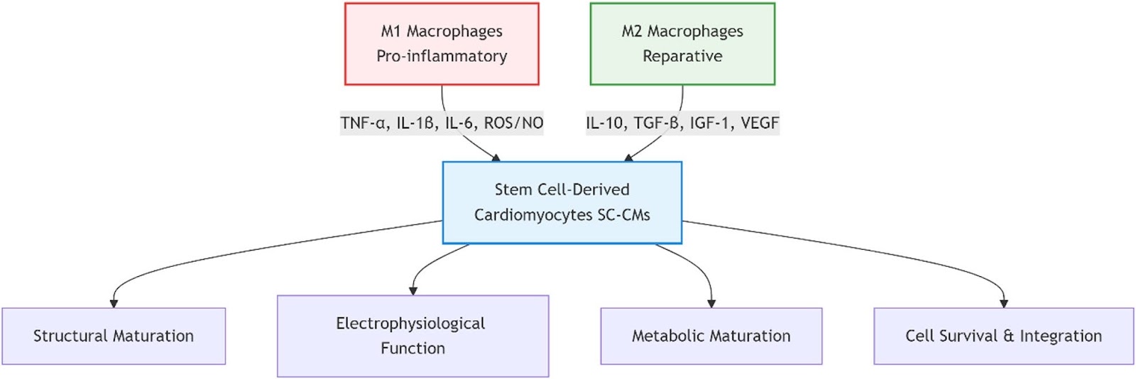
Stem cell-derived cardiomyocytes (SC-CMs) represent a promising cell source for cardiac regenerative medicine, disease modeling, and drug screening. However, their clinical translation faces significant challenges, including functional immaturity, poor long-term survival, and inadequate integration with host tissue following transplantation. The immune microenvironment, particularly the dynamic polarization of macrophages into pro-inflammatory (M1) or reparative (M2) phenotypes, is increasingly recognized as a critical regulator of cardiac repair, yet a systematic understanding of its specific effects on SC-CM fate remains incomplete. This review aims to comprehensively evaluate the dual regulatory roles of M1 and M2 macrophages on the differentiation efficiency, structural and functional maturation, and in vivo transplantation efficacy of SC-CMs. A systematic literature search was conducted in PubMed, Web of Science, Nature, and CNKI for relevant studies published from database inception to July 2025. After screening, 92 articles were included for analysis. The synthesized evidence demonstrates that M1 macrophages and their secreted factors (e.g., TNF-α, IL-1β) impede cardiac differentiation by inhibiting the Wnt/β-catenin pathway, disrupt sarcomeric organization and calcium handling, and maintain SC-CMs in a glycolytic, immature state. In contrast, M2 macrophages enhance SC-CM maturation by providing trophic support (e.g., IGF-1, HGF), promoting electrophysiological maturation and metabolic reprogramming towards oxidative phosphorylation, and facilitating angiogenesis via VEGF. The novelty of this review lies in its integrated perspective on macrophage-driven immunomodulation as a central axis for SC-CM maturation. Furthermore, it discusses emerging therapeutic strategies—such as optimized transplantation timing, co-transplantation with immunomodulatory cells, engineered exosomes, and smart biomaterials—that leverage macrophage polarization to create a favorable microenvironment for SC-CMs. Ultimately, harnessing macrophage-SC-CM crosstalk is a crucial step toward advancing clinically effective and immunologically informed cardiac regeneration therapies.

Research progress on the effects of M1/M2 macrophages on the differentiation and maturation of stem cell-derived cardiomyocytes: a review
Graphical Abstract
Original ResearchVol. 17, None • pp. 155DOI: 10.1186/s13287-026-04974-x

A novel OTUD5 variant disrupts neural progenitor cell homeostasis: mechanistic insights from HEK293T cell-based analyses

Authors: Na Xu, Shihao Wang, Tingting Yang, Meiping Yu, Yu Sun, Yongkun Zhan, Yongguo Yu

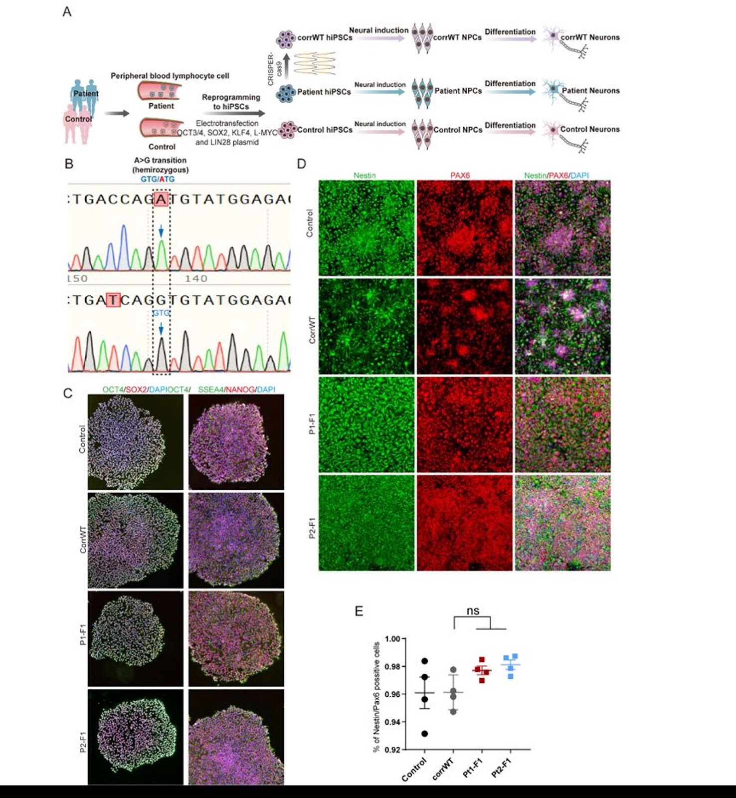
Background Variants in OTUD5 are associated with neurodevelopmental disorders (NDDs), yet the underlying molecular mechanisms remain unclear. This study aimed to investigate the pathogenicity of a novel OTUD5 variant (c.697G > A, p.Val233Met) and elucidate its regulatory role in neural progenitor cell (NPC) proliferation and differentiation, thereby uncovering the function of OTUD5 in neurodevelopment. Methods The OTUD5 variant was identified in two NDD patients via exome sequencing. Patient-derived induced pluripotent stem cells (iPSCs) and CRISPR/Cas9-corrected isogenic controls were generated. NPC proliferative activity was assessed by Ki67 immunofluorescence staining, cell-cycle distribution was analyzed by flow cytometry, and neuronal differentiation was evaluated by Tuj1/MAP2 immunofluorescence staining. Substrate screening was conducted in HEK293T cells using co-immunoprecipitation (Co-IP) and mass spectrometry. Deubiquitination capacity and protein stability were validated through ubiquitination assays and cycloheximide (CHX) chase experiments. Results The p.Val233Met variant, located within the catalytic OTU domain, induced a marked conformational alteration in the OTUD5 protein. Functionally, the variant caused aberrant NPC proliferation (1.8-fold increase in Ki67+ cells, accompanied by release of G1 arrest) and impaired neuronal differentiation (60% reduction in Tuj+ cells). Mechanistically, wild-type OTUD5 stabilized GSK3β by removing K48-linked ubiquitin chains, whereas the mutant isoform exhibited diminished deubiquitinase activity, accelerating GSK3β degradation and shortening its half-life by 40%. Conclusion This study establishes a novel disease mechanism whereby OTUD5 mutations disrupt NPC homeostasis through GSK3β destabilization, highlighting the critical role of ubiquitination regulation in neurodevelopment. Our iPSC model provides a platform for testing GSK3β-targeted therapies in OTUD5-related NDDs.

A novel OTUD5 variant disrupts neural progenitor cell homeostasis: mechanistic insights from HEK293T cell-based analyses
Graphical Abstract
Original ResearchVol. 17, NoneDOI: 10.1186/s13287-026-05080-8

Construction of liver organoid models by hepatobiliary differentiation from human induced pluripotent stem cells: state of the art, challenges and improving strategies

Authors: Yi-Hang Wu, Yang Wang, Wen-Jie Zhang, Li-Li Yuan, Yan Chen

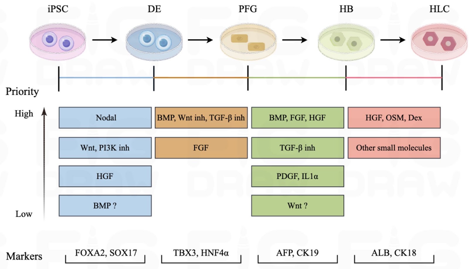
Physiologically relevant liver models are essential for advancing hepatic disorder research, especially for disease modeling and drug development, yet current in vitro systems fail to adequately recapitulate the architecture and function of the liver. Owing to the accessibility, robust proliferation and multilineage differentiation potential of human induced pluripotent stem cells (iPSCs), liver organoids derived from iPSCs have emerged as a promising resource in hepatology. Despite this promise, the field still faces persistent bottlenecks including incomplete hepatic maturation, insufficient incorporation of non-parenchymal cells (notably immune and stromal populations), phenotypic instability, and a lack of consensus on standardized differentiation protocols. Therefore, this review systematically analyzes the challenges and strategies of iPSC differentiation into liver organoids and the related influencing factors by focusing on multidimensional regulation of hepatobiliary development as well as the effects of cellular origin, culture system and liver microenvironment on hepatic differentiation of iPSCs. Moving forward, priority should be given to the following directions: (1) Elucidating the self-assembly mechanism of liver organoids to enable precise control of hepatobiliary differentiation, thereby better governing organoid morphology and improving reproducibility; (2) Replacing exogenous cytokines with small-molecule compounds at different stages of iPSC differentiation to simplify and standardize differentiation protocols; (3) Advancing liver organoid transplantation as a means to validate physiological functionality and shift cell therapy from passive replacement toward active tissue reconstruction; (4) Integrating artificial intelligence to achieve intelligent and precise regulation of hepatic differentiation.

Construction of liver organoid models by hepatobiliary differentiation from human induced pluripotent stem cells: state of the art, challenges and improving strategies
Graphical Abstract
Original ResearchVol. 17, None • pp. 252DOI: 10.1186/s13287-026-05066-6

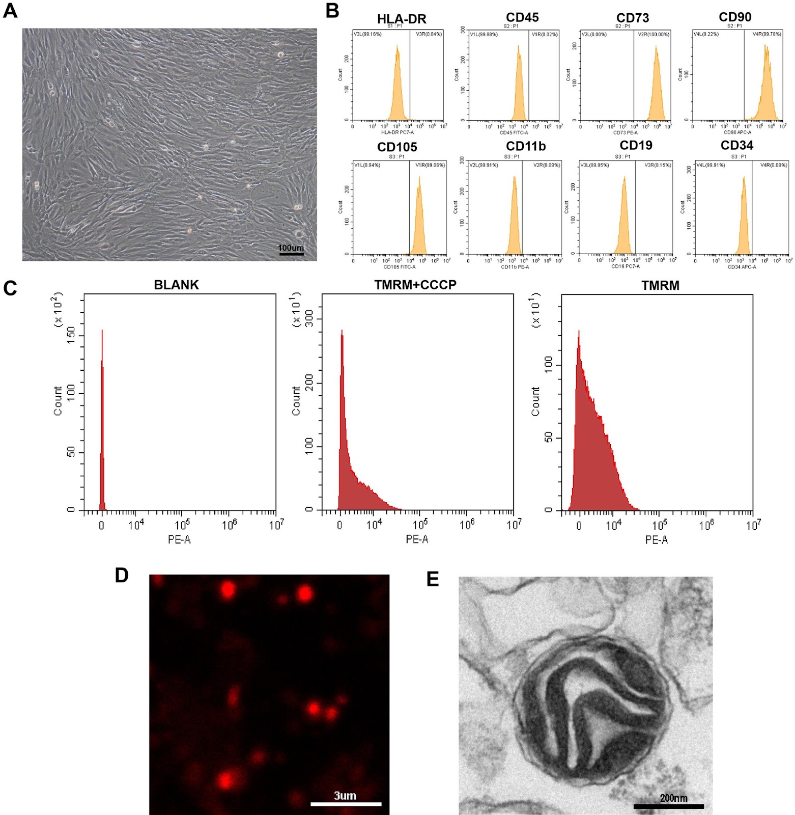
Prevascularization of electrospun PCL/PLA scaffolds using human adipose-derived stem and endothelial cells enhances vascular integration and host angiogenesis in vivo

Authors: Lavinia Grob, Dagmar Brislinger, Marc Mueller, Anja Högler, Kathrin Galistl, Monika Sundl, Daniel Kummer, Nassim Ghaffari-Tabrizi-Wizsy, Hannah Müller, Melanie Pichlsberger, Lars-Peter Kamolz, Ingrid Lang-Olip

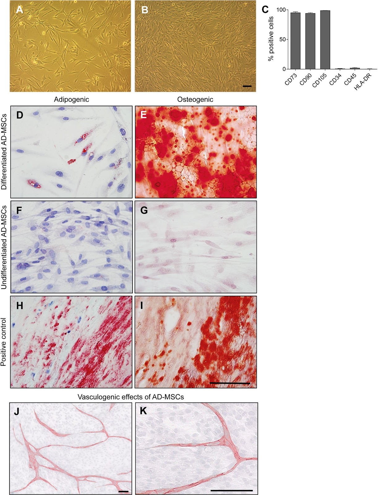
Background Inadequate vascularization remains a major limitation in tissue engineering, often leading to graft failure due to limited oxygen and nutrient supply. Prevascularization, the formation of microvascular networks within scaffolds before implantation, aims to accelerate perfusion and improve graft integration. We developed bilayer electrospun poly(ε-caprolactone)/poly(l-lactide) (PCL/PLA) scaffolds prevascularized by co-culture of human adipose-derived mesenchymal stem cells (AD-MSCs) and human placental arterial endothelial cells (HPAECs). Methods AD-MSCs were isolated from lipoaspirates and characterized by flow cytometry and functional assays. Bilayered PCL/PLA scaffolds were engineered with a wide-meshed layer for cell infiltration and a fine-meshed layer for mechanical stability. Scaffolds were seeded with AD-MSCs, HPAECs, or both (co-culture). Cell viability, adhesion, and apoptosis were analyzed histologically. Angiogenic and vasculogenic potential was evaluated in vitro and in vivo using the chick chorioallantoic membrane (CAM) assay. Results AD-MSCs expressed characteristic markers, demonstrated adipogenic and osteogenic differentiation, and promoted angiogenesis in 2D co-culture. ELISA analyses indicated dynamic secretion of VEGF, HGF, and bFGF, reflecting both paracrine and contact-dependent AD-MSC–HPAEC interactions. On scaffolds, cells primarily adhered to the wide-meshed layer. Co-culture induced vessel-like structures within a multicellular stromal environment; monocultures did not support prevascularization. Five days post-implantation, prevascularized scaffolds exhibited human microvessels at the scaffold–CAM interface and in adjacent tissue, closely associated with AD-MSCs and containing chicken erythrocytes—indicating successful anastomosis and functional perfusion. Quantitative analysis showed a significant increase in vessel branching points in the host CAM tissue in response to AD-MSC-only (2.8-fold)

Prevascularization of electrospun PCL/PLA scaffolds using human adipose-derived stem and endothelial cells enhances vascular integration and host angiogenesis in vivo
Graphical Abstract
Original ResearchVol. 17, None • pp. 42DOI: 10.1186/s13287-025-04865-7

Modeling pathogenesis and progression of metabolic dysfunction-associated steatotic liver disease and therapeutic drug screening using hESC-derived mature polarized hepatocyte organoids

Authors: Changlu Qin, Haibin Wu, Peilin Liao, Wenjiao Yan, Xinyi Shi, Jinghe Xie, Shoupei Liu, Sen Chen, Xiangting Cao, Yongjian Zhou, Yuyou Duan

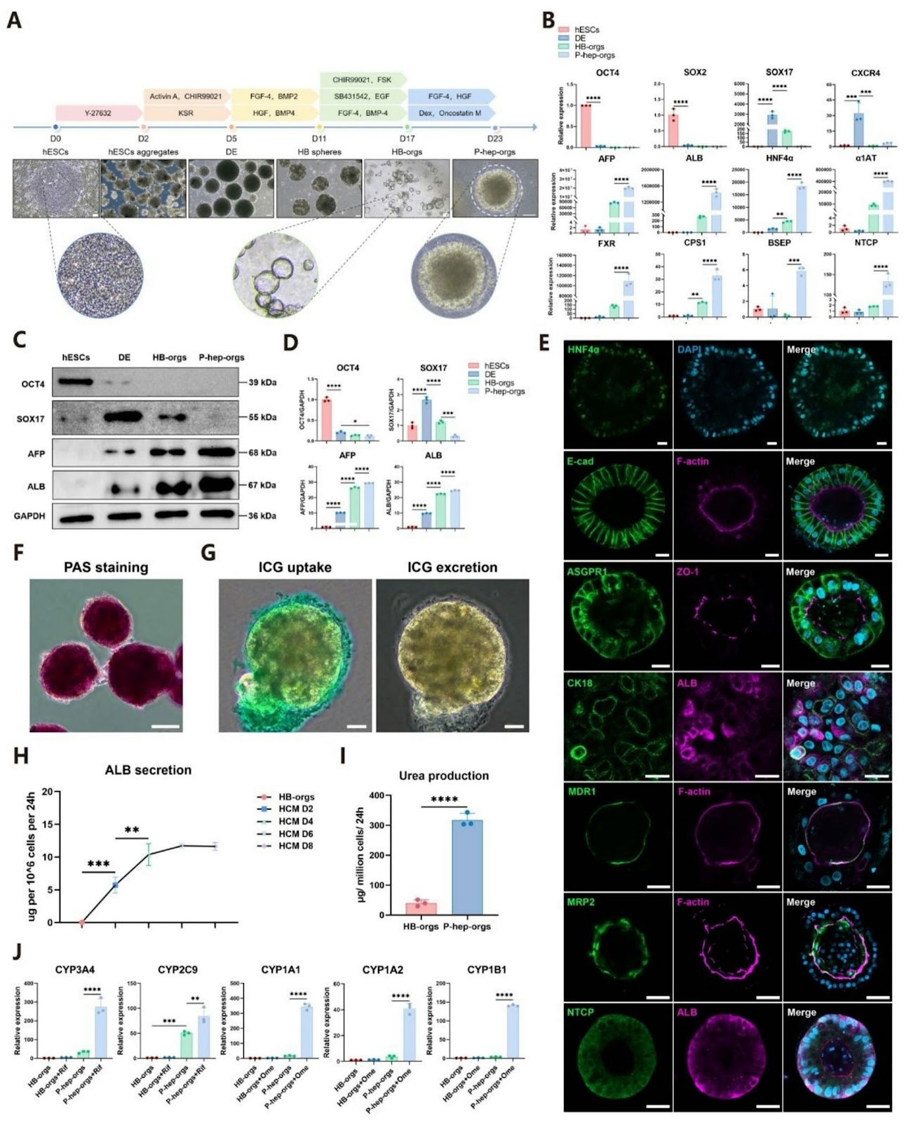
Background: Metabolic dysfunction-associated steatotic liver disease (MASLD), the most prevalent chronic liver disorder worldwide, exhibits complex pathogenesis and lacks effective targeted therapeutics. Existing animal models are limited by prolonged induction periods and interspecies discrepancies, while conventional monolayer hepatocyte cultures fail to recapitulate disease pathology due to inadequate polarization and functional immaturity. Methods: To overcome these limitations, we established an in vitro MASLD model by treating human embryonic stem cell (hESC)-derived mature polarized hepatocyte organoids (P-hep-orgs) with free fatty acids (FFAs). Pathogenesis and progression of MASLD in this model were characterized using multiple assays, and its utility for drug screening was validated with three known antioxidant or lipid-lowering agents. Results: P-hep-orgs derived from hESCs expressed mature hepatocyte markers (e.g., ALB), exhibited polarized architecture (e.g., MRP2) and demonstrated functionalities of mature hepatocytes (e.g., urea production). Moreover, we developed an in vitro MASLD model by treating P-hep-orgs with FFAs. This model recapitulated key pathological progression hallmarks, including disrupted glucose/lipid metabolism, oxidative stress, apoptosis, loss of polarization, impaired liver function, and ductular reaction. Furthermore, transcriptomic analysis revealed that P-hep-orgs treated with FFAs for 10 days shared similar molecular signatures with human MASH liver tissues (581 overlap DEGs). Finally, this model was used to assess the potential efficacy of established antioxidant or lipid-lowering agents (e.g., Vitamin E) in alleviating pathological phenotypes, including lipid accumulation and oxidative stress.

Modeling pathogenesis and progression of metabolic dysfunction-associated steatotic liver disease and therapeutic drug screening using hESC-derived mature polarized hepatocyte organoids
Graphical Abstract
Original ResearchVol. 17, None • pp. 22DOI: 10.1186/s13287-025-04847-9

Device encapsulated MSCs for adaptive secretome therapy to effectively target ischaemic heart injury

Authors: Andrew R. Kompa, David W. Greening, Jarmon G. Lees, Anne M. Kong, Jonathon Cross, Ashley Nowland, Ren J. Phang, Saba Naghipour, Yali Deng, Jack R. T. Darby, Lina Mariana, Cameron Kos, Tanya Hall, Andrew Newcomb, James J. H. Chong, Rebecca H. Ritchie, Janna L. Morrison, Klearchos K. Papas, Kilian Kelly, Derek J. Hausenloy, Thomas Loudovaris, Shiang Y. Lim

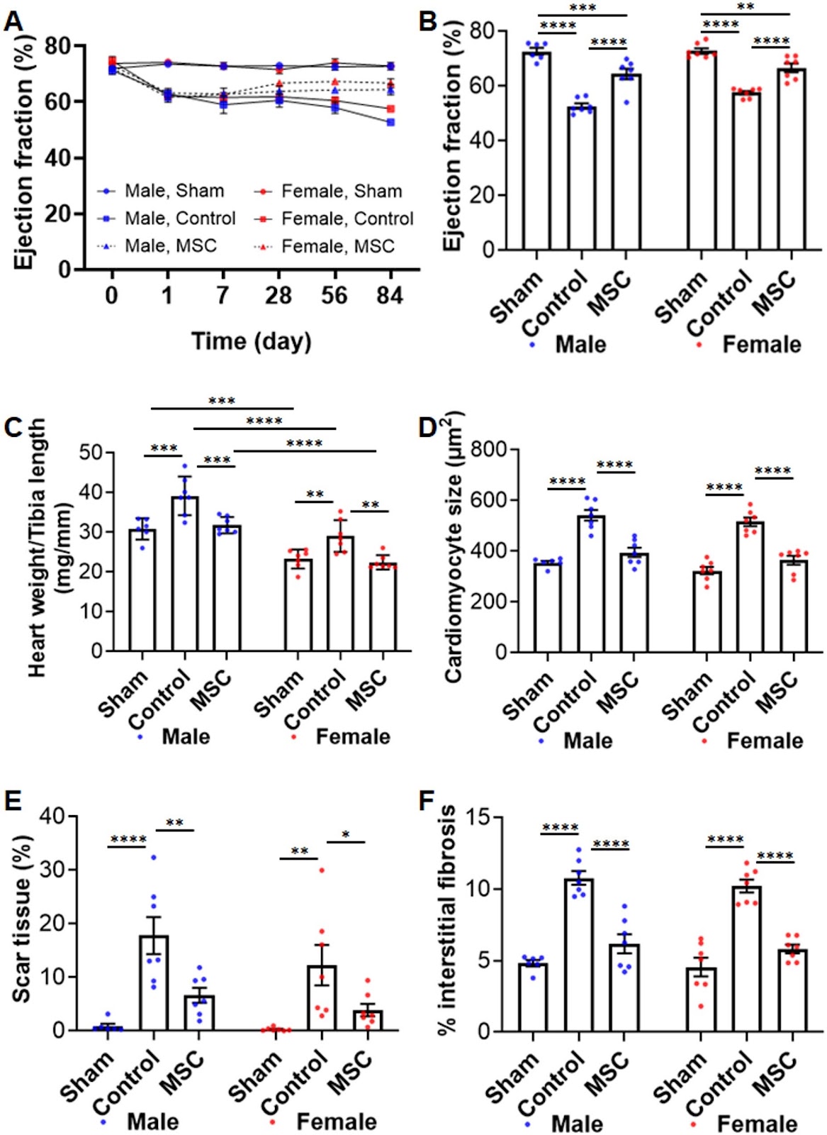
Background: Effective long-term strategies to protect the ischaemic heart remain a significant challenge. Mesenchymal stromal cells (MSCs) offer therapeutic potential primarily through their secretome, a bioactive factor-rich milieu with broad beneficial effects. However, existing delivery methods have not demonstrated sustained cardioprotection. The objective of this study was to evaluate a clinically translatable approach for sustained MSC-secretome delivery to achieve long-term cardioprotection. Methods: Cymerus MSCs, derived from human induced pluripotent stem cells (iPSCs), were encapsulated in a Procyon immunoisolation device and implanted subcutaneously in adult Sprague Dawley rats with chronic myocardial ischaemia-reperfusion injury. A human iPSC-derived engineered cardiac microtissue model was used to simulate ischaemia-reperfusion injury and assess cardioprotective effects in a human context. Proteomic analysis was performed to characterize adaptive changes in MSCs and their secretome post-implantation. Results: The MSC-loaded Procyon device significantly improved cardiac function and reduced adverse left ventricular remodelling over 12 weeks in both young and middle-aged, male and female rats. The encapsulated MSCs remained viable and retained the ability to release therapeutic secretome at 12 weeks post-implantation. In vitro, the MSC secretome protected human engineered cardiac microtissues from simulated ischaemia-reperfusion injury by restoring contractile function, improving cell viability, and reducing oxidative stress. Proteomic profiling of encapsulated MSC identified 179 unique cellular proteins post-implantation, associated with adaptive immune and inflammatory responses as well as wound healing. MSC secretome profiling revealed increased protein diversity associated with tissue repair and immune regulation, suggesting MSCs undergo an adaptive response to ischaemic conditions.

Device encapsulated MSCs for adaptive secretome therapy to effectively target ischaemic heart injury
Graphical Abstract