🧬 SinoBioData Academic Portal
📚 Peer-Reviewed Translated Literature

All Biomedical & Clinical Articles (Page 7)

Browse complete peer-reviewed translations from top Chinese biomedical, oncology, and genomics journals. Read verified previews and download full authentic clinical reports.

Published Research Papers

Showing 24 of 1542 peer-reviewed translated articles (Page 7 of 65)

U-shaped association between magnesium intake and all-cause and cancer mortality in patients with osteoarthritisGraphical AbstractVerified
Chinese Journal of Tissue Engineering Research2026

U-shaped association between magnesium intake and all-cause and cancer mortality in patients with osteoarthritis

BACKGROUND: Clarifying the relationship between dietary magnesium intake and mortality risk in patients with osteoarthritis can provide theoretical basis for optimizing dietary interventions, reducing all-cause mortality and cardiovascular disease mortality, and provide reference value for nutritional management in patients with osteoarthritis. OBJECTIVE: To assess the association between dietary magnesium intake and all-cause and etiological mortality in adults with osteoarthritis in the United States. METHODS: We enrolled data of adults with osteoarthritis from the National Health and Nutrition Examination Survey (conducted by the National Center for Health Statistics under the U.S. Centers for Disease Control and Prevention, it aims to assess the health and nutritional status of the civilian population in the United States) between 2003 and 2020. The association between dietary magnesium intake and mortality in patients with osteoarthritis was evaluated by Cox proportional hazard model and two-stage Cox model. To assess whether the association between dietary magnesium intake and all-cause mortality was consistent across the population and to identify potential high-risk groups, subgroup analyses in terms of age, sex, body mass index, hypertension, diabetes mellitus, physical activity, smoking status, and drinking status were performed and tested for interactions. RESULTS AND CONCLUSION: (1) A total of 2 868 patients with osteoarthritis were included, and 699 all-cause deaths, 281 cardiovascular disease deaths, and 143 cancer deaths were recorded during follow-up. After adjusting for multiple variables, higher dietary magnesium intake was significantly associated with reduced risk of cardiovascular disease mortality; each 1-unit increase in dietary magnesium intake was associated with a 78% reduction in cardiovascular disease mortality (P=0.022 2), and quartile analysis of dietary magnesium intake was consistent with this. Dietary magnesium intake showed a U-shaped association with all-cause mortality and cancer mortality, with thresholds for lowest mortality risk at 0.38 g/d and 0.40 g/d, respectively. When magnesium intake was below the threshold, higher intake was significantly associated with lower all-cause mortality [HR=0.17, 95%CI(0.06, 0.50)] and cancer mortality [HR=0.16, 95%CI(0.01, 1.50)]; but above the threshold, magnesium intake was significantly associated with increased all-cause mortality [HR=2.94, 95%CI(0.55, 15.84)] and cancer mortality [HR=26.30, 95%CI(1.46, 474.73)]. Subgroup analyses further verified the robustness of the results. (2) The results indicate that insufficient dietary magnesium intake may play an adverse role in cardiovascular health, and there is a U-shaped relationship between dietary magnesium intake and all-cause mortality and cancer mortality, with both too low and too high magnesium intake potentially increasing mortality risk. This finding provides new insights into the potential impact of dietary magnesium intake on cardiovascular disease, cancer, and all-cause mortality, provides scientific basis for nutritional intervention in patients with osteoarthritis, and provides theoretical support for the prevention and management of cardiovascular disease and cancer in China, especially in the context of high cardiovascular disease incidence, reasonable magnesium intake helps reduce related mortality.

Read Full Abstract10.12307/2026.21213
Mechanism by which Yougui Pill inhibits pyroptosis of chondrocytes in rats with knee osteoarthritisGraphical AbstractVerified
Chinese Journal of Tissue Engineering Research2026

Mechanism by which Yougui Pill inhibits pyroptosis of chondrocytes in rats with knee osteoarthritis

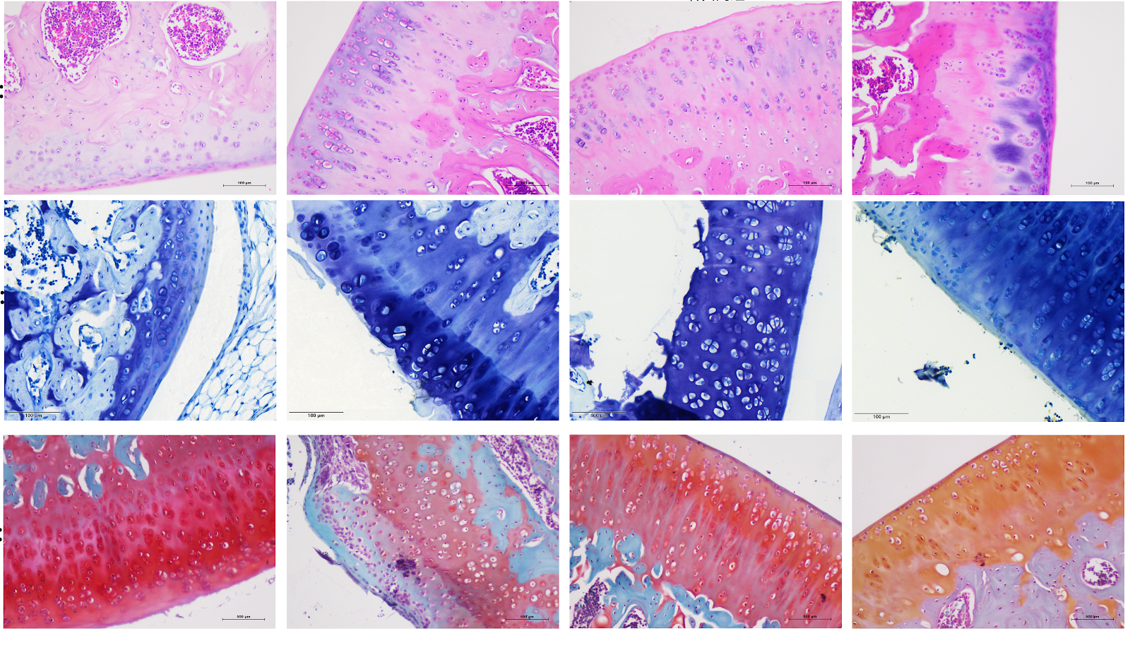
BACKGROUND: Yougui Pill is derived from Jingyue Quanshu. Studies have confirmed that Yougui Pill is highly effective in treating patients with knee osteoarthritis, but its mechanism of action remains unclear. OBJECTIVE: To explore the potential molecular mechanism of Yougui Pill in improving knee osteoarthritis in rats. METHODS: Forty-eight SPF-grade Sprague-Dawley rats were randomly divided into four groups: blank control group, model group, Yougui Pill group and celecoxib group. The latter three groups were subjected to modified Hulth method for surgical modeling of knee osteoarthritis. After wound healing, rats were driven for 8 weeks. After modeling, the celecoxib group was given celecoxib suspension by gavage, the Yougui Pill group was given Yougui Pill decoction by gavage, and the sham operation group and model group were given equal volume of normal saline, once daily for 4 weeks. Hematoxylin-eosin staining, toluidine blue staining, and safranin O-fast green staining were used to observe the pathological changes of rat cartilage tissue; transmission electron microscopy was used to observe the ultrastructure of rat chondrocytes; ELISA was used to detect the levels of interleukin-18, interleukin-1β, and tumor necrosis factor-α in rat serum; western blot was used to detect the protein expression of PI3K, AKT, NF-κB, p-PI3K, p-AKT, p-P65, NLRP3, GSDMD, GSDMD-N, Caspase1, Cleaved-Caspase1, interleukin-18, and interleukin-1β in rat cartilage tissue. RESULTS AND CONCLUSION: Compared with the blank group, the model group showed severe destruction of cartilage edge, cartilage tissue defect, thinning and disordered arrangement of cartilage layer cells, subchondral bone hyperplasia, disordered tide line, severe structural damage of chondrocytes, formation of pyroptotic bodies, significantly increased serum levels of tumor necrosis factor-α, interleukin-18, and interleukin-1β (P < 0.05), and significantly increased protein expression of p-PI3K, p-AKT, p-P65, NLRP3, GSDMD-N, Cleaved-Caspase1, interleukin-18, and interleukin-1β in cartilage tissue (P < 0.01). Compared with the model group, the cartilage structure of rats in the Yougui Pill group and celecoxib group tended to be normal, with deeper cartilage staining, thicker cartilage, more complete chondrocyte membrane, significantly decreased serum levels of tumor necrosis factor-α, interleukin-18, and interleukin-1β (P < 0.05), and significantly decreased protein expression of p-PI3K, p-AKT, p-P65, NLRP3, GSDMD-N, Cleaved-Caspase1, interleukin-18, and interleukin-1β in cartilage tissue (P < 0.01). Compared with the celecoxib group, the Yougui Pill group showed more regular arrangement of chondrocytes, smoother articular cartilage surface, significantly thickened cartilage layer, relatively complete tide line, relatively complete chondrocyte membrane, significantly decreased serum levels of tumor necrosis factor-α, interleukin-18, and interleukin-1β (P < 0.05), and significantly decreased protein expression of p-PI3K, p-AKT, p-P65, NLRP3, GSDMD-N, Cleaved-Caspase1, interleukin-18, and interleukin-1β in cartilage tissue (P < 0.01). These results indicate that Yougui Pill can improve the inflammatory response of chondrocytes in rats with knee osteoarthritis, and the mechanism may be related to inhibiting the activation of PI3K/AKT/NF-κB pathway, thereby regulating NLRP3/Caspase1/GSDMD pathway-mediated pyroptosis.

Read Full Abstract10.12307/2026.21286
Cobalt chloride-induced hypoxic environment accelerates knee cartilage degeneration in New Zealand rabbitsGraphical AbstractVerified
Chinese Journal of Tissue Engineering Research2026

Cobalt chloride-induced hypoxic environment accelerates knee cartilage degeneration in New Zealand rabbits

BACKGROUND: Cobalt chloride solution is commonly used to induce osteoarthritis cell models in vitro. However, its ability to construct animal models of osteoarthritis by intra-articular injection remains unknown. OBJECTIVE: To investigate the effect of intra-articular injection of different concentrations of cobalt chloride solution on cartilage degeneration in the knee joint. METHODS: Thirty-six healthy adult male New Zealand rabbits were randomly divided into four groups: low, medium and high dose cobalt chloride groups and control group. The right hind knee was intra-articularly injected with 100, 200, and 300 μmol/(L·kg) of cobalt chloride, while the left hind knee served as the control knee and was injected with an equal amount of normal saline. At 4, 8 and 12 weeks after operation, four rabbits were killed respectively. The cartilage on the surface of the femur was exposed for gross morphological observation, and then the cartilage tissues were taken for hematoxylin-eosin staining, safranine O-fast green staining, the Osteoarthritis Research Society International scoring, and immunohistochemical staining of interleukin 1 and tumor necrosis factor α, to determine cartilage degeneration in various aspects. RESULTS AND CONCLUSION: (1) Gross observation: At the same postoperative time point, with the increase of cobalt chloride concentration, cartilage degeneration showed a progressive aggravation trend, and the high-dose cobalt chloride group even involved the deep layer of cartilage and subchondral bone; under the same concentration of cobalt chloride, with the prolongation of modeling time, cartilage degeneration progressed progressively. (2) Hematoxylin-eosin staining, safranine O-fast green staining, and Osteoarthritis Research Society International scoring showed that at the same postoperative time point, with the increase of cobalt chloride concentration, the cartilage surface gradually became rough, the superficial layer became thinner, and the destruction aggravated, and the Osteoarthritis Research Society International score gradually increased (P < 0.05); under the same concentration of cobalt chloride, with the prolongation of modeling time, the arrangement of chondrocytes tended to be disordered, polarity was lost, and the destruction of superficial cartilage and subchondral bone progressively aggravated, and the Osteoarthritis Research Society International score gradually increased (P < 0.05). (3) Immunohistochemistry showed that at the same postoperative time point, with the increase of cobalt chloride concentration, cartilage degeneration aggravated, intracellular brown particles increased, and the positive expression of interleukin 1 and tumor necrosis factor α increased (P < 0.01); under the same concentration of cobalt chloride, with the prolongation of modeling time, cartilage destruction and fissures aggravated, and the positive expression of interleukin 1 and tumor necrosis factor α increased (P < 0.01). This experiment successfully established an osteoarthritis model of New Zealand rabbits induced by intra-articular injection of cobalt chloride solution, preliminarily verified the stability and reliability of the animal model, and also proved that with the increase of modeling concentration and the prolongation of modeling time, cartilage degeneration progressed progressively.

Read Full Abstract10.12307/2026.21222
Effects of high-intensity interval training combined with estrogen on satellite cells, myonuclear domain and ribosome function in ovariectomized ratsGraphical AbstractVerified
Chinese Journal of Tissue Engineering Research2026

Effects of high-intensity interval training combined with estrogen on satellite cells, myonuclear domain and ribosome function in ovariectomized rats

BACKGROUND: Estrogen deficiency can lead to a decrease in skeletal muscle mass and muscle strength in postmenopausal women, thereby affecting their quality of life. Muscle mass is maintained by satellite cells, which are regulated by estrogen. Regular exercise, especially high impact exercise (such as resistance training and high-intensity interval training), can induce muscle hypertrophy, but the role and mechanism of estrogen are still unclear. OBJECTIVE: To explore the effects of high-intensity interval training combined with estrogen therapy on skeletal muscle hypertrophy in ovariectomized rats and reveal its possible mechanism. METHODS: Sixty 8-week-old female Sprague-Dawley rats were divided into five groups using a random number table method: sham operation, model sedentary group, model exercise group, model hormone group, or model combined group. Bilateral ovariectomy was used to establish an estrogen deficiency model. Twelve weeks after operation, the model exercise and model combined group performed high-intensity interval training for 8 weeks (3 times/week), and hormone treatment groups received abdominal subcutaneous injection of 17β-estradiol (once a day for 8 weeks). Seventy-two hours after the last training, the grip force of the forelimb was measured by an electronic grip force meter. The gastrocnemius muscle was separated, and muscle mass index was calculated as muscle mass/body mass ratio. Hematoxylin-eosin staining was used to obtain cell cross-sectional area. Immunofluorescence staining was used to classify muscle fiber types and obtain myonuclear number, myonuclear domain size, and activated satellite cell number. BCA method was used to determine total protein concentration. Trizol method was used to extract total RNA. Western blot was used to detect ribosomal protein S6 expression. Real-time quantitative PCR was used to detect ribosomal RNA expression. RESULTS AND CONCLUSION: Compared with the sham operation group, the model sedentary group showed increased body mass and myosin heavy chain type I fiber proportion (P < 0.05), while uterine mass index, gastrocnemius mass index, grip strength, cell cross-sectional area, myosin heavy chain type IIa proportion, satellite cell and myonuclear number, myonuclear domain size, total protein and RNA content, and ribosomal protein S6, 18S rRNA and 28S rRNA expression decreased (P < 0.05). Compared with the model sedentary group, the model exercise group showed decreased body mass and myosin heavy chain type IIb proportion (P < 0.05), while uterine mass index, gastrocnemius mass index, grip strength, cell cross-sectional area, myosin heavy chain type IIa proportion, satellite cell number, myonuclear domain size, total protein and RNA content, and ribosomal protein S6 and 28S rRNA expression increased (P < 0.05). Compared with the model exercise group and model hormone group, the model combined group showed higher gastrocnemius mass index, grip strength, cell cross-sectional area, satellite cell number, myonuclear domain size, total protein and RNA content, and ribosomal protein S6 and 28S rRNA expression (P < 0.05). These results indicate that estrogen can enhance the skeletal muscle hypertrophy response induced by high-intensity interval training in ovariectomized rats, and the mechanism may be related to satellite cell activation, increased myonuclear domain and ribosome biogenesis, and improved ribosome function.

Read Full Abstract10.12307/2026.21239
Gushukang Granule-containing drug serum improves dexamethasone-induced atrophy of C2C12 myotubes via regulating mitochondrial homeostasisGraphical AbstractVerified
Chinese Journal of Tissue Engineering Research2026

Gushukang Granule-containing drug serum improves dexamethasone-induced atrophy of C2C12 myotubes via regulating mitochondrial homeostasis

BACKGROUND: Gushukang Granule is a Chinese herbal compound preparation, commonly used clinically for the treatment of osteoporosis and other bone-related diseases. However, its role in the regulation of muscle metabolism is not clear. OBJECTIVE: To investigate the inhibitory effect of Gushukang Granule-containing serum on dexamethasone-induced C2C12 muscle atrophy. METHODS: (1) In vivo experiment: Thirty-six 3-month-old female Sprague-Dawley rats were randomly divided into model group, blank group and Gushukang group, with 12 rats in each group. The rats in the model group were given 2.5 mg/kg dexamethasone by intragastric administration, once a day, for 1 week, followed by normal feeding for 3 weeks; the Gushukang group was given 0.48 g/kg Gushukang Granule suspension by gavage after modeling, once a day, for 3 weeks; the blank group was not modeled and given equal volume of distilled water by gavage, once a day, for 4 weeks. Two hours after the last administration, left femur and gastrocnemius muscle specimens were taken for hematoxylin-eosin staining, and RNA was extracted from right gastrocnemius and femur tissues; real-time fluorescence quantitative PCR was used to detect mRNA expression levels of mitochondrial function, autophagy and inflammation-related genes. (2) In vitro experiment: C2C12 cells were cultured and induced to differentiate into myotubes with 2% horse serum. Differentiated myotubes were divided into control group, model group and Gushukang-containing serum group. Muscle atrophy model was constructed by incubating with 4 µmol/L dexamethasone for 48 h, and the Gushukang group was treated with 10% Gushukang-containing serum for 24 h after dexamethasone treatment for 24 h. CCK-8 was used to detect cell viability, flow cytometry to detect reactive oxygen species levels, transmission electron microscopy to observe ultrastructural changes, and western blot to detect expression levels of mitochondrial function-related proteins, autophagy-related proteins and antioxidant-related proteins. RESULTS AND CONCLUSION: (1) Hematoxylin-eosin staining showed that compared with the model group, the muscle fiber structure of rats in the Gushukang group recovered better; real-time fluorescence quantitative PCR further verified that Gushukang Granule up-regulated mitochondrial function and autophagy-related genes, supporting its multi-target mechanism to alleviate muscle atrophy. (2) Flow cytometry and CCK-8 results showed that after dexamethasone treatment, reactive oxygen species levels in C2C12 cells were significantly increased (P < 0.05), and cell viability was significantly decreased (P < 0.05); transmission electron microscopy revealed that dexamethasone induced ultrastructural disorder, reduced number of organelles, atrophic and blurred mitochondria, accompanied by a large number of autophagosomes and cytoplasmic vacuoles; western blot results showed that after dexamethasone treatment, the expression of mitochondrial function-related proteins translocase of outer mitochondrial membrane 20 and heat shock protein 60 was significantly decreased, autophagy-related proteins LC3 and Beclin-1 were abnormally expressed (P < 0.05), and silent information regulator 1 expression was significantly decreased (P < 0.05), suggesting that dexamethasone disrupted mitochondrial homeostasis and inhibited autophagy. After treatment with Gushukang Granule-containing serum, reactive oxygen species levels were significantly decreased (P < 0.05), cell viability was significantly increased (P < 0.05); transmission electron microscopy showed that ultrastructure was improved and mitochondrial morphology was relatively restored; western blot results showed that the expression of translocase of outer mitochondrial membrane 20, heat shock protein 60, and silent information regulator 1 was significantly up-regulated (P < 0.05), and LC3 and Beclin-1 expression returned to normal levels (P < 0.05), indicating that Gushukang Granule-containing serum played a positive role in improving mitochondrial function, reducing oxidative stress, and regulating autophagy.

Read Full Abstract10.12307/2026.21233
Effect of blood flow restriction training on the magnitude and temporal characteristics of post-activation performance enhancement: a systematic review and meta-analysisGraphical AbstractVerified
Chinese Journal of Tissue Engineering Research2026

Effect of blood flow restriction training on the magnitude and temporal characteristics of post-activation performance enhancement: a systematic review and meta-analysis

Objective: To systematically compare the acute effects of blood flow restriction combined with preconditioning (to induce post-activation performance enhancement) versus preconditioning alone or sitting, low-intensity preconditioning combined with blood flow restriction versus high-intensity preconditioning, and sitting combined with blood flow restriction versus sitting on sports performance using a multilevel meta-analysis. Methods: Following the PRISMA guidelines, Web of Science, PubMed, SPORTDiscus, and CNKI databases were systematically searched (from inception to May 24, 2025). Inclusion criteria: (1) healthy individuals who were at least physically active; (2) studies with at least one of the following four comparisons: preconditioning + blood flow restriction vs. preconditioning alone; preconditioning + blood flow restriction vs. sitting; low-intensity preconditioning + blood flow restriction vs. high-intensity preconditioning; sitting + blood flow restriction vs. sitting; (3) sports performance (e.g., jump, sprint, bench press throw) as the primary outcome; (4) randomized or non-randomized crossover/parallel designs; (5) published in peer-reviewed Chinese or English journals. Risk of bias was assessed using ROB-2, and evidence quality was evaluated with GRADE. Data were fitted using cluster robust variance estimation and a three-level mixed-effects model, with small-sample corrections. Subgroup analyses and meta-regression explored moderators and sources of heterogeneity. Results: Twelve studies (196 participants, 12 women, 184 men) were included. Main findings: (1) Preconditioning + blood flow restriction was more effective than preconditioning alone in enhancing sports performance (ES=0.21, 95%CI=0.01-0.40, GRADE=low), with the best effect at recovery times of 4-12 min and 50% arterial occlusion pressure (ES=1.49); (2) Preconditioning + blood flow restriction did not significantly differ from sitting (ES=0.52, 95%CI=-0.12-1.15, GRADE=very low), but preconditioning + 140 mmHg blood flow restriction was superior to preconditioning alone (ES=1.21, 95%CI=0.14-2.28); (3) Low-intensity preconditioning + blood flow restriction did not differ from high-intensity preconditioning (ES=-0.10, 95%CI=-0.84-0.64, GRADE=low); (4) Sitting + blood flow restriction did not significantly differ from sitting (ES=0.24, 95%CI=-0.03-0.52, GRADE=very low). Notably, the effects of the latter two comparisons significantly decreased with recovery time (β=-0.04, P < 0.01 and β=-0.04, P=0.02). Conclusion: Preconditioning combined with blood flow restriction is more effective than preconditioning alone in inducing post-activation performance enhancement, preliminarily suggesting the use of 50% arterial occlusion pressure and 4-12 min recovery time. However, preconditioning combined with blood flow restriction does not appear to be more effective than sitting, possibly due to insufficient number of included studies. Additionally, low-intensity preconditioning + blood flow restriction can achieve similar post-activation performance enhancement as high-intensity preconditioning, while the potential benefit of sitting + blood flow restriction on sports performance may diminish over time. Overall, it is preliminarily recommended to use low-intensity preconditioning (e.g., 30% one-repetition maximum squat or bodyweight training) combined with 50% arterial occlusion pressure or 140 mmHg blood flow restriction, with 4-12 min recovery before subsequent performance testing.

Read Full Abstract10.12307/2026.21279
Establishing a diagnostic model for recurrent spontaneous abortion based on the levels of autophagy-related genes in the endometriumGraphical AbstractVerified
Chinese Journal of Tissue Engineering Research2026

Establishing a diagnostic model for recurrent spontaneous abortion based on the levels of autophagy-related genes in the endometrium

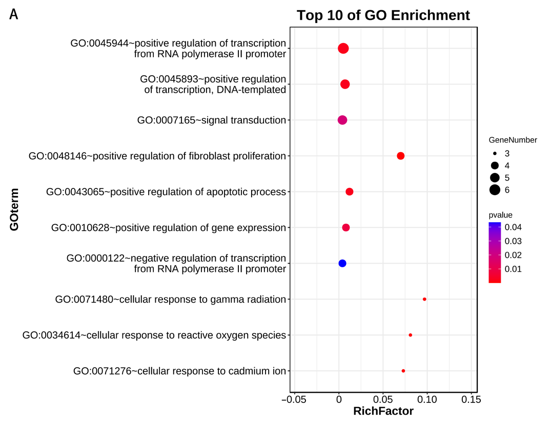
BACKGROUND: The etiology of recurrent spontaneous abortion is complex. With the development of genetics and other fields, it has been found that the abnormal expression of autophagy-related genes may lead to the imbalance of cell homeostasis, thus triggers pathological processes, such as apoptosis, inflammatory response and immunosuppressive response, and affects the endometrial microenvironment, trophoblastic function and immune cell function, thereby leading to recurrent spontaneous abortion. By using the recurrent spontaneous abortion samples in the Gene Expression Omnibus database, the expression changes and regulatory mechanisms of autophagy-related genes were analyzed, which is helpful to reveal the mechanism of recurrent spontaneous abortion and develop new therapeutic strategies. However, the specific mechanism of autophagy-related genes in recurrent spontaneous abortion and their interaction with other biological processes still need to be further studied. OBJECTIVE: To establish a risk score prognostic model for patients with recurrent spontaneous abortion based on autophagy-related genes. METHODS: Endometrial gene expression matrix of patients with recurrent abortion was obtained from the Gene Expression Omnibus database, autophagy-related genes were obtained from the Human Autophagy Database (HADb), and 30 differentially co-expressed autophagy-related genes were identified. The biological functions of autophagy-related genes were analyzed by gene ontology (GO), Kyoto Encyclopedia of Genes and Genomes (KEGG) and DisGeNET enrichment. LASSO and logistic regression analysis identified 16 autophagy-related genes as potential biomarkers. A nomogram model was then constructed, and the predictive accuracy was evaluated using receiver operating characteristic curves. Six machine learning models (random forest, support vector machine, generalized linear model, etc.) were compared to select the optimal model. Nomogram, calibration curves, and decision curve analysis were used to validate the predictive efficacy. RESULTS AND CONCLUSION: GO analysis showed that autophagy-related genes in recurrent spontaneous abortion patients were mainly enriched in autophagy regulation, catabolic processes, and formation of mitochondrial or other organelle membranes. KEGG analysis showed enrichment in autophagy regulation, phosphatidylinositol 3-kinase/protein kinase B signaling pathway, human papillomavirus infection, and neurodegeneration pathways. GSEA analysis indicated involvement in copper detoxification, proton transmembrane transport, sarcoplasmic reticulum components, and regulation of nucleoside diphosphate phosphatase activity. A predictive risk model was constructed, identifying 16 specific autophagy-related genes as predictive targets. Based on the detection efficacy of machine models, the optimal neural network model was selected, and five most important autophagy-related gene variables (MAP2K7, CALCOCO2, SAR1A, TUSC1, and STK11) were identified. Using recurrent spontaneous abortion samples from the European population in the GEO database, the gene expression patterns, differentially expressed genes, and signaling pathways in the endometrium were analyzed from the genetic single nucleotide polymorphism level. The predictive model and machine learning model with good predictive efficacy can screen new therapeutic targets and potential biomarkers for recurrent spontaneous abortion, which has certain guiding significance for clinical work and mechanism research.

Read Full Abstract10.12307/2026.21238
Effects of protein kinase C on the expression of myocardial SarcKATP channels in model rats during exercise preconditioningGraphical AbstractVerified
Chinese Journal of Tissue Engineering Research2026

Effects of protein kinase C on the expression of myocardial SarcKATP channels in model rats during exercise preconditioning

BACKGROUND: Exercise preconditioning produces early and late myocardial protective effects, in which protein kinase C and myocardial ATP-sensitive potassium channels (SarcKATP) are mediators and effectors, respectively. Protein kinase C regulates the expression of myocardial SarcKATP channels. OBJECTIVE: To compare the effects of protein kinase C on the expression of myocardial SarcKATP channel subunits, inward recirculating potassium channel 6.2 (Kir6.2) and sulfonylurea receptor 2A (SUR2A), in exercise preconditioning. METHODS: Forty-eight Sprague-Dawley rats were randomly divided into five groups: control group (no intervention), early exercise preconditioning group, protein kinase C inhibitor (pre-exercise intraperitoneal injection) + early exercise preconditioning group, late exercise preconditioning group, and protein kinase C inhibitor + late exercise preconditioning group. After preconditioning, the distribution and expression changes of Kir6.2 and SUR2A mRNAs in the rat myocardium were observed and detected using real-time fluorescent quantitative PCR. The distribution and expression changes of Kir6.2 and SUR2A proteins were observed and detected using western blot. RESULTS AND CONCLUSION: (1) Compared with the control group, the mRNA expression of Kir6.2 and SUR2A showed no significant difference in the early and late exercise preconditioning group. (2) Compared with the early exercise preconditioning group, the protein kinase C inhibitor + early exercise preconditioning group showed decreased Kir6.2 mRNA expression but increased Kir6.2 protein expression; both SUR2A mRNA and protein expression decreased. (3) Compared with the late exercise preconditioning group, the protein kinase C inhibitor + late exercise preconditioning group showed decreased Kir6.2 mRNA and SUR2A mRNA expression, decreased Kir6.2 protein expression, but increased SUR2A protein expression. (4) These results indicate that for the same subunit (Kir6.2 or SUR2A), protein kinase C exerts coordinated and complementary regulatory effects in early and late exercise preconditioning; for different subunits (Kir6.2 and SUR2A), protein kinase C also exerts coordinated and complementary regulatory effects on their expression in early and late exercise preconditioning.

Read Full Abstract10.12307/2026.21240
Articular cartilage lesions at different stages of steroid-induced osteonecrosis of the femoral head: characteristics and mechanisms of crescent sign formationGraphical AbstractVerified
Chinese Journal of Tissue Engineering Research2026

Articular cartilage lesions at different stages of steroid-induced osteonecrosis of the femoral head: characteristics and mechanisms of crescent sign formation

BACKGROUND: The crescent sign is a significant radiological feature in the progression of steroid-induced osteonecrosis of the femoral head (SIONFH), indicating the separation and defect of articular cartilage and subchondral bone. The appearance of the crescent sign is associated with the mid-to-late stages of the disease and poor prognosis. However, studies on the specific pathological characteristics and progression patterns of articular cartilage in SIONFH remain unclear. OBJECTIVE: To observe the pathological features of articular cartilage in specimens from different stages of SIONFH, explore the progression and pathological mechanisms, and elucidate the formation mechanism of the crescent sign, providing a theoretical basis for optimizing hip-preserving strategies. METHODS: Femoral head specimens were collected from patients with SIONFH who underwent total hip arthroplasty at the First Affiliated Hospital of Guangzhou University of Chinese Medicine from 2021 to 2024. According to the ARCO staging, they were divided into mild, moderate, and severe collapse groups, with fresh femoral neck fracture specimens as controls. All specimens were cut coronally, and the folded cartilage surface in the necrotic area was taken; control group took corresponding area. Hematoxylin-eosin staining and Safranin O-fast green staining were used for morphological observation, immunohistochemistry and western blot for biomarker expression, and apoptosis kit for apoptosis level. RESULTS AND CONCLUSION: (1) Gross observation: The control group showed smooth cartilage surface without folds or hyperplasia, no separation or defect between articular cartilage and subchondral bone, and tough texture. In SIONFH specimens, obvious folds were visible on the cartilage surface, with separation and defects between articular cartilage and subchondral bone, and a loose sensation on pressing. (2) Pathological observation: In the control group, chondrocytes in each layer were arranged neatly, cartilage matrix stained uniformly, tidemark was intact and continuous, calcified cartilage layer and subchondral bone connection was clear and complete, and bone trabeculae were arranged neatly. In SIONFH specimens, chondrocytes were disorganized, empty lacunae increased, matrix staining loss of varying degrees, tidemark duplication and loss, calcified cartilage layer showed numerous cavities and sclerosis, with granulation tissue invasion into cavities, separation and defects between calcified cartilage and subchondral bone, and abundant proliferative granulation tissue in subchondral bone trabecular spaces. (3) Immunohistochemistry: In SIONFH specimens, positive staining of Runt-related transcription factor 2, matrix metalloproteinase 13, matrix metalloproteinase 3, and collagen type I alpha 2 chain increased in calcified cartilage layer and deep cartilage; vascular endothelial growth factor A, hypoxia-inducible factor 1 alpha, interleukin-1 beta, and tumor necrosis factor alpha positive staining increased in subchondral bone trabecular spaces and deep cartilage granulation and scar tissue. (4) Western blot results showed decreased expression of collagen type II alpha 1 chain and SOX9, and increased expression of Runt-related transcription factor 2, matrix metalloproteinase 13, hypoxia-inducible factor 1 alpha, and vascular endothelial growth factor A in SIONFH specimens. (5) Caspase3/7 activity in SIONFH samples was significantly higher than that in the control group, positively correlated with the degree of collapse. (6) These results indicate that articular cartilage lesions in SIONFH mainly concentrate in the deep cartilage and calcified cartilage around the necrotic area. Necrosis of subchondral bone leads to changes in local microenvironment and elastic modulus, causing sclerosis of calcified cartilage. With continued weight-bearing, stress concentration at the necrosis-sclerosis junction leads to brittle fracture, which is the starting point of fracture. Bone and cartilage fracture leads to destruction of the subchondral cortical bone barrier, and invasion of granulation tissue from subchondral bone trabecular spaces directly stimulates calcified cartilage and deep cartilage, resulting in terminal differentiation, apoptosis, matrix degradation, and cavity formation of chondrocytes, leading to decreased repair capacity of articular cartilage. The diseased cartilage cannot properly interlock with subchondral bone, and with disease progression, extensive separation and defects eventually appear between bone and cartilage, manifesting as the crescent sign on imaging.

Read Full Abstract10.12307/2026.21225
Prostaglandin E1 pretreatment inhibits ferroptosis in endothelial cells in a rat model of spinal cord ischemia-reperfusion injuryGraphical AbstractVerified
Chinese Journal of Tissue Engineering Research2026

Prostaglandin E1 pretreatment inhibits ferroptosis in endothelial cells in a rat model of spinal cord ischemia-reperfusion injury

BACKGROUND: Ferroptosis is an important pathological mechanism in spinal cord ischemia-reperfusion injury. Although studies have confirmed that prostaglandin E1 attenuates cerebral microvascular endothelial cell injury in the hippocampus induced by chronic cerebral hypoperfusion, its effect on ferroptosis of endothelial cells after spinal cord ischemia-reperfusion injury remains poorly studied. OBJECTIVE: To investigate whether prostaglandin E1 pretreatment attenuates spinal cord ischemia-reperfusion injury by inhibiting ferroptosis in endothelial cells and to elucidate possible mechanisms. METHODS: (1) Cell experiment: Rat spinal cord microvascular endothelial cells were divided into four groups. Control group was cultured under normoxia (20% O2) with complete medium. Model group was subjected to oxygen-glucose deprivation (OGD) for 3 hours (hypoxia chamber with 95% N2 and 5% CO2, glucose-free serum-free medium) followed by reoxygenation for 12 hours (normoxia, complete medium) to simulate spinal cord ischemia-reperfusion injury. Pretreatment group received prostaglandin E1 for 2 hours after OGD and before reoxygenation. Inhibitor group received ML385 (Nrf2 inhibitor) for 2 hours after OGD, then prostaglandin E1 for 2 hours, followed by reoxygenation for 12 hours. After treatment, intracellular malondialdehyde, glutathione, and Fe2+ levels were measured; cell viability was assessed by CCK-8; immunofluorescence staining and western blot were used to detect ACSL4 and GPX4 expression; flow cytometry measured reactive oxygen species; western blot detected Nrf2 and HO-1 protein expression. (2) Animal experiment: 45 rats were randomly divided into three groups: sham group (n=15) underwent laparotomy without aortic occlusion; model group (n=15) underwent occlusion of abdominal aorta for 30 minutes followed by tail vein injection of saline, then reperfusion; pretreatment group (n=15) underwent occlusion for 30 minutes followed by tail vein injection of prostaglandin E1, then reperfusion. At 24 hours after reperfusion, motor function and neuronal injury were assessed by BBB score, inclined plane test, and Nissl staining; blood-spinal cord barrier integrity and microvascular density were evaluated by spinal cord water content, immunofluorescence staining of ZO-1, and CD34 immunohistochemistry; ferroptosis in spinal cord tissue was assessed by immunofluorescence, Prussian blue staining, western blot, and biochemical assays. RESULTS AND CONCLUSION: (1) Cell experiment: OGD/reoxygenation reduced cell viability, induced ferroptosis, and downregulated Nrf2 and HO-1 protein expression in rat spinal cord microvascular endothelial cells. Prostaglandin E1 pretreatment inhibited these effects; ML385 partially reversed the protective effect of prostaglandin E1. (2) Animal experiment: Prostaglandin E1 pretreatment alleviated motor dysfunction, neuronal injury, and blood-spinal cord barrier damage, improved microvascular density, and inhibited ferroptosis in spinal cord tissue after spinal cord ischemia-reperfusion injury. (3) These results indicate that prostaglandin E1 pretreatment protects against spinal cord ischemia-reperfusion injury by activating the Nrf2/HO-1 signaling pathway to inhibit ferroptosis in endothelial cells.

Read Full Abstract10.12307/2026.21237
Mechanism by which the paraventricular nucleus of the hypothalamus is involved in chronic pain and anxiety in mice with lumbar disc herniationGraphical AbstractVerified
Chinese Journal of Tissue Engineering Research2026

Mechanism by which the paraventricular nucleus of the hypothalamus is involved in chronic pain and anxiety in mice with lumbar disc herniation

BACKGROUND: Patients with lumbar disc herniation (LDH) often experience comorbid anxiety due to chronic pain and functional limitations, significantly affecting their quality of life. However, the mechanisms underlying the pain-anxiety comorbidity remain unclear. OBJECTIVE: To investigate the neural regulatory mechanisms of the paraventricular nucleus in the hypothalamus in a mouse model of lumbar disc herniation with chronic pain-anxiety comorbidity. METHODS: A total of 100 C57BL/6 mice were randomly divided into a normal group (24 mice) and a model group (76 mice). The lumbar disc herniation model was established in the model group using a needle puncture method. Seventy-two successfully modeled mice were randomly divided into the model group, oxytocin group, and oxytocin+Vasotocin group, with 24 mice in each group. Mice in the oxytocin group received a 200 nL injection of oxytocin (0.5 μg/μL) into the paraventricular nucleus of the hypothalamus. Mice in the oxytocin+Vasotocin group received a 200 nL injection of oxytocin into the paraventricular nucleus and a 20 μL intraperitoneal injection of Vasotocin (an oxytocin antagonist, 0.15 μg/μL). Anxiety-like behavioral changes were evaluated via the elevated plus maze and open field tests on day 20 after modeling. Mechanical paw withdrawal threshold and thermal paw withdrawal latency experiments were conducted for all groups before modeling and 21 days after modeling. On day 21 post-modeling, immunofluorescence staining was used to observe c-FOS expression in the paraventricular nucleus of the hypothalamus; qPCR was used to detect mRNA expression of inflammatory factors prostaglandin E2, tumor necrosis factor α, and interleukin-1β in dorsal root ganglion tissue; Western blot was used to detect oxytocin receptor and p-ERK1/2 protein expression in the paraventricular nucleus. RESULTS AND CONCLUSION: Compared with the normal group, the model group showed significantly decreased mechanical and thermal pain thresholds (P < 0.05), significantly reduced time and entries in the open arms of the elevated plus maze (P < 0.05), significantly reduced time and entries in the open field (P < 0.05), significantly increased mRNA expression of prostaglandin E2, tumor necrosis factor α, and interleukin-1β in dorsal root ganglion tissue (P < 0.05), significantly increased c-FOS expression in the paraventricular nucleus (P < 0.05), significantly decreased oxytocin receptor protein expression, and significantly increased p-ERK1/2 protein expression (P < 0.05). Compared with the model group, the oxytocin group showed significantly increased mechanical and thermal pain thresholds (P < 0.05), significantly increased time and entries in the open arms of the elevated plus maze (P < 0.05), significantly increased time and entries in the open field (P < 0.05), significantly decreased mRNA expression of prostaglandin E2, tumor necrosis factor α, and interleukin-1β in dorsal root ganglion tissue (P < 0.05), significantly decreased c-FOS expression in the paraventricular nucleus (P < 0.05), significantly increased oxytocin receptor protein expression, and significantly decreased p-ERK1/2 protein expression (P < 0.05). Compared with the oxytocin group, the use of Vasotocin reversed the beneficial effects of oxytocin on pain and anxiety, increased inflammatory factor expression, significantly decreased oxytocin receptor protein expression, and significantly increased p-ERK1/2 protein expression. These results indicate that oxytocin can significantly improve chronic pain and anxiety-like behavior in mice with lumbar disc herniation, inhibit dorsal root ganglion inflammation, and the mechanism may be related to activation of the ERK signaling pathway in the paraventricular nucleus and downregulation of inflammatory factor expression.

Read Full Abstract10.12307/2026.21236
Mechanisms by which mitochondria-endoplasmic reticulum interaction stress mediates activation of inflammatory vesicles in nerve roots of lumbar intervertebral disc herniation rabbits modulated by acupotomyGraphical AbstractVerified
Chinese Journal of Tissue Engineering Research2026

Mechanisms by which mitochondria-endoplasmic reticulum interaction stress mediates activation of inflammatory vesicles in nerve roots of lumbar intervertebral disc herniation rabbits modulated by acupotomy

BACKGROUND: Acupotomy, as one of the representative therapies of minimally invasive interventional therapy, has been applied to the clinical treatment of lumbar disc herniation, which can antagonize nerve root inflammatory response in lumbar disc herniation with precise curative effects, but its potential mechanism of action remains to be explored. OBJECTIVE: To investigate how acupotomy intervention affects the mitochondrial-mitochondria-associated endoplasmic reticulum membranes-endoplasmic reticulum interaction stress-mediated NLRP3 inflammasome activation in the microenvironment of nerve roots in the model rabbits of lumbar disc herniation. METHODS: Forty healthy adult New Zealand white rabbits were randomly divided into a blank control group (10 rabbits) and a model group (30 rabbits). Animal models were established in the model group through a standard autologous nucleus pulposus transplantation method. Once the model was successfully established, the model rabbits were randomly divided into model control group (10 rabbits), electroacupuncture intervention group (10 rabbits), and acupotomy intervention group (10 rabbits). One week after modeling, acupotomy and electroacupuncture interventions were performed. After 3 weeks of intervention, rabbit dorsal root ganglion cells were isolated and cultured. TUNEL staining was used to detect cell apoptosis. Western blot was used to detect the expression levels of NLRP3, MAMs-related proteins, endoplasmic reticulum stress marker protein GRP78, specific marker protein CHOP, and TXNIP protein in the key PERK axis. Mito Tracker and ER Tracker fluorescent probe staining and transmission electron microscopy were used to observe the structural coupling of mitochondria and endoplasmic reticulum. Flow cytometry was used to analyze Ca2+ levels. Reactive oxygen species probe staining was used to detect reactive oxygen species content. RESULTS AND CONCLUSION: Compared with the model control group, the apoptosis rate in the acupotomy intervention group was significantly decreased (P=0.000 2); the protein expressions of NLRP3 (P=0.014 4), IP3R (P=0.013 2), GRP75 (P=0.009 9), VDAC1 (P=0.000 3), GRP78 (P=0.006 5), CHOP (P=0.008 5), and TXNIP (P=0.001 5) were significantly downregulated, while MFN2 (P=0.010 8) protein expression was significantly upregulated; Ca2+ level (P < 0.000 1) and reactive oxygen species level (P=0.039 2) were significantly decreased. Mito Tracker and ER Tracker fluorescent probe staining results showed that the co-localization level of mitochondria-endoplasmic reticulum was highest in the model control group, lowest in the normal control group, and higher in the acupotomy intervention group than in the normal control group but lower than in the model control group. Transmission electron microscopy observation showed that the contact/association between mitochondria and endoplasmic reticulum was enhanced in the model control group, less in the normal control group, and enhanced in the acupotomy intervention group compared with the normal control group but lower than in the model control group. These results indicate that acupotomy intervention can regulate the interaction stress between mitochondria and endoplasmic reticulum, modulate mitochondria-associated endoplasmic reticulum membranes, reduce Ca2+ influx, reactive oxygen species generation, and mitochondrial dysfunction, thereby inhibiting the formation of NLRP3 inflammasome. This elucidates the upstream mechanism by which acupotomy inhibits NLRP3 inflammasome assembly by regulating mitochondria-endoplasmic reticulum interaction stress, reveals the deep-level therapeutic targets of acupotomy for lumbar disc herniation, and provides a theoretical basis for acupotomy treatment of lumbar disc herniation.

Read Full Abstract10.12307/2026.21275
Visual analysis of research hotspots in the field of gut microbiota in the elderly at home and abroadGraphical AbstractVerified
Chinese Journal of Tissue Engineering Research2026

Visual analysis of research hotspots in the field of gut microbiota in the elderly at home and abroad

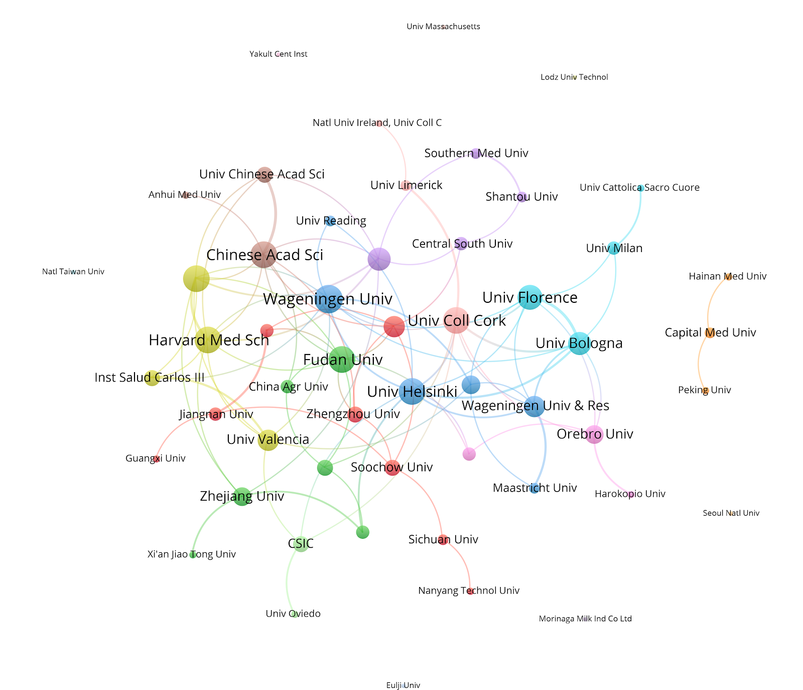
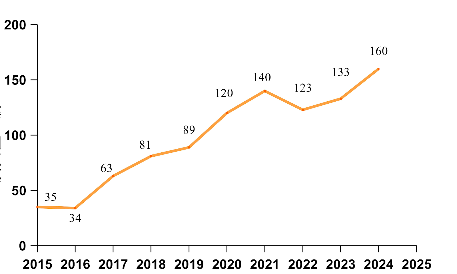
BACKGROUND: As the population ages, research on gut microbiota in the elderly is gaining attention. However, bibliometric analysis in this field is still lacking. OBJECTIVE: To comprehensively analyze literature on gut microbiota in older adults from multiple databases, identify current research hotspots, predict future trends, and provide potential directions for subsequent research. METHODS: CNKI was searched using the subject terms “gut microbiota in older adults,” “gut microecology in older adults,” and “intestinal flora in older adults.” The Web of Science database was searched using the search strategy of “TS=(elderly gut microbe OR elderly gut microbiome OR elderly gut microbiota OR elderly intestinal microbiome OR elderly intestinal microbiota).” Bibliometric tools VOSviewer and CiteSpace were employed to systematically analyze publication years, country distribution, research institutions, authors, and keywords in the retrieved literature. RESULTS AND CONCLUSION: A total of 250 and 604 eligible articles were obtained from the CNKI and Web of Science databases, respectively. From 2014 to 2023, the global publication volume in the field of gut microbiota in older adults showed a steady upward trend. Research interest and discussions in this field have increased significantly worldwide, with expanding depth and breadth through interdisciplinary collaboration. Notably, COVID-19, oxidative stress, depression, and cognitive impairment emerged as prominent keywords in the past 2 years. This bibliometric analysis visually demonstrated the research status and development trends in the field of gut microbiota in the elderly over the past decade. The field is currently in a rising phase, and further exploration of the mechanisms of gut microbiota and intervention strategies for related diseases is still needed.

Read Full Abstract10.12307/2026.21271
Feng's spinal manipulation for cervical spondylosis: kinematic changesGraphical AbstractVerified
Chinese Journal of Tissue Engineering Research2026

Feng's spinal manipulation for cervical spondylosis: kinematic changes

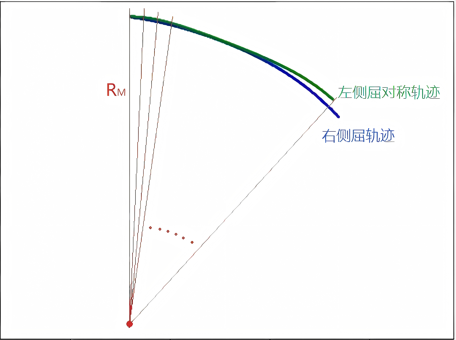
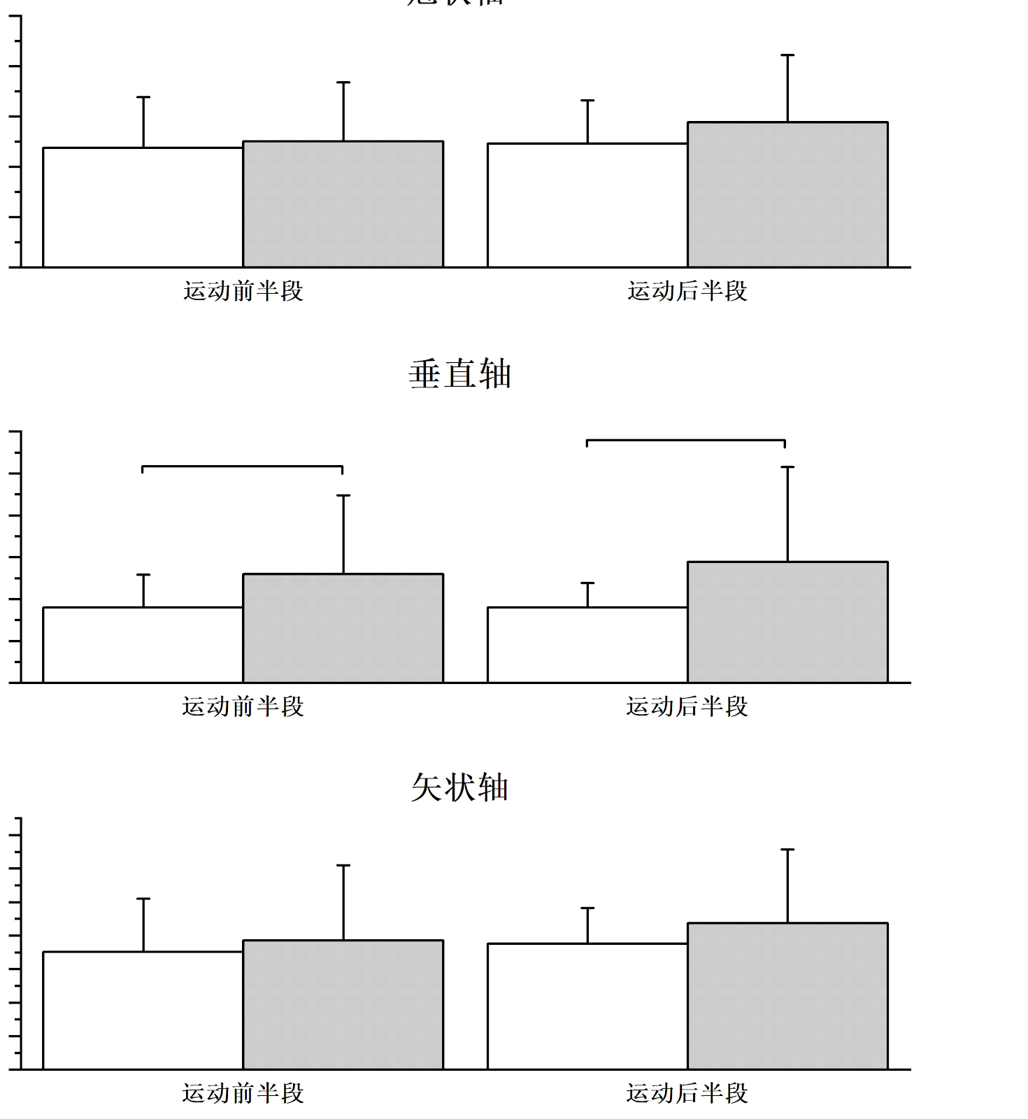
BACKGROUND: Patients with cervical spondylosis often exhibit varying kinematic abnormalities due to degenerative structural changes and biomechanical imbalances in the cervical spine. Although previous studies have compared specific kinematic parameters between healthy individuals and cervical spondylosis patients, research on coupled motions and their associated ratios remains limited. OBJECTIVE: To investigate changes in kinematic parameters in cervical spondylosis patients before and after Feng's spinal manipulation therapy. METHODS: Thirty patients with cervical spondylosis and 30 healthy controls were enrolled. Participants completed three standardized motion tasks: lateral flexion, flexion-extension, and axial rotation. Three-dimensional cervical spine kinematics were quantified using stereophotogrammetry upon admission and discharge. The following kinematic parameters were analyzed: primary range of motion, coupled motion range, coupled motion patterns, motion symmetry, motion smoothness, and motion velocity. RESULTS AND CONCLUSION: Compared with healthy controls, patients with cervical spondylosis showed significantly reduced maximal angles in lateral flexion, flexion-extension, and axial rotation (P < 0.05), and significantly increased ratios of coupled flexion-extension during lateral flexion, coupled rotation during lateral flexion, coupled lateral flexion during extension, and coupled lateral flexion during rotation (P < 0.05). After treatment, patients showed significant improvements in visual analog scale score and cervical dysfunction index (P < 0.05). Significant differences were found in maximal lateral flexion angle, lateral flexion symmetry, maximum and average lateral flexion velocity, maximal flexion-extension angle, maximum and average flexion-extension velocity, maximal rotation angle, rotation symmetry, maximum rotation velocity, and average left rotation velocity before and after treatment (P < 0.05). No significant differences were observed in coupled motion patterns before and after treatment (P > 0.05). Significant differences were found in the ratios of coupled flexion-extension during right lateral flexion, coupled rotation during lateral flexion, coupled flexion-extension during right rotation, and coupled lateral flexion during rotation before and after treatment (P < 0.05). In conclusion, patients with cervical spondylosis exhibit increased ratios of some coupled motions relative to primary motions. Feng's spinal manipulation can significantly improve clinical symptoms and effectively restore cervical motor function.

Read Full Abstract10.12307/2026.21272
Burn and multi-omic biomarkers: causal relationships with 41 inflammatory factors and 35 blood and urine markersGraphical AbstractVerified
Chinese Journal of Tissue Engineering Research2026

Burn and multi-omic biomarkers: causal relationships with 41 inflammatory factors and 35 blood and urine markers

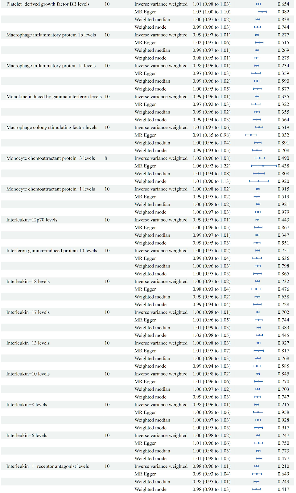
BACKGROUND: Conventional observational studies are inadequate to reveal the potential causal relationship of biomarkers in burns patients. Mendelian randomization, leveraging genetic variation as an instrumental variable to mimic the advantages of randomized controlled trials, has emerged as a crucial tool for dissecting causal associations in complex diseases. OBJECTIVE: To explore the relationship of burn injury with 41 inflammatory cytokines and 35 blood and urinary biomarkers using Mendelian Randomization. METHODS: (1) Burn-related data of genome-wide association studies were obtained from the IEU open GWAS project database, constructed by The University of Bristol, UK, including 218 131 samples and 16 380 465 single nucleotide polymorphisms were included in the study. (2) Data for 41 types of inflammatory cytokines were derived from a study involving 8 293 participants in the Finnish Young Cardiovascular Risk Study database, which is constructed by the Research Centre for Applied and Preventive Cardiovascular Medicine, University of Turku. (3) Data for 35 types of blood and urinary biomarkers were derived from a study involving 363 228 participants from the UK Biobank, which is a large biomedical database project jointly initiated by the UK government, the Wellcome Trust, and the Medical Research Council of the UK. Single nucleotide polymorphisms were employed as instrumental variables, and analyses were conducted using inverse variance weighting, MR Egger, weighted median, and weighted mode methods. Cochrane's Q test was used to identify heterogeneity, and MR Egger intercept test, MR-PRESSO test, and leave-one-out analysis were used to assess the reliability of exposure-outcome associations. RESULTS AND CONCLUSION: Burn injury reduced levels of interleukin-9 (OR=0.97; 95%CI, 0.949 to 0.997; P=0.030) and testosterone (OR=0.997; 95%CI, 0.995 to 0.999; P=0.025), with no heterogeneity or horizontal pleiotropy, demonstrating robustness. The Mendelian randomization analysis indicates that burn injury leads to decreased levels of interleukin-9 and testosterone, suggesting that increasing these levels may aid in tissue repair and improve protein breakdown rate after burn.

Read Full Abstract10.12307/2026.21270
Performance of unstable barbell bench press and changes in electromyographic activity after transcranial direct current stimulationGraphical AbstractVerified
Chinese Journal of Tissue Engineering Research2026

Performance of unstable barbell bench press and changes in electromyographic activity after transcranial direct current stimulation

BACKGROUND: Transcranial direct current stimulation can enhance human motor performance by modulating cortical excitability, but its impact on the performance of unstable resistance exercise remains unclear. OBJECTIVE: To investigate the effects of transcranial direct current stimulation on unstable barbell bench press performance and electromyographic activity. METHODS: A randomized, self-controlled crossover design was employed. Twenty-two male college students were randomly assigned to receive either active anodal transcranial direct current stimulation or sham stimulation. After transcranial direct current stimulation interventions, subjects performed a fatigue test involving the unstable barbell bench press. Performance during the unstable barbell bench press task was assessed by recording both the number of completed repetitions and barbell acceleration, serving as indicators of load capacity and movement stability control. In addition, surface electromyographic signals were collected from the right biceps brachii, triceps brachii, anterior deltoid, posterior deltoid, and pectoralis major during the exercise. A paired t-test was used to examine differences in repetition counts between true and sham stimulation conditions. Repeated measures analysis of variance was applied to analyze differences in triaxial acceleration, agonist muscle activation levels, and antagonist co-activation levels. RESULTS AND CONCLUSION: (1) No significant difference in the number of barbell bench press repetitions was observed between the true and sham stimulation conditions. However, the mean amplitude of Y-axis acceleration was substantially lower in the true stimulation group than the sham stimulation group. (2) True transcranial direct current stimulation markedly increased the activation level of the anterior deltoid and the co-activation level of the posterior deltoid. (3) There was no significant interaction between transcranial direct current stimulation and exercise phase on any measured variable. (4) These findings suggest that transcranial direct current stimulation does not significantly affect muscle endurance performance during unstable barbell bench press, but it can improve movement stability, possibly by increasing anterior deltoid activation and posterior deltoid co-activation, thereby enhancing shoulder joint stiffness and stability. In competitive sports and clinical rehabilitation, transcranial direct current stimulation may be considered to modulate performance in unstable load-bearing tasks according to training objectives.

Read Full Abstract10.12307/2026.21274
Visual analysis of shear wave elastography in skeletal muscle researchGraphical AbstractVerified
Chinese Journal of Tissue Engineering Research2026

Visual analysis of shear wave elastography in skeletal muscle research

BACKGROUND: Shear-wave elastography is valuable for rehabilitation diagnosis and treatment, but it has not been sufficiently promoted in clinical practice. OBJECTIVE: To explore the trends and hotspots of ultrasound shear wave elastography in skeletal muscle research by visualizing and analyzing the international literature from the past 10 years, thereby providing a reference for clinical diagnosis and follow-up research. METHODS: Based on the Web of Science Core Collection database (2015-2024), the number of publications, countries/regions, institutions, authors, journals, cited literature, and key words from the 978 included articles were visualized and analyzed using CiteSpace software. RESULTS AND CONCLUSION: (1) With a 16.2% average annual growth in global publications, China has the highest number of publications worldwide (197), but its international collaborative network is relatively weak. The University of Nantes in France has the highest number of publications (50), and the University of Queensland has the most influential collaborative network. (2) Ultrasound in Medicine and Biology is the journal with the most publications (33). Noriaki Ichihashi is the most prolific author. (3) The gastrocnemius muscle is one of the most frequently examined sites. Shear wave elastography shows significant clinical potential in central nervous system diseases and sports injuries. (4) The research focus has shifted from basic biomechanics to dynamic clinical assessment and therapeutic interventions. (5) Future diagnostic techniques should be more standardized and refined, establishing normative data ranges for muscle tissue, while considering individual biological variability in elasticity values.

Read Full Abstract10.12307/2026.21266
Association between sarcopenia and osteoporosis: a genome-wide data analysis in European populationsGraphical AbstractVerified
Chinese Journal of Tissue Engineering Research2026

Association between sarcopenia and osteoporosis: a genome-wide data analysis in European populations

BACKGROUND: Sarcopenia and osteoporosis have attracted significant attention in the academic community due to their high prevalence and severe adverse outcomes. Although existing studies have suggested a potential causal relationship between sarcopenia and osteoporosis, the evidence remains insufficient. OBJECTIVE: Based on large-scale genome-wide data, to explore the causal relationship between genetically predicted sarcopenia and osteoporosis through a bidirectional Mendelian randomization approach. METHODS: Genome-wide significant loci (P < 5×10⁻⁸) associated with sarcopenia-related traits were selected from the UK Biobank database (an open database jointly developed by the UK government, the Medical Research Council, and the Wellcome Trust), followed by linkage disequilibrium analysis. Osteoporosis data were obtained from the GEnetic Factors for OSteoporosis Consortium (GEFOS; an open database funded by the EU Framework Program for Research and Development, jointly led by Erasmus University Medical Center in the Netherlands), including 28,498 European ancestry subjects, with a focus on data from osteoporosis-prone fracture sites. The study used inverse variance weighting as the primary analysis method, supplemented by MR-Egger regression, weighted median method, and MR-RAPS for multiple validation. To ensure the reliability of the results, multiple sensitivity analyses were performed. RESULTS AND CONCLUSION: Inverse variance weighting analysis showed a bidirectional causal relationship between whole-body fat-free mass and bone mineral density (P < 0.05). Forward causal analysis indicated that whole-body fat-free mass was positively associated with lumbar spine bone mineral density (OR=1.124, 95%CI: 1.008-1.253, P=0.035) and negatively associated with forearm bone mineral density (OR=0.821, 95%CI: 0.699-0.966, P=0.017). Reverse causal analysis showed that forearm bone mineral density (OR=1.033, 95%CI: 1.002-1.066, P=0.036), lumbar spine bone mineral density (OR=1.054, 95%CI: 1.025-1.084, P < 0.001), and femoral neck bone mineral density (OR=1.059, 95%CI: 1.008-1.113, P=0.021) were all positively associated with whole-body fat-free mass. A reduction in whole-body fat-free mass can lead to decreased lumbar spine bone mineral density, and a decrease in bone mineral density at various sites further exacerbates the loss of whole-body fat-free mass. Although the data in this study mainly come from European populations, due to the universality of genome-wide association analysis methods and the commonality of genetic backgrounds, the results still have important reference value for exploring the pathogenesis of sarcopenia and osteoporosis in the Chinese population, formulating clinical intervention strategies, and assessing genetic risk.

Read Full Abstract10.12307/2026.21267
Application of patch-clamp technique in traditional Chinese medicine: a visual analysis of relevant literatureGraphical AbstractVerified
Chinese Journal of Tissue Engineering Research2026

Application of patch-clamp technique in traditional Chinese medicine: a visual analysis of relevant literature

BACKGROUND: In recent years, the interdisciplinary application potential of patch-clamp technique in traditional Chinese medicine research has gradually emerged, but a systematic summary of its applications in this field has not yet been conducted. OBJECTIVE: To visualize the application of patch-clamp technology in traditional Chinese medicine field through CiteSpace knowledge map analysis, and to reveal the progress and trends of this technology in the modernization research of traditional Chinese medicine. METHODS: The literature sources included CNKI, VIP, WanFang, PubMed and Web of Science Core Collection database. The computer-assisted literature search was conducted to build a database of patch-clamp technology applications in traditional Chinese medicine field from database inception to September 2024. The authors, institutions, and keywords were subjected to visual analysis and knowledge map drawing using CiteSpace 6.3.R1 software and bibliometric methods. RESULTS AND CONCLUSION: (1) A total of 819 articles were included, with 968 authors. The First Affiliated Hospital of Henan University of Chinese Medicine was the institution with the most publications in the Chinese database, while Harbin Medical University was the institution with the most publications in the English database. (2) The research directions, keyword clustering, and emergence analysis of each institution showed that the application of patch-clamp technique in traditional Chinese medicine mainly focused on cardiovascular electrophysiology, pharmacology of Chinese materia medica, and nervous system electrophysiology. (3) The development of patch-clamp technique in traditional Chinese medicine generally presented an evolutionary path of 'basic mechanism → target deepening → clinical translation', reflecting a transformation from a single technical tool to a multidisciplinary intersection platform. (4) It is suggested that the core influence of authors needs further improvement, and cross-regional cooperation among research institutions is insufficient, so cross-regional cooperation should be strengthened. Current research techniques are single and research content is thin; future research should integrate multiple technologies, cross disciplines, enrich research content, and expand research directions, providing evidence support for in-depth exploration of the internal mechanisms of traditional Chinese medicine.

Read Full Abstract10.12307/2026.21265
Intervention with Compound Kidney-Invigorating Granules in a mouse model of osteoporosis: role of the TRIB3/beta-catenin axisGraphical AbstractVerified
Chinese Journal of Tissue Engineering Research2026

Intervention with Compound Kidney-Invigorating Granules in a mouse model of osteoporosis: role of the TRIB3/beta-catenin axis

BACKGROUND: Previous studies have shown that knockdown of β-catenin can inhibit the osteogenic differentiation of human bone marrow mesenchymal stem cells and reduce the expression of TRIB3. Serum containing Compound Kidney-Invigorating Granules can promote the expression of β-catenin and TRIB3 in human bone marrow mesenchymal stem cells, and induce human bone marrow mesenchymal stem cells to differentiate into osteogenic cells. OBJECTIVE: To further explore the mechanism of Compound Kidney-Invigorating Granules in a mouse model of osteoporosis based on the TRIB3/β-catenin axis. METHODS: 8-week-old female C57BL/6 mice were randomly divided into the following experimental groups: blank control, sham operation, model, and low-, medium-, and high-dose Compound Kidney-Invigorating Granules groups, and positive drug group. Except for the blank control and sham operation groups, bilateral ovariectomy was performed to establish an osteoporosis mouse model. One week after modeling, mice in the low-, medium-, and high-dose groups were intragastrically administered 7.05, 14.1, and 28.2 g/kg Compound Kidney-Invigorating Granules, respectively; the blank control, sham operation, and model groups received an equal volume of normal saline once daily; the positive control group received 1.53 mg/kg alendronate sodium once weekly. After 12 weeks of administration, Micro-CT was used to detect changes in femoral bone microarchitecture; hematoxylin-eosin staining and Masson staining were used to detect pathological changes in the femur; Western blot was used to detect the expression levels of TRIB3, β-catenin, alkaline phosphatase, and osteopontin in bone tissue. RESULTS AND CONCLUSION: Compared with the blank control and sham operation groups, the model group showed sparse bone trabeculae and significantly increased empty lacunae; the protein expression levels of TRIB3, β-catenin, alkaline phosphatase, and osteopontin were significantly decreased (P < 0.05). Compared with the model group, the medium- and high-dose Compound Kidney-Invigorating Granules groups showed more complete and regular bone trabeculae, and the protein expression levels of TRIB3, β-catenin, alkaline phosphatase, and osteopontin were significantly upregulated (P < 0.05). These results indicate that Compound Kidney-Invigorating Granules exert a therapeutic effect on osteoporosis model mice, suggesting that the formula may act through the TRIB3/β-catenin axis.

Read Full Abstract10.12307/2026.21278
Osteoporotic vertebral compression fractures: a visual analysis of current status and emerging trendsGraphical AbstractVerified
Chinese Journal of Tissue Engineering Research2026

Osteoporotic vertebral compression fractures: a visual analysis of current status and emerging trends

BACKGROUND: Current literature primarily focuses on comparing the clinical efficacy and surgical safety of various operative approaches for osteoporotic vertebral compression fractures, while there is a notable lack of research addressing the current status, hotspots, and future trends in the treatment and prognosis of osteoporotic vertebral compression fractures. OBJECTIVE: To analyze the current status, hotspots and trends of research on osteoporotic vertebral compression fractures by bibliometric methods. METHODS: English articles related to the treatment and prognosis of osteoporotic vertebral compression fractures published between 1994 and 2023 were retrieved from the Web of Science Core Collection. Bibliometric and visualization analyses were conducted using CiteSpace and VOSviewer to evaluate publication volume, geographic distribution, institutional contributions, journal sources, author contributions, keyword frequency, research hotspots, and the most influential literature. RESULTS AND CONCLUSION: A total of 2,275 articles were included. Global research output on osteoporotic vertebral compression fractures increased annually, with the highest number of publications in 2022 (215 articles). The literature spanned 68 countries/regions, with China leading with 644 articles. Soochow University was the most productive institution (90 articles). European Spine Journal had the highest number of publications (n=146), while Spine had the highest citation count (n=1,923), average citations per article (58.5), and H-index (45), ranking second in publication volume (n=106). Yang, Huilin was the most prolific author (n=65), and French physician Galibert, P had the highest citations (n=300). Early research hotspots focused on 'vertebroplasty', 'balloon kyphoplasty', and 'polymethylmethacrylate', which later shifted to 'surgical management' and 'nonoperative treatment', and more recently expanded to 'safety' and 'risk factors', which are expected to remain future research trends.

Read Full Abstract10.12307/2026.21268
Molecular mechanism and natural drug screening for ferroptosis-targeted therapy in rheumatoid arthritisGraphical AbstractVerified
Chinese Journal of Tissue Engineering Research2026

Molecular mechanism and natural drug screening for ferroptosis-targeted therapy in rheumatoid arthritis

BACKGROUND: Current research in rheumatoid arthritis focuses on iron metabolism-related proteins and the effects of ferroptosis on immune cells. This study proposes new approaches to target ferroptosis in the treatment of rheumatoid arthritis from the perspective of traditional Chinese medicine. These approaches include developing new traditional Chinese medicine therapies, creating individualized treatment plans based on patients’ genes and biomarkers, optimizing therapeutic strategies, and improving symptoms. These strategies aim to facilitate early treatment and improve prognosis. OBJECTIVE: Bioinformatics was applied to investigate the molecular mechanism of treating rheumatoid arthritis from the perspective of ferroptosis, and to screen potential traditional Chinese medicines and active ingredients, opening up a new way for the treatment of rheumatoid arthritis. METHODS: The Gene Expression Omnibus database, maintained by the National Center for Biotechnology Information, is primarily used to store and share high-throughput gene expression, microarray, and sequencing data. The Gene Expression Omnibus database enables researchers to search and analyze genomic data related to various diseases. This study is based on publicly available summary statistics databases and does not require ethical approval. The Gene Expression Omnibus database was searched for datasets related to rheumatoid arthritis that met the screening criteria. The Sanger sequencing platform was then used to obtain the transcriptome data of rheumatoid arthritis. Finally, the limma algorithm was applied to screen the differentially expressed genes. Meanwhile, the ferroptosis-related gene set was extracted from the FerrDb database. Through integration analysis, we obtained the intersection of the differentially expressed genes and the ferroptosis-related genes. We constructed a protein interaction network and performed network topology analysis. Then, using DAVID Bioinformatics Resources 6.8, we conducted Gene Ontology and Kyoto Encyclopedia of Genes and Genomes pathway enrichment analyses to explore biological functions and signaling pathways. Finally, the SymMap platform was used to identify natural medicines, and the Traditional Chinese Medicine Systems Pharmacology Database and Analysis Platform was used to find small molecule compounds and corresponding targets for molecular docking analysis. RESULTS AND CONCLUSION: Two datasets, GSE55457 and GSE55235, were identified from the GEO database, and 340 common differentially expressed targets were obtained via limma analysis. A total of 487 ferroptosis-related targets were collected from the FerrDb platform, and 17 common targets related to ferroptosis and rheumatoid arthritis were obtained after intersection. A protein interaction network was constructed using the 17 common targets, containing 16 target proteins and 33 interactions. Core targets such as EGFR, AR, MAPK8, CDKN1A, JUN, ATM, and EGR1 occupied important positions in the network. Five core targets (EGFR, AR, MAPK8, CDKN1A, and JUN) were identified as key ferroptosis-related targets in rheumatoid arthritis. GO and KEGG enrichment analyses showed that treating rheumatoid arthritis via the ferroptosis pathway may be related to DNA binding processes and SMAD2/3 signaling pathways. Molecular docking results showed that natural small molecule compounds such as progesterone, estradiol, and quercetin could form good binding with core targets, providing new directions for drug development and research in rheumatoid arthritis.

Read Full Abstract10.12307/2026.21269
Tetramethylpyrazine improves iron metabolism disorders in a rat model of spinal cord injury via the Keap-1/Nrf2 signaling pathwayGraphical AbstractVerified
Chinese Journal of Tissue Engineering Research2026

Tetramethylpyrazine improves iron metabolism disorders in a rat model of spinal cord injury via the Keap-1/Nrf2 signaling pathway

BACKGROUND: The clinical management of spinal cord injury remains a global medical challenge, with no currently available ideal treatment. Traditional Chinese medicine has therapeutic advantages for spinal cord injury. Notably, tetramethylpyrazine, an active component of Chuanxiong rhizome, has been shown to significantly suppress pathological responses including neuroinflammation and apoptosis after spinal cord injury, exhibiting promising therapeutic potential. However, its mechanisms require further elucidation. OBJECTIVE: To investigate the regulatory effects of tetramethylpyrazine on the Kelch-like ECH-associated protein 1/nuclear factor-erythroid 2-related factor 2 (Keap-1/Nrf2) signaling pathway and iron metabolism following spinal cord injury, and to elucidate its neuroprotective mechanisms. METHODS: Thirty-six Sprague-Dawley rats were randomly allocated into: sham group (laminectomy+saline, n=12), model group (spinal cord injury+saline, n=12), and tetramethylpyrazine group (spinal cord injury+tetramethylpyrazine, n=12). After 4 weeks, neuronal morphology was assessed by Nissl staining; iron deposition by Prussian blue staining; iron content by iron assay kit; expression of Keap-1, ferritin heavy chain 1 (FTH1), and ferritin light chain (FTL) by immunohistochemistry; Nrf2 expression by immunofluorescence; protein levels of Nrf2, FTH1, and FTL by western blot; and mRNA levels of Nrf2, Keap-1, FTH1, and FTL by RT-PCR. RESULTS AND CONCLUSION: Compared with the sham group, the model group showed disrupted neuronal structure, increased iron deposition and iron content, increased Keap-1 expression, and decreased Nrf2, FTH1, and FTL expression at both protein and mRNA levels (P < 0.01). Tetramethylpyrazine treatment significantly ameliorated these changes, as evidenced by improved neuronal structure, reduced iron deposition and content, decreased Keap-1 expression, and increased Nrf2, FTH1, and FTL expression compared with the model group (P < 0.01). These findings indicate that tetramethylpyrazine can regulate the Keap-1/Nrf2 signaling pathway and its downstream targets FTH1 and FTL, thereby improving iron metabolism disorders and facilitating spinal cord injury repair.

Read Full Abstract10.12307/2026.21277
Potential targets of glucagon-like peptide 1 receptor agonist ticagrelor in the treatment of Alzheimer’s diseaseGraphical AbstractVerified
Chinese Journal of Tissue Engineering Research2026

Potential targets of glucagon-like peptide 1 receptor agonist ticagrelor in the treatment of Alzheimer’s disease

BACKGROUND: Glucagon-like peptide 1 receptor agonists, as novel drug candidates for the treatment of neurodegenerative diseases, have achieved breakthrough progress in clinical research on Alzheimer’s disease, with drugs such as Semaglutide advancing to phase III clinical trials. However, there remains a significant knowledge gap regarding the molecular mechanism of neuroprotective effects of these drugs. OBJECTIVE: To innovatively integrate multi-omics analysis techniques and network pharmacology methods, to systematically analyze the intersection network between the gene lineage related to Alzheimer’s disease pathology and the potential targets of ticagrelor, to identify key regulatory genes, and to verify their molecular mechanisms through in vitro and in vivo experiments. METHODS: A multi-dimensional research strategy was adopted: (1) Constructing the differential expression gene profile of Alzheimer’s disease using the DisGeNET database that covers various disease-related genomics. (2) Obtaining the structure of Tirzepatide from PubChem database with bioactive molecules and screening potential targets. (3) Conducting Gene Ontology (GO) functional annotation and Kyoto Encyclopedia of Genes and Genomes (KEGG) pathway enrichment analysis using DAVID database. (4) Constructing protein-protein interaction network using STRING database and Cytoscape 3.9.1, and screening key genes via topological network analysis. (5) Cell-level verification: HT22 cells were divided into control group, model group (treated with β-amyloid 1-42 oligomers for 36 h to establish an in vitro AD model), and treatment group (pretreated with β-amyloid 1-42 oligomers for 24 h, then co-treated with ticagrelor for 12 h). Western blot was used to analyze the protein expression of angiotensin II type 2 receptor (AGTR2), and ELISA was used to detect the expression levels of synaptic markers such as synaptophysin 1 and postsynaptic density protein 95. (6) Animal experiments: Three groups were used: control group (WT C57BL/6 mice, intraperitoneal injection of saline), model group (3xTg mice, intraperitoneal injection of saline), and treatment group (3xTg mice, intraperitoneal injection of 20 nmol/L ticagrelor), all administered every other day for a total of 15 doses. Morris water maze was used to analyze cognitive behavioral improvements in AD model mice; Western blot was used to quantitatively analyze the expression of β-amyloid (6E10) and phosphorylated Tau protein (P-tau-181). RESULTS AND CONCLUSION: (1) A total of 3,397 AD-related genes were screened from DisGeNET database; 10 key genes with the highest connectivity were identified based on protein association: AGTR2, NTSR1, NTSR2, GHSR, C5AR1, C3AR1, OPRM1, SSTR2, OPRD1, STAT3. GO enrichment and KEGG pathway analysis suggested that ticagrelor may improve AD by enhancing neuroreceptor-ligand function. (2) Cell experiments suggested that ticagrelor may exert therapeutic effects by improving synaptic function in AD, and AGTR2 may be a potential target of ticagrelor in treating AD. (3) Animal experiments indicated that ticagrelor improved cognitive ability in 3xTg mice, and ameliorated abnormal β-amyloid deposition and Tau protein phosphorylation in the brain of 3xTg mice. (4) Conclusion: The study reveals that AGTR2 is a key molecular target of ticagrelor in the pathological process of AD, and ticagrelor may treat AD by regulating AGTR2-mediated synaptic function improvement.

Read Full Abstract10.12307/2026.21276