🧬 SinoBioData Academic Portal
📚 Peer-Reviewed Translated Literature

All Biomedical & Clinical Articles (Page 5)

Browse complete peer-reviewed translations from top Chinese biomedical, oncology, and genomics journals. Read verified previews and download full authentic clinical reports.

Published Research Papers

Showing 24 of 1542 peer-reviewed translated articles (Page 5 of 65)

Development of an in vitro TurboID labeling assay for the detection of protoRAG-interacting proteins in the amphioxusGraphical AbstractVerified
Acta Biochimica et Biophysica Sinica2026

Development of an in vitro TurboID labeling assay for the detection of protoRAG-interacting proteins in the amphioxus

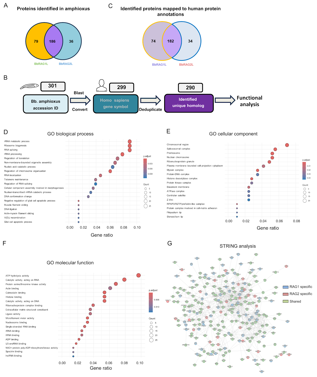
The recombination-activating gene (RAG)-mediated V(D)J rearrangement is essential for adaptive immunity in jawed vertebrates. RAG evolved from an invertebrate RAG-like (RAGL) transposase, with the amphioxus protoRAG (BbRAG1L/BbRAG2L) serving as a key model. However, the regulatory mechanisms of protoRAG remain unclear. Here, we developed an in vitro proximity labeling assay using TurboID fused to BbRAG1L or BbRAG2L to identify interacting proteins from amphioxus hepatic cecum and colon lysates. The fusion proteins were expressed in Expi293F cells, purified, and incubated with amphioxus lysates in the presence of biotin and ATP. Biotinylated proteins were enriched via streptavidin beads and analyzed by LC-MS/MS. This approach enables the identification of protoRAG-interacting proteins without the need for transgenic amphioxus, providing a valuable tool to study the evolution of RAG regulation.

Read Full Abstract10.3724/abbs.2026030
Super-resolution imaging reveals higher-order structures within common fragile sites in human mitotic chromosomesGraphical AbstractVerified
Acta Biochimica et Biophysica Sinica2026

Super-resolution imaging reveals higher-order structures within common fragile sites in human mitotic chromosomes

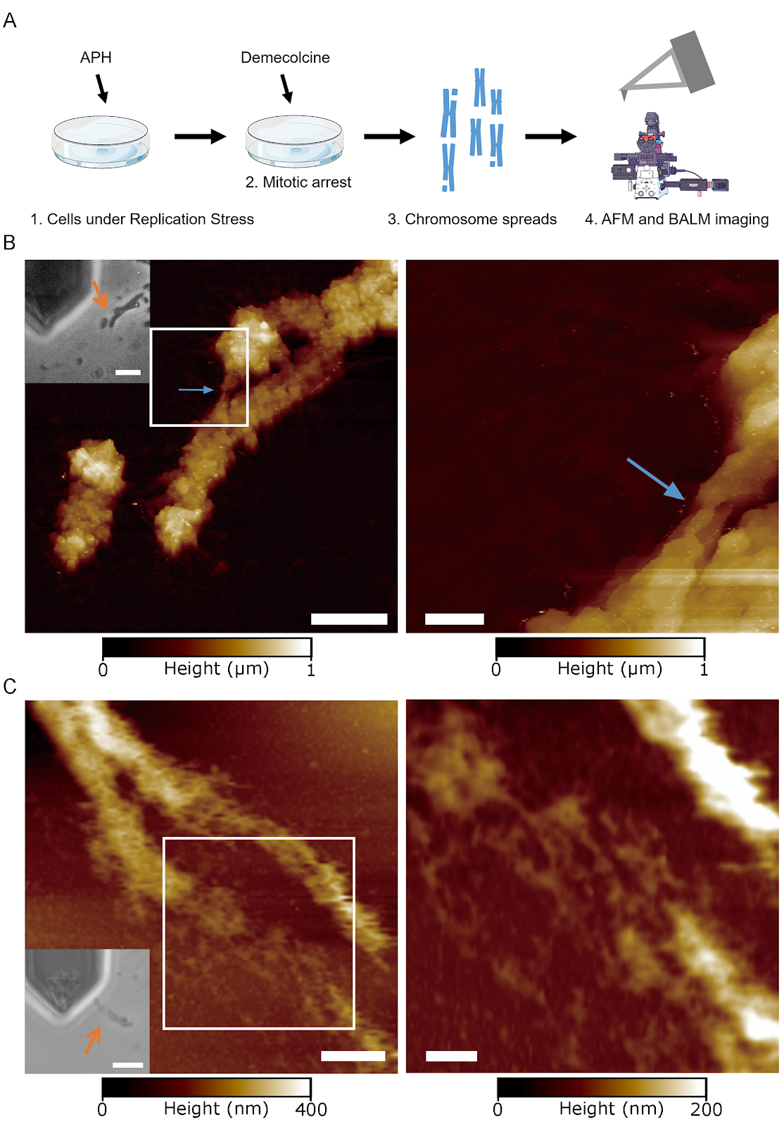
Common fragile sites (CFSs) are large genomic loci that are frequently deleted under replication stress and are thought to play a role in carcinogenesis as well as developmental disorders during early embryogenesis. They often appear as gaps or breaks in mitotic chromosomes by conventional optical microscopy and are also loci that are replicated in mitosis in a process called mitotic DNA synthesis (MiDAS). However, we still have a very poor understanding of the potential mechanisms underlying their genomic instability. We recently found that in normal mitotic chromosomes, there is a two-level hierarchy in the higher-order chromatin structure, with mechanically resistant ~90 nm Mitotic NanoDomains (MNDs) assembling into ~125 nm compact granules (CGs). In this work, we combine multiple super-resolution imaging techniques, including binding-activated localization microscopy (BALM), atomic force microscopy (AFM), and stochastic optical reconstruction microscopy (STORM), to characterize CFSs. Our super-resolution data indicate that sites that appear as gaps by conventional microscopy can be filled with chromatin of the size and shape of MNDs but not CGs. Moreover, we find that MiDAS loci only form an array of MNDs with no CGs. Taken together, our work suggests that under replication stress, CFS loci can not only be replicated but also assembled into higher-order chromatin. However, this organization is limited to the level of MNDs, which suggests that a failure to progress beyond MNDs to form CGs constitutes a key structural deficiency at these loci.

Read Full Abstract10.3724/abbs.2026014
Identification and experimental validation of core genes associated with breast cancer brain metastasis via machine learningGraphical AbstractVerified
Acta Biochimica et Biophysica Sinica2026

Identification and experimental validation of core genes associated with breast cancer brain metastasis via machine learning

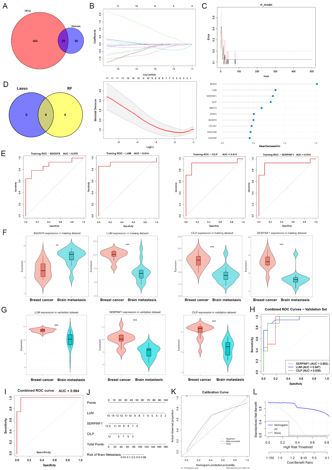
Breast cancer (BC) is the most common malignancy among women, with approximately 2.3 million new cases diagnosed annually. Brain metastasis is a significant cause of mortality, particularly in HER2-positive and triple-negative subtypes. Current therapies are limited by the blood-brain barrier. This study aimed to identify core genes associated with breast cancer brain metastasis (BCBM) using bioinformatics and machine learning. We analyzed the GSE43837 dataset (19 nonmetastatic primary breast tumors and 19 brain metastases) using differential expression analysis and weighted gene coexpression network analysis (WGCNA). We identified 245 upregulated and 188 downregulated genes. WGCNA revealed key modules (midnightblue and black) with 89 candidate genes. Intersection with differentially expressed genes yielded 29 overlapping genes. LASSO regression and random forest identified four core genes: B3GNT9, SERPINF1, LUM, and CILP. ROC analysis showed strong discriminatory power (AUC > 0.87). External validation in GSE125989 confirmed downregulation of SERPINF1, LUM, and CILP in brain metastases, with a combined model achieving AUC 0.984. Experimental validation in zebrafish and mouse models confirmed the role of these genes in BCBM. These findings suggest that SERPINF1, LUM, CILP, and B3GNT9 are potential biomarkers and therapeutic targets for BCBM.

Read Full Abstract10.3724/abbs.2026037
Distinct miR319a Identified from Persicaria chinensis Mediates Cross-Kingdom Suppression of Cervical Cancer by Targeting ITGA3Graphical AbstractVerified
Acta Biochimica et Biophysica Sinica2026

Distinct miR319a Identified from Persicaria chinensis Mediates Cross-Kingdom Suppression of Cervical Cancer by Targeting ITGA3

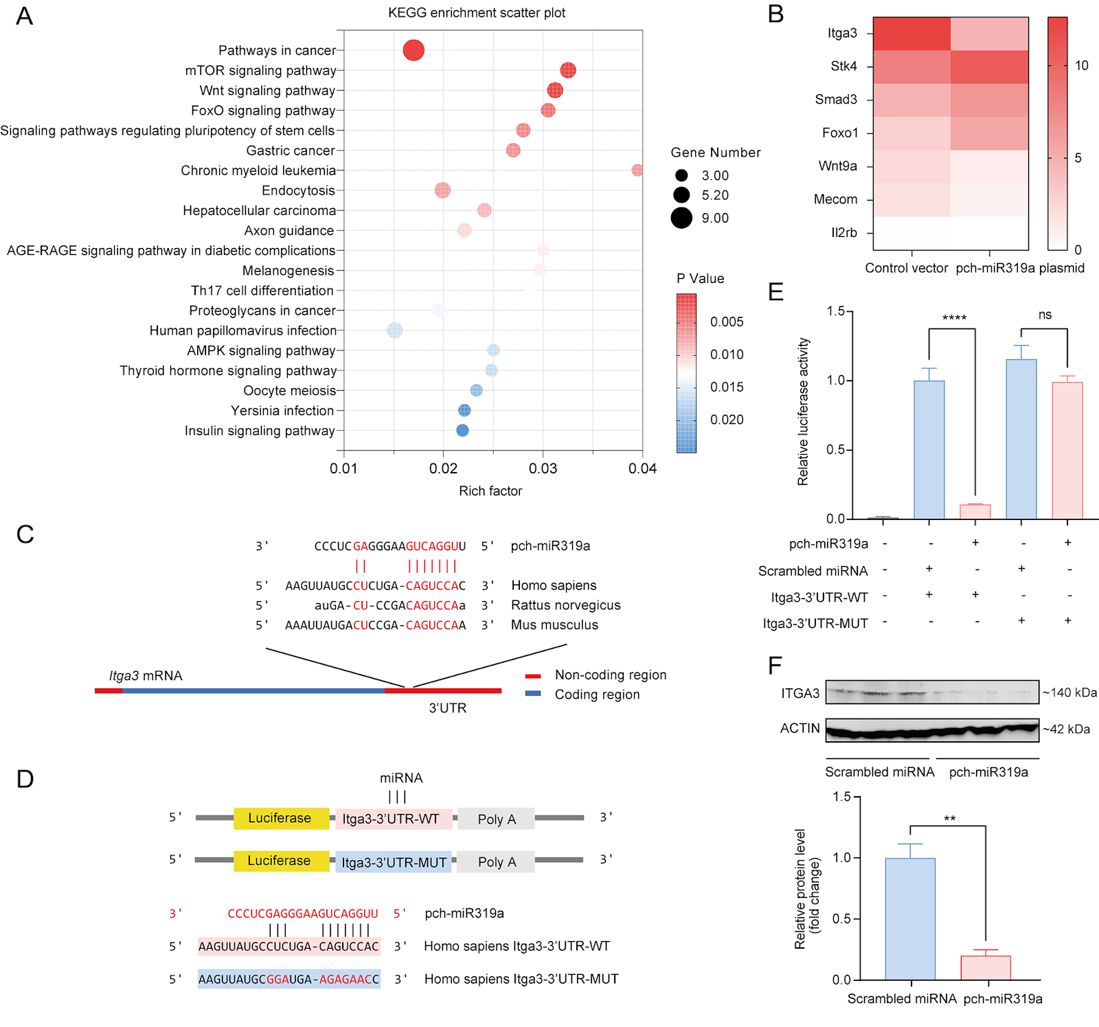
Persicaria chinensis, a well-known traditional Chinese medicinal herb that is both edible and medicinal, has been widely acknowledged for its therapeutic effects, such as anti-inflammatory, antioxidant, and antitumor activities. However, the role of miRNAs from this plant in the cross-kingdom regulation of human diseases has not been investigated. In this study, we analyze the miRNA expression profile of P. chinensis using high-throughput sequencing and identify a total of 673 miRNAs, including 422 novel miRNAs that are unique to this plant and 251 conserved miRNAs. Among the conserved miRNAs, pch-miR319a is found to be the most abundant. Moreover, food-oriented pch-miR319a accumulates in the uterus and tumors and exhibits a rich repertoire of target genes within cancer-related pathways, demonstrating significant cross-kingdom regulatory potential. Utilizing the dual-luciferase reporter gene assay, we demonstrate that pch-miR319a from P. chinensis targets the Itga3 gene, which is associated with cervical cancer progression. Overexpression of pch-miR319a significantly decreases the viability, migration, and induces apoptosis of HeLa cervical cancer cells in vitro. Moreover, in a syngeneic mouse tumor model of cervical cancer, treatment with pch-miR319a effectively inhibits tumor growth and downregulates the expressions of ITGA3 and the proliferation marker Ki-67. Our study highlights the potential of pch-miR319a from P. chinensis as a novel therapeutic agent for cervical cancer by targeting ITGA3 and provides new insights into the cross-kingdom regulatory mechanisms of plant miRNAs in human diseases.

Read Full Abstract10.3724/abbs.2026010
Biochemical and structural studies reveal the substrate specificity and catalytic mechanism of MYG1 as a two-metal ion-dependent 3′→5′ exonucleaseGraphical AbstractVerified
Acta Biochimica et Biophysica Sinica2026

Biochemical and structural studies reveal the substrate specificity and catalytic mechanism of MYG1 as a two-metal ion-dependent 3′→5′ exonuclease

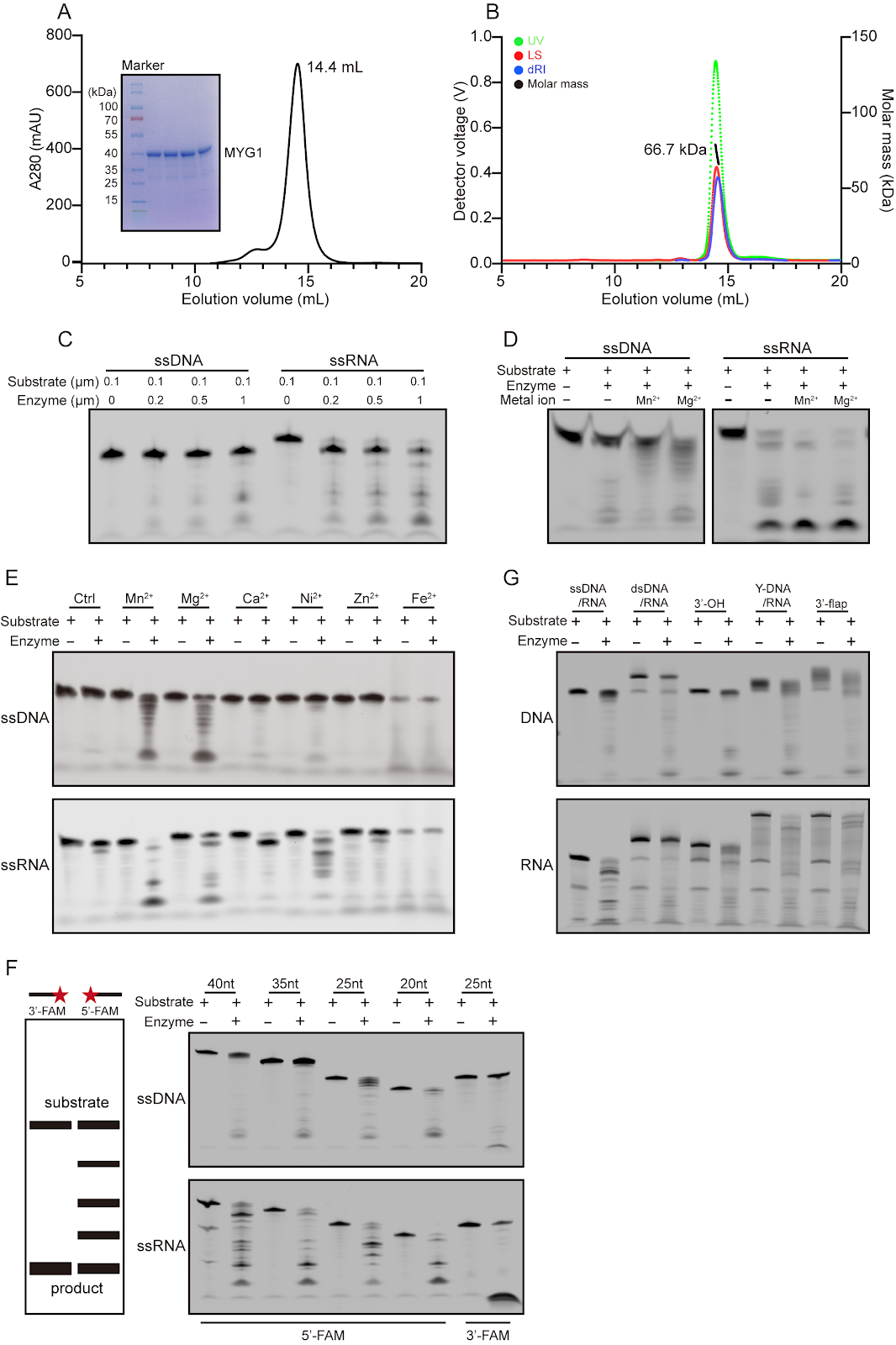
Nucleases are a class of enzymes that specifically cleave nucleic acids in all living organisms. They play crucial roles in essential biological processes, including the regulation of gene expression, DNA damage repair, and RNA processing and degradation. MYG1 (melanocyte proliferating gene 1) is a highly conserved eukaryotic protein that exhibits 3′→5′ exonuclease activity. This study systematically characterizes the enzymatic properties of MYG1 and determines its structures in complexes with metal ions and various mono- and poly-(deoxy)nucleotides. The functional roles of key residues involved in metal ion binding and substrate binding in the catalytic reaction are examined through site-directed mutagenesis, enzymatic activity assay, and structure determination. Our biochemical and structural data together demonstrate that MYG1 is a Mn2+- or Mg2+-dependent 3′→5′ exonuclease capable of cleaving a variety of nucleic acids with different structures. It exhibits the highest activity for single-stranded RNA and a nucleotide preference for U in single-stranded RNA and dT in single-stranded DNA. Mechanistically, MYG1 functions as a dimer, with the active site formed by the catalytic domain of monomer 1 and the substrate-binding domain of monomer 2, and cleaves nucleic acids through a two-metal ion-mediated catalytic mechanism. These findings establish a molecular basis for further investigations into the biological functions and molecular mechanisms of MYG1 within cells and its potential roles in human diseases.

Read Full Abstract10.3724/abbs.2026058
HDAC6 promotes osimertinib resistance evolution in non-small cell lung cancer by activating EGFR degradation through the ubiquitin-proteasome pathwayGraphical AbstractVerified
Acta Biochimica et Biophysica Sinica2026

HDAC6 promotes osimertinib resistance evolution in non-small cell lung cancer by activating EGFR degradation through the ubiquitin-proteasome pathway

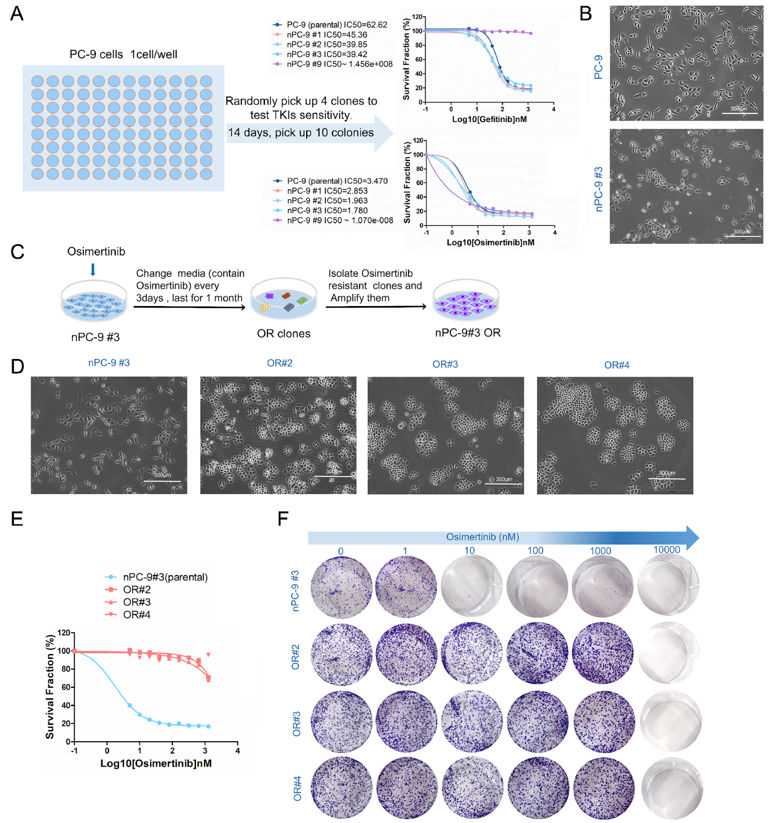
Osimertinib is the standard first-line treatment for patients with non-small cell lung cancer (NSCLC) harboring EGFR-sensitive mutations. However, drug resistance inevitably develops, highlighting the critical need for strategies to overcome this resistance and prolong therapeutic efficacy. Understanding the mechanisms underlying drug resistance is essential, and drug-resistant cell models serve as valuable tools for investigating acquired resistance. In this study, we establish an osimertinib resistance evolution model in vitro by continuous high-dose drug induction and identify cell lines exhibiting “permanent” resistance to osimertinib (osimertinib resistant, OR). Transcriptome sequencing (RNA-seq), gain- and loss-of-function assay, including lentiviral-mediated overexpression and RNAi knockdown, pharmacological inhibition, and protein degradation analysis reveal significant alterations in genes associated with epigenetic regulation, notably a marked upregulation of histone deacetylase 6 (HDAC6) in OR cells. Knockdown of HDAC6 or pharmacological inhibition of HDAC6 restores the sensitivity of OR cells to osimertinib, whereas overexpression of HDAC6 in sensitive cells reduces drug efficacy and accelerates the onset of resistance. Furthermore, we find that HDAC6 upregulation promotes EGFR degradation, thereby contributing to resistance. Collectively, our findings demonstrate the utility of drug resistance evolution models in identifying key resistance factors. HDAC6 plays a pivotal role in osimertinib resistance, and targeting HDAC6 may represent a novel therapeutic strategy to overcome resistance and enhance treatment efficacy.

Read Full Abstract10.3724/abbs.2026084
Radiomics Analysis of Intracranial Aneurysms: A Systematic Review and Meta-Analysis of Radiomics Quality Score and Clinical ApplicationsGraphical AbstractVerified
Chinese Journal of New Drugs2026

Radiomics Analysis of Intracranial Aneurysms: A Systematic Review and Meta-Analysis of Radiomics Quality Score and Clinical Applications

Background: Intracranial aneurysms (IAs) are a significant cause of subarachnoid hemorrhage, with high morbidity and mortality. Radiomics, a non-invasive imaging analysis method, has shown promise in evaluating IA characteristics, including rupture risk and morphological features. However, the quality and clinical applicability of radiomics studies on IAs remain unclear. Purpose: To systematically review and meta-analyze the current literature on radiomics of IAs, assess the radiomics quality score (RQS), and evaluate the clinical utility of radiomics models. Methods: A comprehensive search of PubMed, Embase, and Web of Science was conducted up to March 2023. Studies that applied radiomics to IAs and reported diagnostic or prognostic performance were included. Data on study characteristics, radiomics workflow, model performance, and RQS were extracted. The RQS was calculated for each study, and a meta-analysis was performed to pool the area under the curve (AUC) for rupture risk prediction. Results: A total of 23 studies met the inclusion criteria. The median RQS was 10 (range 2-18), indicating overall moderate quality. The pooled AUC for rupture risk prediction was 0.86 (95% CI: 0.82-0.90), demonstrating good discriminative ability. However, significant heterogeneity was observed (I² = 78%). Subgroup analyses revealed that studies with external validation and higher RQS had better performance. Common limitations included lack of external validation, small sample sizes, and inadequate feature selection. Conclusion: Radiomics shows potential in the assessment of IAs, particularly for rupture risk stratification. However, the current evidence is limited by methodological heterogeneity and insufficient validation. Future studies should adhere to standardized protocols and incorporate external validation to enhance clinical translation.

Read Full Abstractpub_80__articleID_520
Comprehensive Analysis of Fingerprint Patterns and Their Association with Genetic Markers in a Chinese PopulationGraphical AbstractVerified
Chinese Journal of New Drugs2026

Comprehensive Analysis of Fingerprint Patterns and Their Association with Genetic Markers in a Chinese Population

Fingerprint patterns are complex quantitative traits that have been used for personal identification and are hypothesized to be influenced by genetic factors. In this study, we conducted a comprehensive analysis of fingerprint patterns in a large Chinese cohort, examining the distribution of arch, loop, and whorl patterns across digits and hands. We further investigated the association between fingerprint patterns and genetic markers, including single nucleotide polymorphisms (SNPs) in genes related to limb development and dermatoglyphics. Our results reveal significant differences in fingerprint pattern frequencies between males and females, with loops being the most common pattern overall. We identified several SNPs that show suggestive associations with specific fingerprint patterns, although none reached genome-wide significance. Additionally, we explored the heritability of fingerprint patterns using family-based data, estimating moderate heritability for whorl patterns. Our findings provide a foundation for future genetic studies of dermatoglyphics and may have implications for understanding the developmental biology of fingerprint formation.

Read Full Abstractpub_80__articleID_540
Development and Application of a Renal Function Monitoring System Based on Fluorescence Lifetime Imaging TechnologyGraphical AbstractVerified
Chinese Journal of New Drugs2026

Development and Application of a Renal Function Monitoring System Based on Fluorescence Lifetime Imaging Technology

Renal function monitoring is critical for the diagnosis and management of chronic kidney disease (CKD). Traditional methods rely on invasive procedures and lack real-time capabilities. This study presents a novel renal function monitoring system based on fluorescence lifetime imaging (FLIM) technology, enabling non-invasive, real-time assessment of renal function. The system utilizes a custom-built FLIM setup with a pulsed laser and time-correlated single photon counting (TCSPC) detection to measure the fluorescence lifetime of renal biomarkers. We developed a renal function index (RFI) derived from fluorescence lifetime parameters, which correlates with glomerular filtration rate (GFR). In vitro and in vivo experiments were conducted using a rat model of CKD. Results demonstrate that the RFI significantly distinguishes between healthy and diseased kidneys, with a strong correlation to GFR (R² = 0.89). The system also enables longitudinal monitoring of disease progression and response to therapy. Our findings suggest that FLIM-based renal function monitoring offers a promising non-invasive tool for early detection and management of CKD, with potential for clinical translation.

Read Full Abstractpub_80__articleID_524
Research Progress on Multidrug-Resistant Bacteria and Antimicrobial Resistance MechanismsGraphical AbstractVerified
Chinese Journal of New Drugs2026

Research Progress on Multidrug-Resistant Bacteria and Antimicrobial Resistance Mechanisms

Antimicrobial resistance (AMR) poses a significant global health threat, with multidrug-resistant (MDR) bacteria emerging as a major concern. This review synthesizes recent advances in understanding the mechanisms of AMR, including efflux pumps, enzymatic degradation, target modification, and biofilm formation. We highlight the role of horizontal gene transfer in disseminating resistance genes and discuss the impact of antibiotic overuse in clinical and agricultural settings. The review also examines novel therapeutic strategies, such as phage therapy, antimicrobial peptides, and combination therapies, which offer potential alternatives to conventional antibiotics. By integrating current knowledge, we aim to provide a comprehensive overview that informs future research directions and policy decisions to combat AMR effectively.

Read Full Abstractpub_80__articleID_534
circ_0006156 promotes esophageal squamous cell carcinoma progression via activation of the TGFβ/Smad pathwayGraphical AbstractVerified
Acta Biochimica et Biophysica Sinica2026

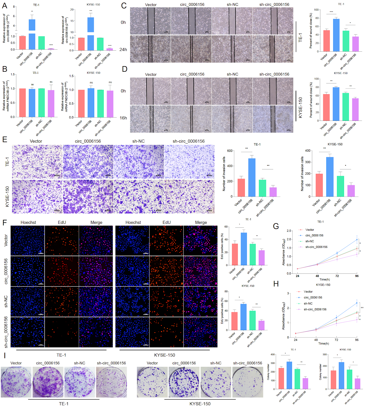
circ_0006156 promotes esophageal squamous cell carcinoma progression via activation of the TGFβ/Smad pathway

Esophageal squamous cell carcinoma (ESCC) represents a common malignancy of the digestive system. Circular RNAs (circRNAs) are a distinct class of single-stranded non-coding RNAs that are essential in the progression of various tumors given that they can act as microRNA (miRNA) sponges in a manner similar to that of mRNAs. In this study, circ_0006156 is screened to be highly expressed in ESCC tissues through high-throughput sequencing and quantitative real-time polymerase chain reaction. Subsequent in vitro and in vivo experiments are conducted to validate its biological functions. Furthermore, the regulatory relationships among circ_0006156, miR-202-5p, and TGFBR1 are investigated using RNA antisense purification, miRNA sequencing, RNA immunoprecipitation, fluorescence in situ hybridization, dual-luciferase reporter assay, and bioinformatics analyses. The results show significant overexpression of circ_0006156 in ESCC tissues, with relatively shorter overall survival observed in patients with high circ_0006156 expression. circ_0006156 is further identified to directly bind to miR-202-5p. miR-202-5p inhibits the proliferation, migration, and invasion of ESCC cells in vitro and partially rescues the effects induced by circ_0006156. Consistent results are reported by subcutaneous xenograft tumor experiments in nude mice. In addition, circ_0006156 is confirmed to act as an endogenous sponge for miR-202-5p, which results in a relieved suppression of its target gene TGFβR1. In summary, circ_0006156 can regulate TGFβR1 expression by sponging miR-202-5p, which may further activate the TGFβ/Smad pathway and promote ESCC progression. Collectively, circ_0006156 functions as a novel oncogenic RNA in ESCC and may serve as a potential tumor marker.

Read Full Abstract10.3724/abbs.2026049
SPP1 facilitates sorafenib resistance in hepatocellular carcinoma by upregulating aerobic glycolysis in endothelial cellsGraphical AbstractVerified
Acta Biochimica et Biophysica Sinica2026

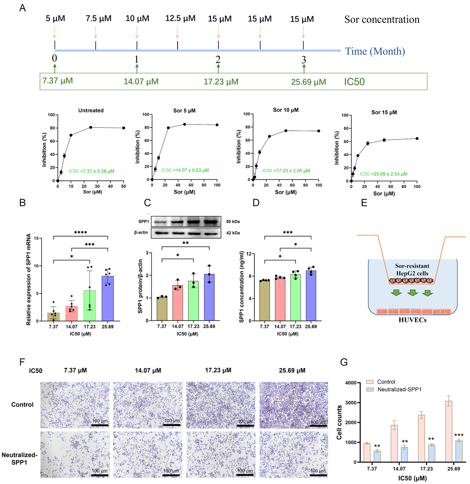
SPP1 facilitates sorafenib resistance in hepatocellular carcinoma by upregulating aerobic glycolysis in endothelial cells

The occurrence of resistance to sorafenib, a first-line treatment for hepatocellular carcinoma (HCC), significantly limits its clinical efficacy. Therefore, investigating the potential mechanism of sorafenib resistance in HCC is highly important for developing HCC treatment strategies. In the present study, we identify that SPP1 (encoding osteopontin; OPN) is significantly elevated in sorafenib-resistant HCC. Furthermore, the upregulation of SPP1 is related to vascular invasion, advanced disease stage and poor prognosis in HCC patients. As the IC50 value of sorafenib increases in HepG2 cells, the SPP1 protein secreted by the cells is significantly upregulated, which subsequently facilitates the proliferation of human umbilical vein endothelial cells (HUVECs) and resistance to sorafenib. Further studies reveal that SPP1 induces resistance to sorafenib in HepG2 cells by upregulating glycolysis in HUVECs and further producing lactate. Mechanistically, SPP1 increases the expressions of the glucose transporter GLUT1 and the key glycolytic enzymes PFK1 and PKM2 in HUVECs, resulting in lactate accumulation, which in turn promotes the phosphorylation levels of BRAF and ERK as well as HIF-1α expression in HepG2 cells, leading to sorafenib resistance in HCC. Notably, SPP1 silencing can inhibit the proliferation and invasion of sorafenib-resistant HepG2 cells both in vitro and in vivo. Importantly, lactate derived from HUVECs plays a more dominant role in sorafenib resistance than does SPP1 in HepG2 cells. In summary, SPP1 enhances sorafenib resistance in HepG2 cells through promoting aerobic glycolysis in HUVECs, suggesting that the SPP1-aerobic glycolysis axis might be a prognostic biomarker as well as a potential therapeutic target for sorafenib-resistant HCC.

Read Full Abstract10.3724/abbs.2025245
Altered gut microbial dynamics and the antivascular remodeling effect of carnosine in hypobaric hypoxic pulmonary hypertension ratsGraphical AbstractVerified
Acta Biochimica et Biophysica Sinica2026

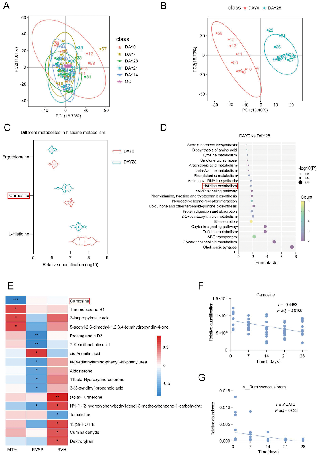
Altered gut microbial dynamics and the antivascular remodeling effect of carnosine in hypobaric hypoxic pulmonary hypertension rats

Exposure to chronic hypobaric hypoxia provokes marked alterations in the gut microbiota and its metabolome, yet the functional significance of histidine-derived metabolites in hypobaric hypoxic pulmonary hypertension (PH) remains underexplored. Here, we employ 16S rDNA, metagenomic, and untargeted metabolomic sequencing to characterize longitudinal shifts in the fecal microbiota and metabolites during hypobaric hypoxic PH development in Sprague-Dawley rats. Fecal carnosine levels and the abundance of its producer, Ruminococcus bromii, both decrease significantly over 28 days of hypobaric hypoxia (P < 0.05). Spearman correlation shows that carnosine is inversely correlated with the percentage of pulmonary arteriole media thickness (MT%; r = –0.8741, P < 0.001). Therapeutic supplementation with carnosine restores systemic and pulmonary antioxidant defenses and attenuates vascular remodeling without altering right ventricular pressures. In vitro, carnosine inhibits hypoxia-induced pulmonary artery smooth muscle cell (PASMC) proliferation and migration and suppresses nuclear factor erythroid 2-related factor 2 (Nrf2) accumulation. These findings reveal dynamic gut-lung crosstalk in hypobaric hypoxic PH and nominate carnosine as a metabolite-based intervention to mitigate hypoxia-driven pulmonary vascular remodeling.

Read Full Abstract10.3724/abbs.2025237
Development of a Colloidal Gold Immunochromatographic Strip Based on GAPDH for Pentatrichomonas hominis in DogsGraphical AbstractVerified
Acta Biochimica et Biophysica Sinica2026

Development of a Colloidal Gold Immunochromatographic Strip Based on GAPDH for Pentatrichomonas hominis in Dogs

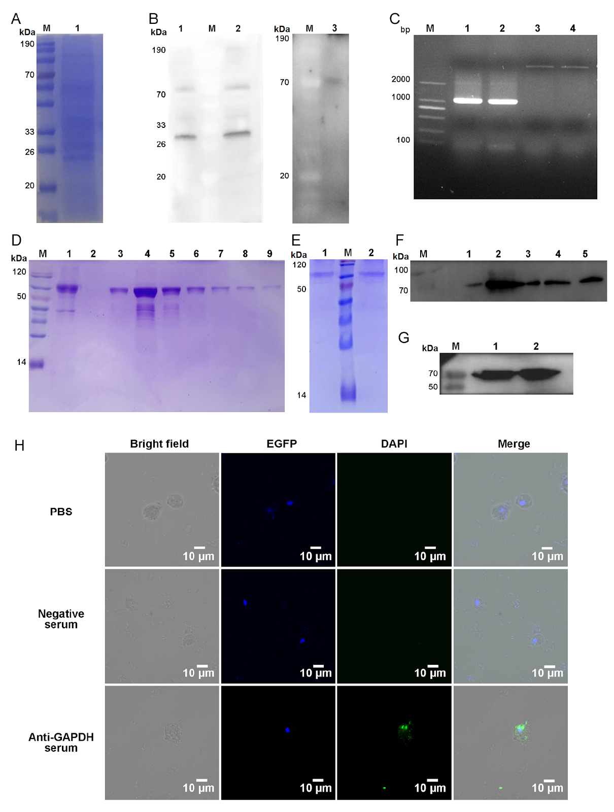
Pentatrichomonas hominis is a zoonotic protozoan that primarily inhabits the cecum and colon of dogs, with infection rates as high as 47.4%, posing a significant public health risk due to close human contact. Current detection methods, including direct smear microscopy and PCR, have limitations in sensitivity, equipment requirements, and operational complexity. To address the need for a practical on-site detection method, we employed liquid chromatography-tandem mass spectrometry (LC-MS/MS) to identify specific antigens from P. hominis excretory-secretory (ES) proteins. Glyceraldehyde 3-phosphate dehydrogenase (GAPDH) was selected as a candidate antigen due to its high immunogenicity and species specificity. The GAPDH gene was cloned and expressed in E. coli, and the recombinant protein was purified. Mouse anti-GAPDH serum was generated, and its reactivity was confirmed by Western blot and indirect ELISA (titer 1:102,400). Immunofluorescence localization showed GAPDH in the cytoplasm of P. hominis trophozoites. Based on this antigen, we developed a colloidal gold immunochromatographic strip for rapid detection of P. hominis in dogs. The strip demonstrated high sensitivity and specificity, providing a practical tool for veterinary diagnosis and epidemiological surveillance. This study is the first to report an immunochromatographic strip for P. hominis detection, offering a rapid, user-friendly alternative to existing methods.

Read Full Abstract10.3724/abbs.2026043
A bifunctional aptamer-siRNA chimera targeting ACE2 for the inhibition of SARS-CoV-2 S pseudovirus entry and replicationGraphical AbstractVerified
Acta Biochimica et Biophysica Sinica2026

A bifunctional aptamer-siRNA chimera targeting ACE2 for the inhibition of SARS-CoV-2 S pseudovirus entry and replication

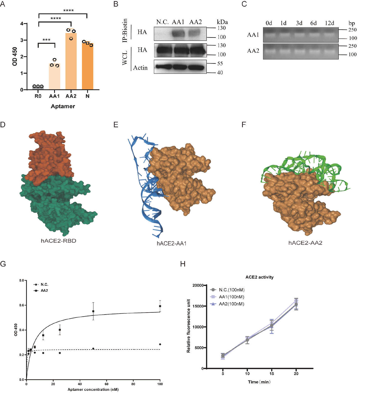
The relentless evolution of severe acute respiratory syndrome coronavirus 2 (SARS-CoV-2) and the emergence of immune-evasive variants underscore an urgent need for novel therapeutic strategies that are resilient to viral mutations. Targeting conserved host factors essential for viral entry represents a promising approach to overcome this challenge. Here, we report the development of a bifunctional therapeutic platform targeting the primary human receptor for SARS-CoV-2, angiotensin-converting enzyme 2 (ACE2). Using systematic evolution of ligands by exponential enrichment (SELEX), we isolate a high-affinity DNA aptamer, designated AA2, that binds to human ACE2 with a dissociation constant (Kd) of 5.41 ± 1.23 nM. Molecular docking and competitive binding assays confirm that AA2 sterically hinders the interaction between the viral spike receptor-binding domain (RBD) and ACE2. Consequently, AA2 demonstrates potent neutralization of SARS-CoV-2 S pseudovirus entry into host cells. To achieve a synergistic antiviral effect, we engineer an aptamer-siRNA chimera (AsiC) by conjugating AA2 to a short interfering RNA (siRNA) targeting the GFP coding region of the pseudovirus genome. This AsiC construct significantly represses viral replication compared to aptamer or siRNA treatment alone, validating a dual mechanism of action that combines receptor blockade with targeted gene silencing. This study establishes a robust proof-of-concept for an ACE2-targeted AsiC, representing a new class of dual-function antiviral therapeutics with the potential to effectively combat current and future ACE2-dependent coronaviruses.

Read Full Abstract10.3724/abbs.2026087
PPIA as a central regulator in a novel cell death pathway activated by iron homeostasis and redox disruption in multiple myelomaGraphical AbstractVerified
Acta Biochimica et Biophysica Sinica2026

PPIA as a central regulator in a novel cell death pathway activated by iron homeostasis and redox disruption in multiple myeloma

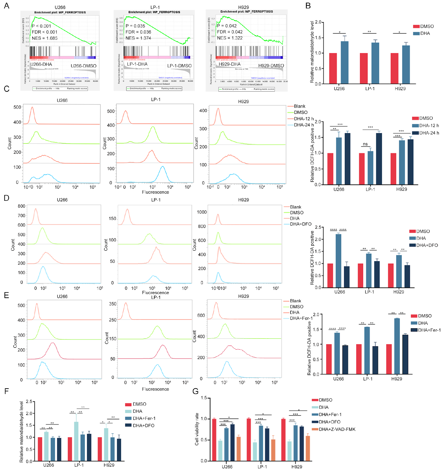
Multiple myeloma (MM) patients frequently experience relapse, disease progression, and drug resistance, necessitating novel therapeutic strategies. This study investigates the role of peptidylprolyl isomerase A (PPIA) in mediating dihydroartemisinin (DHA)-induced ferroptosis in MM. Building on our previous work establishing the prognostic relevance of ferroptosis in MM, we elucidate the mechanism by which DHA triggers this form of cell death through the disruption of iron metabolism and redox homeostasis. DHA significantly reduces the viability of MM cell lines and primary CD138+ cells derived from patient bone marrow samples and attenuates tumor burden in orthotopic MM models. Mechanistically, DHA upregulates the iron-regulatory genes transferrin receptor 1 (TFRC) and heme oxygenase-1 (HMOX1), thereby perturbing iron homeostasis and promoting ferroptosis. Crucially, DHA targets the oxidized form of PPIA, a redox-sensitive chaperone, binding to it and preventing its reduction, which elevates intracellular reactive oxygen species (ROS). Combined treatment with DHA and erastin, which concurrently disrupt iron and amino acid metabolism, exerts synergistic cytotoxicity and enhances MM inhibition. Furthermore, in a syngeneic mouse model, DHA promotes T-cell activation and augments tumor suppression. Collectively, these findings underscore PPIA's pivotal role in a novel ferroptotic cell death pathway and reveal new therapeutic opportunities for MM.

Read Full Abstract10.3724/abbs.2026081
Efficient production of recombinant mAbs mediated by a MAR-enhanced transposon vector combined with blasticidin selection in CHO cellsGraphical AbstractVerified
Acta Biochimica et Biophysica Sinica2026

Efficient production of recombinant mAbs mediated by a MAR-enhanced transposon vector combined with blasticidin selection in CHO cells

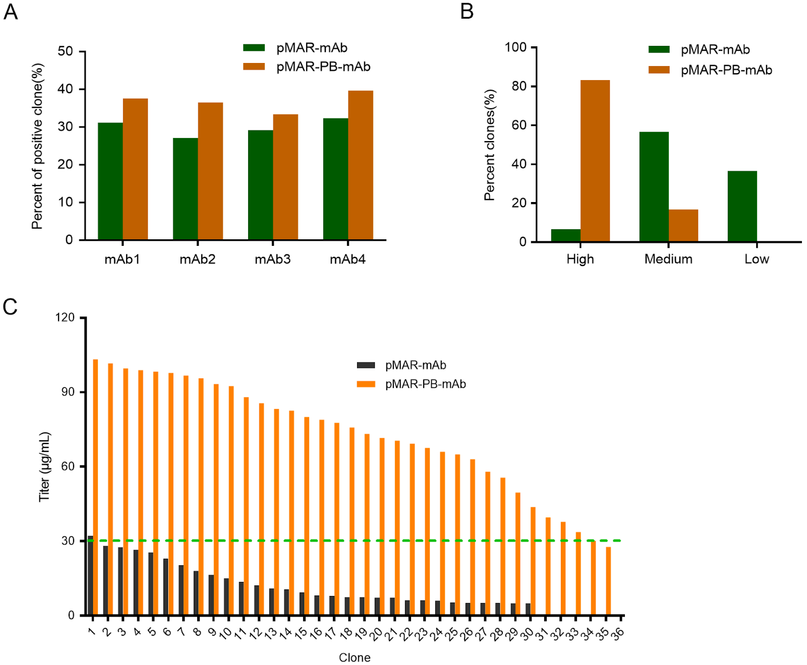
Recombinant antibodies, primarily produced in Chinese hamster ovary (CHO) cells, are widely used to treat various diseases. For industrial production, a rapid and efficient method to screen stable, high-expressing clones is essential. However, conventional screening based on random integration is often cumbersome and labor intensive. This study establishes a novel strategy for generating stable, high-yielding clones by combining a MAR-based piggyBac (PB) transposon semitargeted integration system with blasticidin (BSD) selection. Compared to the random integrated vector pMAR-mAb, the MAR-PB system increases the titers (3.95- to 5.61-fold) and specific protein productivity (Qp; 4.28- to 6.07-fold) of four monoclonal antibodies in stable cell pools. When compared to PB-only vectors, the MAR-PB transposon system enhances the titers (by up to 2.50-fold) and Qp (1.96- to 2.77-fold), respectively. The increased antibody production correlates with elevated mRNA expression. Furthermore, this approach increases the proportion of high-expressing clones by more than 10-fold and significantly improves volumetric yield. Importantly, this approach promotes the long-term stability of recombinant mAb expression for over 60 generations. Transcriptome analysis reveals that the system modulates genes involved in DNA binding, transcriptional regulation, and protein binding. In conclusion, the MAR-based PB transposon system combined with BSD selection presents a significant improvement for efficiently generating high-yielding and stable CHO cell clones, offering a valuable tool for recombinant antibody production.

Read Full Abstract10.3724/abbs.2025251
OAZ1/CASP8AP2 Double Knockout Enhances Recombinant Protein Production in HEK293 Cells through Metabolic Reprogramming and Antiapoptotic EffectsGraphical AbstractVerified
Acta Biochimica et Biophysica Sinica2026

OAZ1/CASP8AP2 Double Knockout Enhances Recombinant Protein Production in HEK293 Cells through Metabolic Reprogramming and Antiapoptotic Effects

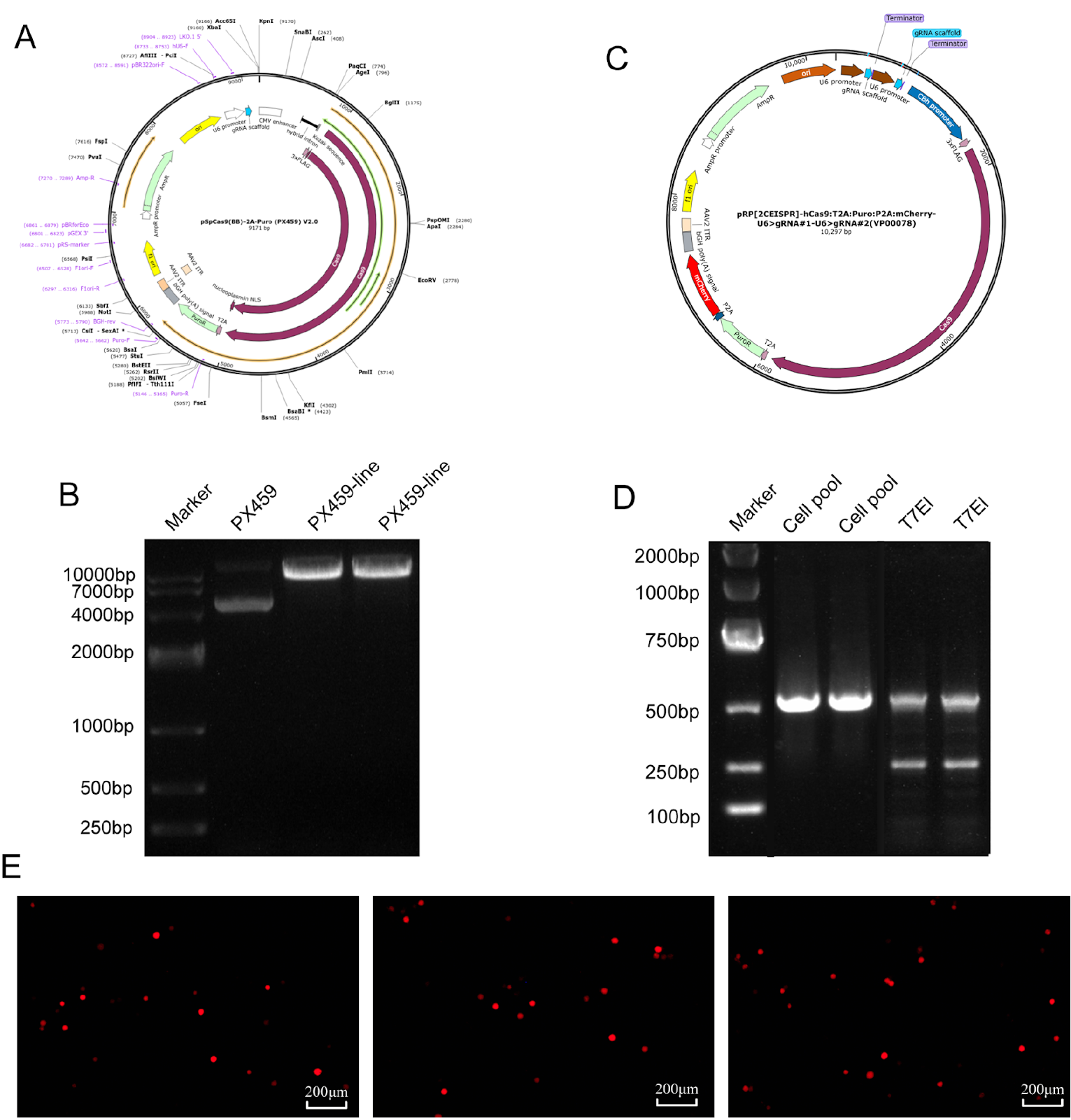
Human embryonic kidney (HEK) 293 cells are widely used for recombinant protein production because of their efficient posttranslational modification capabilities. However, their large-scale culture is often limited by metabolic stress and early apoptosis, leading to insufficient protein yields. In this study, we aim to increase protein expression through the coordinated modulation of metabolic and apoptotic pathways. Using CRISPR/Cas9 technology, we target and knockout the genes of ornithine decarboxylase antizyme 1 (OAZ1), which regulates polyamine metabolism, and caspase 8-associated protein 2 (CASP8AP2), an apoptosis-related protein. We successfully construct an OAZ1/CASP8AP2 double-knockout HEK293 cell line. Following transfection with the knockout vector and screening of single-cell clones, multiple levels of validation confirm the successful gene knockout. The results show that the double-knockout cells exhibit significantly reduced apoptosis rates. Furthermore, the production of recombinant secreted alkaline phosphatase (SEAP) and vitronectin (VN) increases by 2.1 folds and 2.9 folds, respectively, compared with those in wild-type cells. Metabolic profiling reveals that the cell cycle is arrested in the G1/G0 phase, accompanied by increased specific consumption and production rates of key metabolites. This study demonstrates that concurrent inhibition of apoptosis and optimization of metabolism effectively enhances recombinant protein production in HEK293 cells, suggesting a novel strategy for improving HEK293 cell-based expression.

Read Full Abstract10.3724/abbs.2025196
PCIF1 Modulates Glioblastoma Cell Migration and Invasion by Altering PI(3,4)P2 Levels through the PI5-Phosphatase INPP5BGraphical AbstractVerified
Acta Biochimica et Biophysica Sinica2026

PCIF1 Modulates Glioblastoma Cell Migration and Invasion by Altering PI(3,4)P2 Levels through the PI5-Phosphatase INPP5B

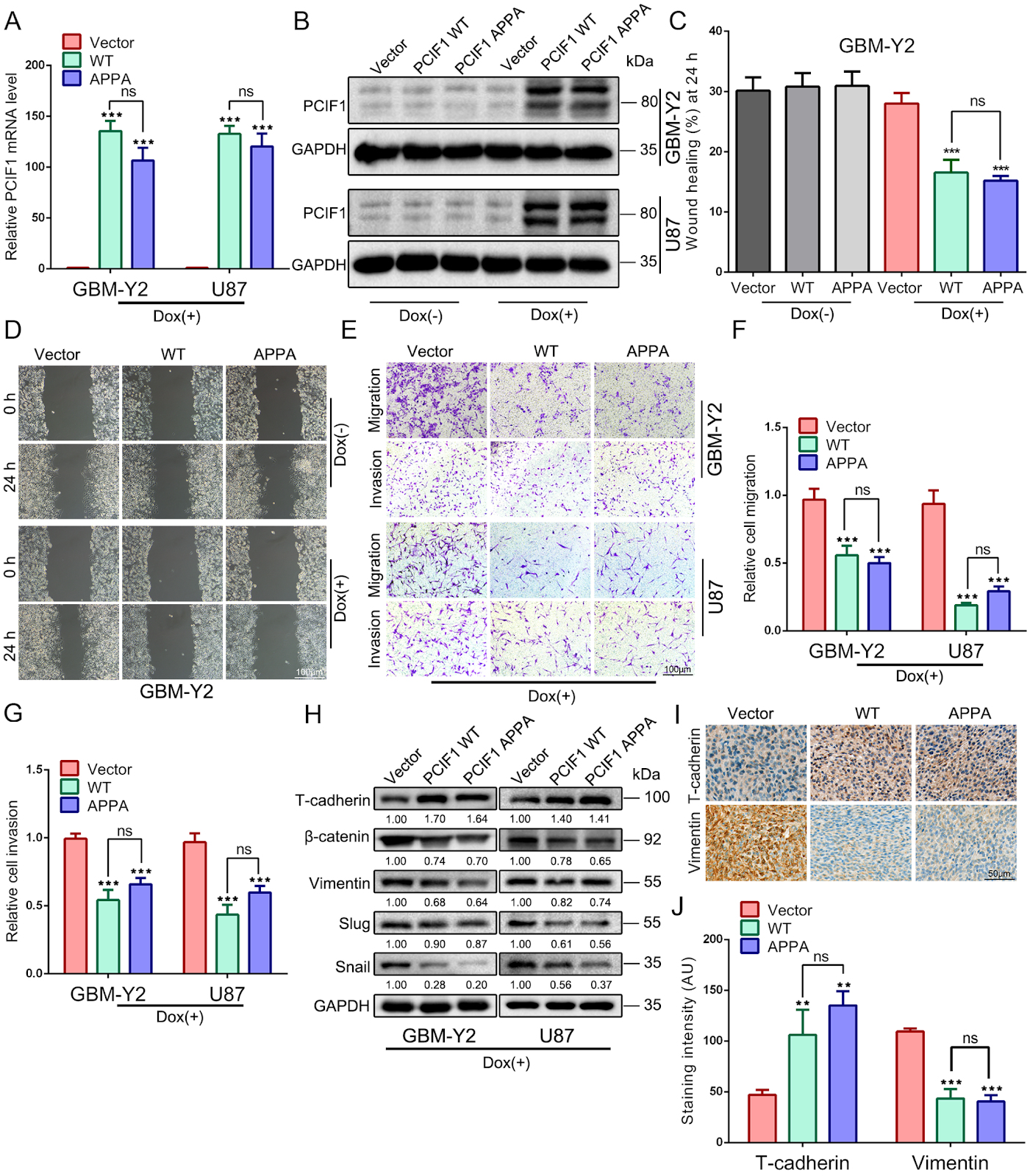
Phosphorylated CTD Interacting Factor 1 (PCIF1) is the exclusive methyltransferase responsible for the N6,2-O-dimethyladenosine (m6Am) modification in mammalian mRNA. Our previous research identified PCIF1 as a potent tumor suppressor in glioma, demonstrating its ability to impair cell proliferation, induce G2/M phase arrest, and promote apoptosis. However, its role in glioma cell migration and invasion remains unclear. In this study, we investigate how PCIF1 regulates glioma cell migration and invasion. Overexpression of PCIF1 inhibits migration and invasion, whereas PCIF1 knockdown enhances these behaviors. Corresponding changes are observed in mesenchymal markers (Vimentin, β-catenin, Snail, Slug) and the epithelial marker T-cadherin, indicating that PCIF1 suppresses epithelial-to-mesenchymal transition (EMT)-mediated glioma invasion. Mechanistically, PCIF1 modulates the AKT pathway by promoting proteasomal degradation of AKT while increasing phosphorylated AKT (p-AKT) levels, revealing a complex regulatory mechanism. PCIF1 knockdown upregulates INPP5B, a lipid phosphatase, causing accumulation of PI(3,4)P2 and enhanced AKT activation. Conversely, PCIF1 overexpression increases PI(3,4,5)P3 production, elevating p-AKT levels. This bidirectional regulation suggests that PCIF1 influences phosphoinositide signaling and AKT activation. Our findings highlight PCIF1 as a key modulator of glioblastoma cell migration and invasion through phosphoinositide signaling, positioning it as a potential biomarker and therapeutic target in glioma.

Read Full Abstract10.3724/abbs.2026027
P300-mediated H3K18 acetylation triggers necroptosis via modulation of KRT18 transcription in diabetic nephropathyGraphical AbstractVerified
Acta Biochimica et Biophysica Sinica2026

P300-mediated H3K18 acetylation triggers necroptosis via modulation of KRT18 transcription in diabetic nephropathy

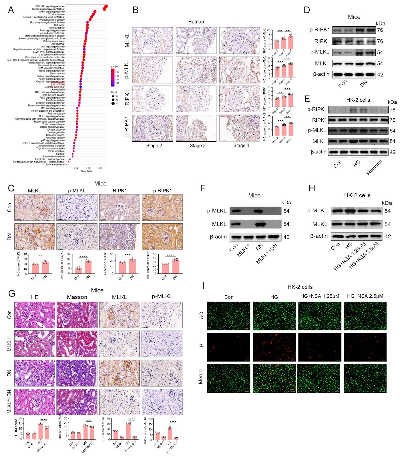
Diabetic nephropathy (DN) is a major cause of end-stage renal disease. While glomerular damage is a known aspect of its pathology, tubular epithelial cell necroptosis also plays a crucial role in disease progression. Epigenetic modifications, particularly histone acetylation, have garnered attention for their role in the regulation of kidney damage-related gene expression. This study explores whether the histone acetyltransferase P300 regulates KRT18 expression via histone H3 lysine 18 acetylation (H3K18ac), driving tubular epithelial cell necroptosis and accelerating DN progression. We establish an STZ-induced diabetic nephropathy mouse model and a high glucose-treated HK-2 cell model. Western blot analysis, qPCR, immunohistochemistry, and AO/PI staining are employed to assess the expression levels of P300, H3K18ac, KRT18, and necroptosis-related proteins (RIPK1 and MLKL). Functional validation of the P300-KRT18 axis is performed using shRNA interference, overexpression, and the small molecule inhibitor C646. Both in vivo and in vitro models show significant upregulation of P300, H3K18ac, and KRT18, coupled with RIPK1/MLKL pathway activation and increased cell death. P300 knockdown or C646 treatment effectively inhibits H3K18ac and KRT18 expression, reducing necroptosis; KRT18 knockdown also alleviates P300 overexpression-induced cell death. Co-transfection with P300 overexpression and KRT18 interference demonstrates that KRT18 is a key downstream effector of P300-mediated necroptosis. In conclusion, P300 upregulates KRT18 expression through H3K18 acetylation, subsequently activating the RIPK1/MLKL pathway and promoting tubular epithelial cell necroptosis. The P300-KRT18 axis may serve as a novel epigenetic therapeutic target for DN, suggesting that epigenetic regulation could be a viable intervention strategy to delay DN progression.

Read Full Abstract10.3724/abbs.2026015
Sanguinarine exerts anti-hepatocellular carcinoma activity by targeting FDX1 to induce FDX1/LIAS/DLAT/HSP70 axis-dependent cuproptosisGraphical AbstractVerified
Acta Biochimica et Biophysica Sinica2026

Sanguinarine exerts anti-hepatocellular carcinoma activity by targeting FDX1 to induce FDX1/LIAS/DLAT/HSP70 axis-dependent cuproptosis

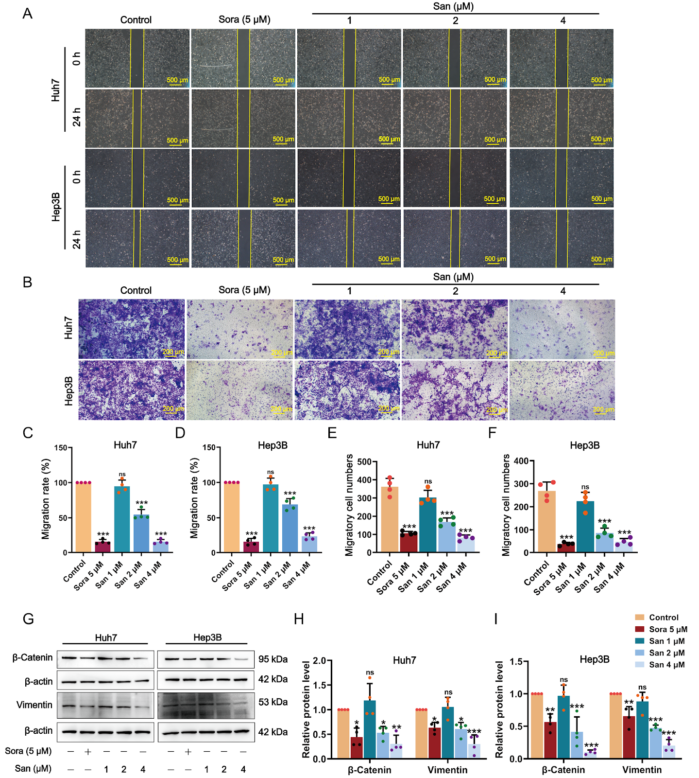
Hepatocellular carcinoma (HCC), the predominant type of primary liver cancer, represents an extremely aggressive malignancy. The induction of cuproptosis has developed into a favorable therapeutic direction for HCC, considering its strong association with HCC. Sanguinarine (San), a benzophenanthridine alkaloid derived from traditional herbs such as Chelidonium majus L., demonstrates broad-spectrum anticancer activities against various cancer cell types. However, the precise molecular mechanisms underlying its effects in the treatment of HCC remain largely undefined. This investigation seeks to examine the anti-HCC effects of San and to explore the mechanisms underlying these effects through the induction of cuproptosis. In vitro experiments demonstrate that San markedly inhibits the proliferation, movement, and epithelial-mesenchymal transition of HCC cells while enhancing their apoptosis. In vivo, San notably impedes tumor growth and upregulates the cuproptosis signature markers ferredoxin 1 (FDX1), oligomeric dihydrolipoamide S-acetyltransferase (DLAT), and heat shock protein 70 (HSP70) in HCC xenograft tumor models. Mechanistically, San induces proteotoxic stress and cuproptosis in HCC cells by increasing copper concentration, upregulating the expression of FDX1, lipoic acid synthetase (LIAS), HSP70, and lipoylated DLAT aggregation, and simultaneously reducing mitochondrial membrane potential and intracellular glutathione and pyruvate levels. Moreover, the combination of San with copper ionophores (Elesclomol-CuCl2) exhibits synergistic effects in promoting cuproptosis. FDX1 silencing markedly diminishes San-induced suppression of cell proliferation and FDX1 and HSP70 levels in HCC cells. Additionally, molecular docking analysis predicts that San exhibits the highest potential for binding with FDX1. Surface plasmon resonance experiments and cellular thermal shift assay confirm that San strongly interacts with FDX1 and markedly enhances the thermostability of FDX1. In conclusion, our findings indicate that San substantially inhibits the progression of HCC by targeting FDX1/LIAS/DLAT/HSP70 axis-dependent cuproptosis.

Read Full Abstract10.3724/abbs.2026025
Integrating genetically encoded fluorescent sensors to elucidate the spatiotemporal choreography of necrosis by sodium overloadGraphical AbstractVerified
Acta Biochimica et Biophysica Sinica2026

Integrating genetically encoded fluorescent sensors to elucidate the spatiotemporal choreography of necrosis by sodium overload

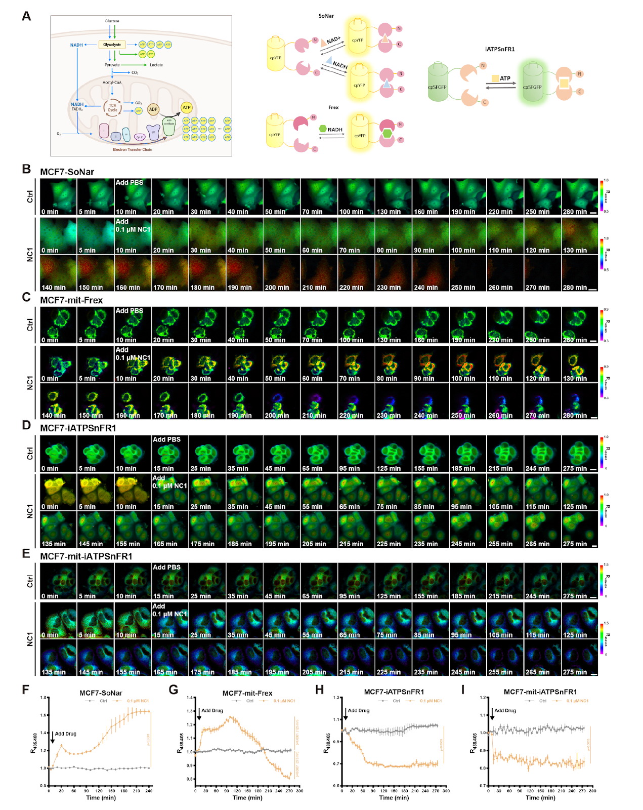
Necrosis by sodium overload (NECSO) is a distinct cell death modality induced by the chemical regulator necrocide 1 (NC1), which targets the transient receptor potential melastatin 4 (TRPM4) channel to drive excessive sodium influx and potassium efflux. This unique necrosis is characterized by the exchange of monovalent ions, a restrained ATP supply, redox disorder and a final membrane rupture as main features. Real-time monitoring of metabolic and redox causality during the whole process in living cells has been technically challenging. To bridge this gap, we integrate an advanced toolset of genetically encoded fluorescent sensors to monitor monovalent ions, energy metabolites, and redox equivalents with high spatiotemporal resolution. By directing these sensors to specific subcellular compartments, we successfully capture the real-time choreography of potassium loss specifically induced by NC1 via TRPM4. Furthermore, we establish a precise detection paradigm for evaluating energy currency by integrating sensors for NADH and ATP that are capable of subcellular imaging. We observe general and rapid NADH accumulation along with an ATP shortage in the mitochondria and cytosol. A concomitant reduction in mitochondrial oxidative stress is observed. This study not only elucidates the metabolic progression of a peculiar type of necrosis but also establishes a robust methodological framework for applying genetically encoded sensors to broader physiological and toxicological research.

Read Full Abstract10.3724/abbs.2026102
Ginsenoside Rh2 alleviates osteoporosis by attenuating oxidative stress-induced osteoblast dysfunction via the FoxO1/β-catenin pathwayGraphical AbstractVerified
Acta Biochimica et Biophysica Sinica2026

Ginsenoside Rh2 alleviates osteoporosis by attenuating oxidative stress-induced osteoblast dysfunction via the FoxO1/β-catenin pathway

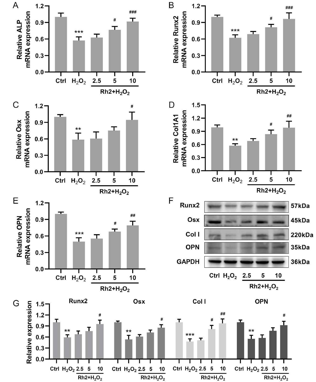
The degree of oxidative stress decreases osteoblast function with age, which leads to a decline in bone compressive capacity. Ginsenoside Rh2 is a known clinical or adjuvant therapy for various tissues. In this study, we investigate the pharmacological effects of Rh2 against oxidative stress-induced osteoblasts. Osteoblasts are pretreated with Rh2 for 48 h and then exposed to hydrogen peroxide (H2O2), which results in significantly decreased ROS levels, increased antioxidant enzyme activity, and enhanced mitochondrial function. Functionally, Rh2 increases alkaline phosphatase (ALP) expression, together with enhanced mineralization and expression of osteogenesis-associated genes. Rh2 also promotes the nuclear translocation of FoxO1 and β-catenin, whereas it does not reverse reduced mineralization caused by decreased FoxO1 or β-catenin activity, indicating that its effect is mediated through the functional interaction between FoxO1 and β-catenin. In a mouse model of lipopolysaccharide (LPS)-induced bone loss, Rh2 administration improves trabecular microstructure, increases osteoblast numbers, and upregulates serum metabolites associated with bone formation. Immunofluorescence analysis further reveals that Rh2 promotes the nuclear co-localization of FoxO1 and β-catenin in femurs, indicating their coordinated action within this signaling axis. These findings indicate that Rh2 mitigates oxidative stress-induced osteoblast dysfunction via the FoxO1/β-catenin pathway, highlighting the pivotal role of redox balance in bone remodeling and suggesting a promising therapeutic strategy for osteoporosis.

Read Full Abstract10.3724/abbs.2026065
Phillyrin protects against myocardial ischemia/reperfusion injury by promoting KNL1 K605 acetylation to inhibit the p53/p21 pathwayGraphical AbstractVerified
Acta Biochimica et Biophysica Sinica2026

Phillyrin protects against myocardial ischemia/reperfusion injury by promoting KNL1 K605 acetylation to inhibit the p53/p21 pathway

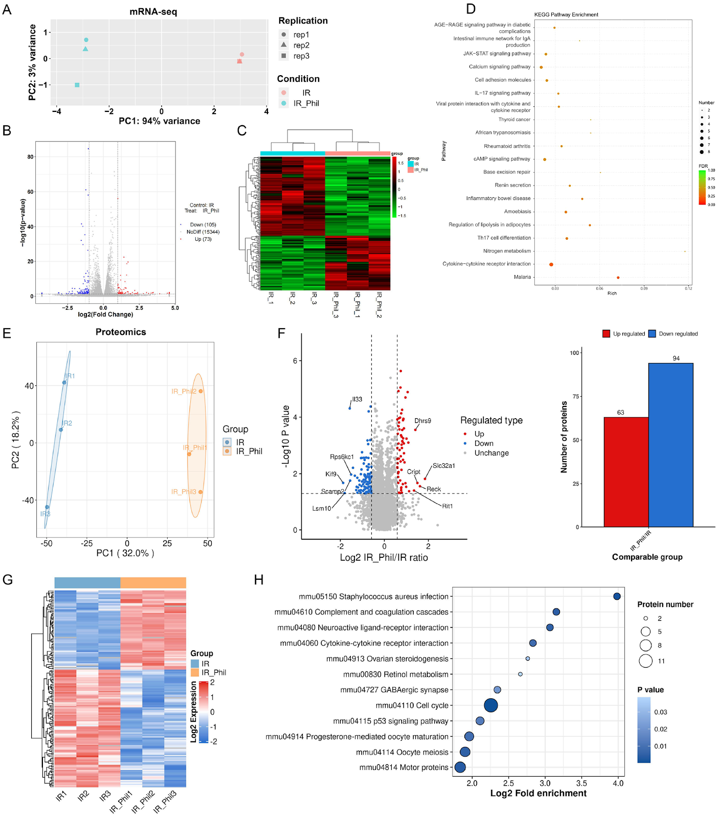
Reperfusion therapy is critical for acute myocardial infarction but is often accompanied by myocardial ischemia/reperfusion injury (MIRI). Phillyrin, a natural lignan from Forsythia suspensa, exerts anti-inflammatory and antioxidant effects; however, its role and mechanism in MIRI remain unclear. In this study, HL-1 cardiomyocytes are subjected to oxygen-glucose deprivation/reperfusion (OGD/R). Cell viability, apoptosis, oxidative stress, and inflammation are measured after phillyrin treatment. Multiomics (mRNA-seq, proteomics, and acetylproteomics) is used to identify key targets and pathways. Molecular docking, co-immunoprecipitation, site-directed mutagenesis, and western blot analysis are used to validate posttranslational regulation. A mouse MIRI model is established to confirm the in vivo cardioprotective effects of phillyrin. Phillyrin preserves cell viability and reduces apoptosis, oxidative stress, and inflammation in OGD/R-injured HL-1 cells. Multiomics integration reveals that phillyrin acts primarily through posttranslational regulation and highlights kinetochore scaffold 1 (KNL1) as the only protein that is both upregulated and hyperacetylated at lysine 605 (K605). Mechanistically, phillyrin may bind to the KNL1 C-terminus and enhance the interaction between KNL1 and acetyltransferase p300/CBP. KNL1 K605R mutation and Knl1 knockdown reduce KNL1 protein expression and reverse the inhibitory effects of phillyrin on p53 pathway-mediated apoptosis, oxidative stress, and inflammation. In mouse MIRI models, phillyrin reduces infarct size, myocardial damage, and cardiomyocyte apoptosis; these effects are abolished by knockdown of Knl1. Therefore, phillyrin promotes KNL1 acetylation at K605 to increase KNL1 protein expression, thereby inhibiting p53 signaling and alleviating apoptosis, oxidative stress, and inflammation in MIRI. This study identifies KNL1 acetylation at K605 as a novel posttranslational modification target for cardioprotection.
Read Full Abstract10.3724/abbs.2026104