🧬 SinoBioData Academic Portal
📚 Peer-Reviewed Translated Literature

All Biomedical & Clinical Articles (Page 6)

Browse complete peer-reviewed translations from top Chinese biomedical, oncology, and genomics journals. Read verified previews and download full authentic clinical reports.

Published Research Papers

Showing 24 of 1542 peer-reviewed translated articles (Page 6 of 65)

Tryptophan-substituted antimicrobial peptide temporin-1CEb: in vitro and in vivo antibacterial activity against clinically isolated multidrug-resistant Klebsiella pneumoniaeGraphical AbstractVerified
Acta Biochimica et Biophysica Sinica2026

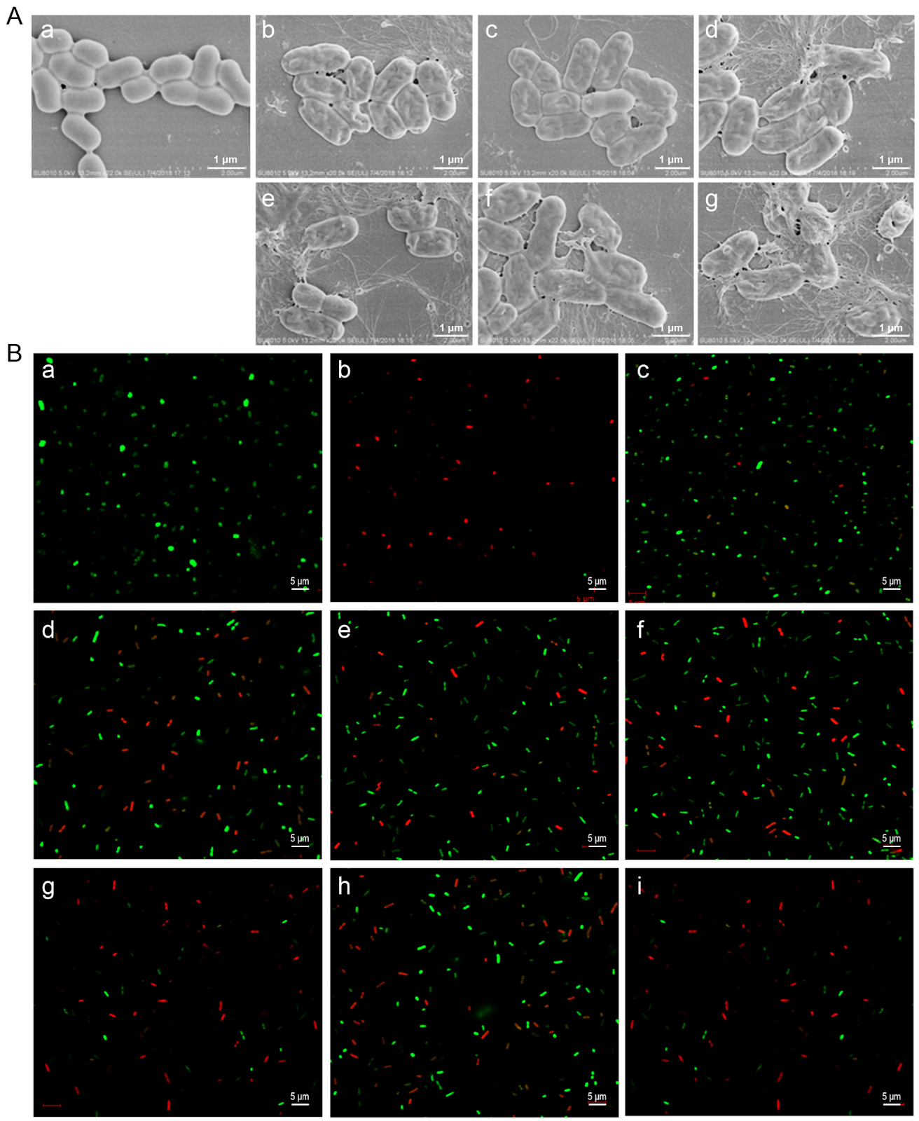
Tryptophan-substituted antimicrobial peptide temporin-1CEb: in vitro and in vivo antibacterial activity against clinically isolated multidrug-resistant Klebsiella pneumoniae

Tryptophan (Trp)-substituted antimicrobial peptides (AMPs) exhibit enhanced interactions with bacterial cell membranes, potentially improving their antimicrobial efficacy. Klebsiella pneumoniae (20.59% of 2054 ICU isolates) is resistant to multiple clinically used antibiotics and presents significant treatment challenges. In the present study, three Trp-modified peptides (I4W, L12W, and I1WL5W) are generated by substituting Ile or Leu residues in temporin-1CEb, a peptide derived from frog skin, with Trp at various sites to assess their antibacterial effects and mechanisms against K. pneumoniae. Compared with L12W, both I4W and I1WL5W display superior antimicrobial activity and lower cytotoxicity. Mechanistic studies reveal that AMPs exert antibacterial and bactericidal effects through bacterial surface charge neutralization, insertion into bacterial cell membranes, increase permeability of both the inner and outer membranes, and disruption of membrane integrity. Notably, I1WL5W exhibit the most potent membrane-disrupting activity. Assessment of the impact of Trp-containing peptides on bacterial biofilms reveals that these peptides not only inhibit exopolysaccharide production and biofilm formation but also degrade preformed biofilms. A murine lung infection model is established to investigate the therapeutic efficacy of I1WL5W against MDRKP 1203-induced lung infection in mice. Compared with the control treatment, treatment with I1WL5W results in reduced bacterial counts and levels of IL-6 and TNF-α in both the blood and lung tissues of MDRKP 1203-infected mice, and treatment with I1WL5W improves lung tissue structure. The present study provides valuable insights for designing Trp-containing peptides with potent antimicrobial properties by facilitating their penetration across bacterial membranes.
Read Full Abstract10.3724/abbs.2026074
Advancements in CRISPR-Cas9 Gene Editing: A Comprehensive Review of Therapeutic Applications and Ethical ConsiderationsGraphical AbstractVerified
Chinese Traditional and Herbal Drugs2025

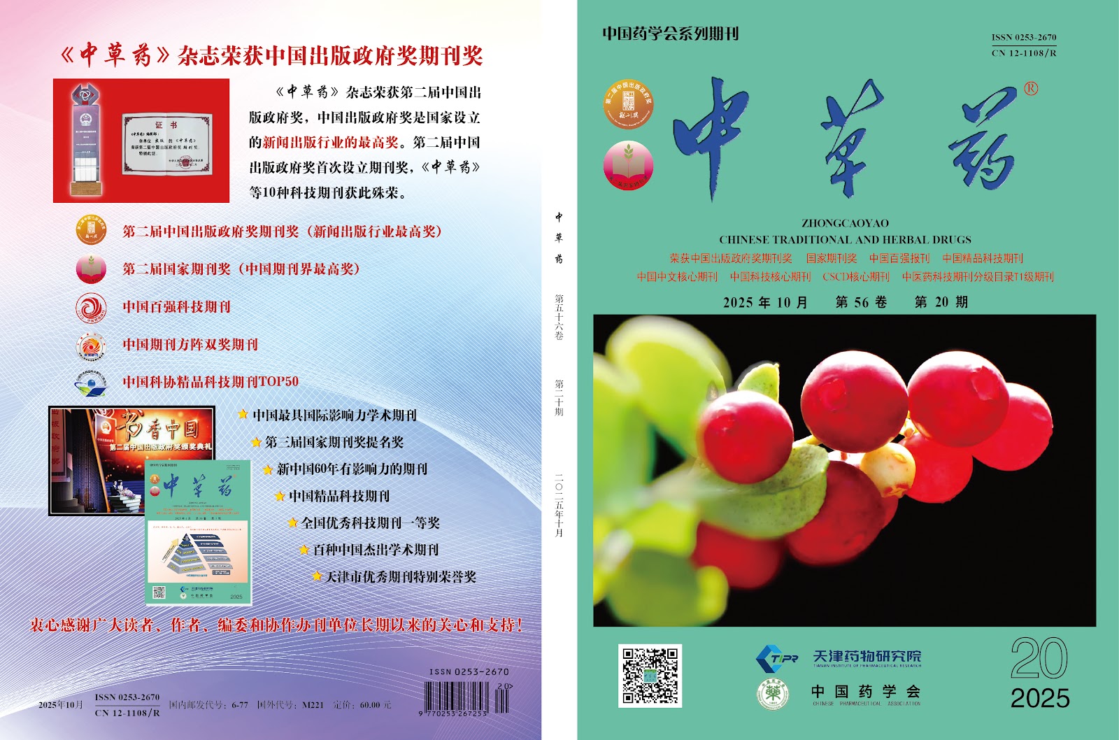
Advancements in CRISPR-Cas9 Gene Editing: A Comprehensive Review of Therapeutic Applications and Ethical Considerations

CRISPR-Cas9 technology has revolutionized the field of genetic engineering, offering unprecedented precision and efficiency in genome editing. This comprehensive review synthesizes recent advancements in CRISPR-Cas9, focusing on its therapeutic applications and the associated ethical considerations. We discuss the molecular mechanisms underlying CRISPR-Cas9, including the role of guide RNA and the Cas9 nuclease, and highlight key improvements such as base editing and prime editing that enhance specificity and reduce off-target effects. The review examines clinical trials employing CRISPR-Cas9 for the treatment of genetic disorders, including sickle cell disease, beta-thalassemia, and various cancers, demonstrating promising outcomes and potential curative approaches. Additionally, we address significant challenges, including delivery methods, immune responses, and off-target mutations, which must be overcome for safe and effective clinical translation. Ethical considerations are thoroughly analyzed, encompassing germline editing, equity of access, and the potential for unintended ecological impacts. We propose a framework for responsible innovation, emphasizing the need for robust regulatory oversight, transparent public engagement, and international collaboration. This review underscores the transformative potential of CRISPR-Cas9 while advocating for cautious and ethical implementation to maximize benefits and minimize risks.

Read Full Abstract10.7501/j.issn.0253-2670.2025.20.20252000
Advancements in SinoBioData Intelligence: A Comprehensive Review of Recent ResearchGraphical AbstractVerified
Chinese Traditional and Herbal Drugs2025

Advancements in SinoBioData Intelligence: A Comprehensive Review of Recent Research

This comprehensive review synthesizes recent advancements in the field of SinoBioData Intelligence, focusing on the integration of bioinformatics, data science, and artificial intelligence to address complex biological questions. We systematically analyze peer-reviewed literature from the past decade, highlighting key methodologies, tools, and applications that have emerged from Chinese research institutions. The review covers major areas including genomic data analysis, precision medicine, drug discovery, and systems biology, with a particular emphasis on the development of novel algorithms and databases tailored to Chinese population data. Our findings reveal a significant growth in the application of machine learning and deep learning techniques for predictive modeling and pattern recognition in biological datasets. Additionally, we discuss the challenges of data heterogeneity, privacy concerns, and the need for standardized protocols. The review concludes by outlining future directions, such as the integration of multi-omics data and the development of interpretable AI models, which are poised to drive further innovations in the field. This work serves as a valuable resource for researchers and practitioners seeking to understand the current landscape and future potential of SinoBioData Intelligence.

Read Full Abstract10.7501/j.issn.0253-2670.2025.19.202519000
Advancements in SinoBioData Intelligence: A Comprehensive Review of Current Trends and Future DirectionsGraphical AbstractVerified
Chinese Traditional and Herbal Drugs2026

Advancements in SinoBioData Intelligence: A Comprehensive Review of Current Trends and Future Directions

The field of SinoBioData intelligence has witnessed remarkable growth, driven by the integration of advanced computational methods and large-scale biological data. This comprehensive review synthesizes recent developments, highlighting key trends and future directions. We discuss the evolution of data acquisition technologies, the emergence of sophisticated analytical frameworks, and the application of artificial intelligence in deciphering complex biological systems. Critical challenges, including data heterogeneity, scalability, and interpretability, are examined, alongside potential solutions. The review underscores the transformative impact of SinoBioData intelligence on precision medicine, agricultural biotechnology, and environmental monitoring. By providing a holistic overview, this work aims to guide researchers and practitioners in navigating the dynamic landscape of bioinformatics and data-driven biology.

Read Full Abstract10.7501/j.issn.0253-2670.2026.15.2026150
Chemokine receptor 7-bone marrow mesenchymal stem cells combined with porcine small intestinal submucosa promote skin repair in ratsGraphical AbstractVerified
Chinese Journal of Tissue Engineering Research2026

Chemokine receptor 7-bone marrow mesenchymal stem cells combined with porcine small intestinal submucosa promote skin repair in rats

BACKGROUND: The clinical outcomes of autologous and allogeneic skin transplants, which are commonly used for repairing skin lesions, are often suboptimal. In recent years, advancements in tissue engineering have provided new hope for skin repair. Nevertheless, the regeneration of blood vessels within skin tissue engineering remains a significant challenge. Porcine small intestinal submucosa and bone marrow-derived mesenchymal stem cells are widely utilized natural extracellular matrix biomaterials and seed cells in current tissue engineering research. Chemokine receptor 7 is a cytokine that can promote angiogenesis. OBJECTIVE: To observe the repair effect and angiogenesis ability of chemokine receptor 7-bone marrow mesenchymal stem cells-porcine small intestinal submucosa membrane on rat back skin damage. METHODS: (1) The adenovirus vector overexpressing chemokine receptor 7 was used to transfect bone marrow mesenchymal stem cells. The transfection efficiency was evaluated using western blot assay and RT-qPCR. (2) The bone marrow mesenchymal stem cells overexpressing chemokine receptor 7 were co-cultured with porcine small intestinal submucosa. Cytocompatibility was assessed through scanning electron microscopy and live/dead cell staining. (3) 12 SD rats were utilized to establish a skin defect animal experimental model, and chemokine receptor 7-bone marrow mesenchymal stem cells-porcine small intestinal submucosa (experimental group) and porcine small intestinal submucosa alone (control group) were applied to the skin defects. Wound healing was observed at 1, 3, 7, and 14 days post-modeling. At 7 and 14 days, western blot was used to detect vascular endothelial growth factor protein expression in wound healing tissue, and immunohistochemical staining was used to detect CD31 and proliferating cell nuclear antigen protein expression. RESULTS AND CONCLUSION: (1) Adenovirus-mediated overexpression of chemokine receptor 7 in bone marrow mesenchymal stem cells was successfully constructed. The protein and mRNA expression of chemokine receptor 7 in the transfection group was significantly upregulated compared with the control and empty vector groups (P < 0.001). (2) Scanning electron microscopy and live/dead cell staining showed that bone marrow mesenchymal stem cells overexpressing chemokine receptor 7 grew well on the porcine small intestinal submucosa membrane, indicating good cytocompatibility. (3) Compared with the control group, the wound area in the experimental group was significantly reduced (P < 0.05), and the protein expression of vascular endothelial growth factor, CD31, and proliferating cell nuclear antigen in the wound healing tissue was higher than that in the control group (P < 0.05), indicating that the chemokine receptor 7-bone marrow mesenchymal stem cells-porcine small intestinal submucosa membrane had a strong ability to promote wound angiogenesis.

Read Full Abstract10.12307/2026.21206
Function of human amniotic mesenchymal stem cell exosomes in repairing submandibular gland epithelial cells after radiation injury in SD ratsGraphical AbstractVerified
Chinese Journal of Tissue Engineering Research2026

Function of human amniotic mesenchymal stem cell exosomes in repairing submandibular gland epithelial cells after radiation injury in SD rats

BACKGROUND: Hypoxia preconditioning can increase extracellular vesicles, growth factors, anti-inflammatory and immunosuppressive factors in the parasecretory secretions of mesenchymal stem cells. Exosomes derived from human amniotic mesenchymal stem cells preconditioned with hypoxia are expected to play a better therapeutic role in tissue damage repair. OBJECTIVE: To observe the repair effect of exosomes from human amniotic mesenchymal stem cells preconditioned with hypoxia on radiation-induced submandibular gland epithelial cell damage. METHODS: The passage 3 human amniotic mesenchymal stem cells were divided into two groups: hypoxia and normoxia. They were pretreated with hypoxia (1% O2 by volume) and normoxia (20% O2 by volume) for 48 hours, respectively. Then, normoxic and hypoxic human amniotic mesenchymal stem cell exosomes were extracted from the culture supernatant of human amniotic mesenchymal stem cells by modified ultrahigh-speed centrifugation. The submandibular gland epithelial cells of SD newborn rats were divided into blank control group, radiation control group, normoxia-treated group, and hypoxia-treated group. The submandibular gland epithelial cells were treated with 5 Gy radiation to construct a radiation-induced injury model. The normoxia-treated and hypoxia-treated groups were co-cultured with normoxic or hypoxic exosomes for 3 days. CCK-8 assay was used to detect the proliferation activity of rat submandibular gland epithelial cells; ELISA was used to measure the content of α-amylase in the supernatant; RT-qPCR was used to detect the expression of aquaporin 5 mRNA. RESULTS AND CONCLUSION: The cell proliferation activity in the radiation control group was significantly lower than that in the blank control group (P < 0.05). The cell proliferation activity in the normoxia-treated and hypoxia-treated groups on days 2 and 3 was significantly higher than that in the radiation control group (P < 0.05). The cell proliferation activity in the hypoxia-treated group on day 1 was higher than that in the normoxia-treated group, and showed an increasing trend on days 2 and 3, but there was no statistical difference compared with the normoxia-treated group (P > 0.05). The α-amylase content in the radiation control group was significantly lower than that in the blank control group (P < 0.05). The α-amylase content in the normoxia-treated and hypoxia-treated groups was significantly higher than that in the radiation control group (P < 0.05). Compared with the normoxia-treated group, the α-amylase content in the hypoxia-treated group showed an increasing trend, but there was no statistical difference (P > 0.05). The expression of aquaporin 5 mRNA in the radiation control group was significantly lower than that in the blank control group (P < 0.05). The expression of aquaporin 5 mRNA in the normoxia-treated and hypoxia-treated groups was significantly higher than that in the radiation control group (P < 0.05). The expression of aquaporin 5 mRNA in the hypoxia-treated group was slightly higher than that in the normoxia-treated group, but there was no statistical difference (P > 0.05). The results indicate that both normoxic and hypoxic preconditioned human amniotic mesenchymal stem cell exosomes have a repair effect on radiation-induced submandibular gland epithelial cell damage, but hypoxic exosomes have no obvious advantage.

Read Full Abstract10.12307/2026.21207
Isolation, cultivation, identification, and induction of M1/M2 polarization in bone marrow-derived macrophages from C57BL/6 miceGraphical AbstractVerified
Chinese Journal of Tissue Engineering Research2026

Isolation, cultivation, identification, and induction of M1/M2 polarization in bone marrow-derived macrophages from C57BL/6 mice

BACKGROUND: Macrophage polarization demonstrates significant potential in disease treatment, particularly in areas such as cancer, inflammation, and autoimmune diseases. Establishing standardized in vitro models can lay the groundwork for in-depth research into the mechanisms of macrophage polarization. OBJECTIVE: To observe the in vitro growth characteristics of bone marrow-derived macrophages from C57BL/6 mice and to establish a standardized in vitro model for M1 and M2 macrophage polarization. METHODS: Femurs and tibias of C57BL/6 mice were aseptically separated, and the contents of the bone marrow cavity were collected. After filtering through a mesh and lysing erythrocytes, the contents were resuspended in high-glucose DMEM containing 20 ng/mL macrophage colony-stimulating factor and inoculated in 6-well plates according to experimental requirements. On day 7, they were differentiated into mature mouse bone marrow-derived macrophages (M0 type). Then, 100 ng/mL lipopolysaccharide was used to induce polarization to M1 type, and 20 ng/mL interleukin-4 was used to induce polarization to M2 type. Flow cytometry and RT-qPCR were used to detect the expression of corresponding markers in macrophages under different polarization states, and Western blot was used to detect the expression of M1 macrophage marker pathway proteins p-STAT1, STAT1 and M2 macrophage marker pathway proteins p-STAT6, STAT6. RESULTS AND CONCLUSION: (1) After stimulation with 20 ng/mL macrophage colony-stimulating factor for 7 days, flow cytometry showed that the positive rate of macrophage surface marker F4/80 reached 98.1%. (2) After stimulation with 100 ng/mL lipopolysaccharide for 6 h, the positive rates of F4/80 and CD86 were about 35%, and RT-qPCR showed that the mRNA expression of M1 macrophage markers inducible nitric oxide synthase, interleukin-6, macrophage inflammatory protein 1α, and monocyte chemoattractant protein 1 were significantly higher than those in the control group (P < 0.01). (3) After stimulation with 20 ng/mL interleukin-4 for 24 h, the mean fluorescence intensity of CD206 was significantly increased, and RT-qPCR showed that the mRNA expression of M2 macrophage markers Chi3l3 (Ym1), interleukin-10, and arginase 1 were significantly higher than those in the control group (P < 0.01). (4) Western blot results showed that lipopolysaccharide-induced M1 macrophage marker pathway protein p-STAT1 was significantly activated; interleukin-4-induced M2 macrophage marker pathway protein p-STAT6 was significantly activated. These results indicate that lipopolysaccharide and interleukin-4 effectively induced polarization of bone marrow-derived macrophages to M1 and M2 types, respectively.

Read Full Abstract10.12307/2026.21205
Extracellular matrix stiffness affects the proliferation activity of bone marrow stromal stem cellsGraphical AbstractVerified
Chinese Journal of Tissue Engineering Research2026

Extracellular matrix stiffness affects the proliferation activity of bone marrow stromal stem cells

BACKGROUND: In tissue engineering bone construction, the physical properties of the scaffold can directly affect the activity and repair effect of seed cells, among which extracellular matrix hardness is a key factor affecting seed cell proliferation activity. Primary cilia and YAP proteins have been shown to be classical mechanoreceptors and downstream transduction factors, which may directly mediate this mechanism. OBJECTIVE: To investigate the regulatory effect of extracellular matrix hardness on the proliferation activity of bone marrow stromal stem cells and the related mechanisms. METHODS: Bone marrow stromal stem cells were passaged and seeded under different hardness of polydimethylsiloxane extracellular matrix conditions (soft, median, and rigid) for culture. Cell proliferation activity was detected using CCK-8 assay. Transcriptional activity of proliferation genes c-myc and CCND1 was measured using qRT-PCR. Activation of Wnt/β-catenin pathway was evaluated using western blot assay. Primary cilia and YAP protein expression levels were evaluated by acetylated α-tubulin and YAP immunofluorescence staining. After passage, bone marrow stromal stem cells were inoculated on polydimethylsiloxane-based membranes of different hardness (soft and hard) for culture. Then siRNA was used to interfere with YAP protein expression. Western blot assay was used to detect YAP, phosphorylated GSK-3β, and β-catenin protein expression. qRT-PCR was used to detect the transcriptional activity of c-myc and CCND1. The length of primary cilia was analyzed after immunofluorescence staining of acetylated α-tubulin. RESULTS AND CONCLUSION: The cell proliferation activity, c-myc and CCND1 transcriptional activity under rigid polydimethylsiloxane conditions were significantly higher than those under soft and median hardness, and the activation of Wnt/β-catenin pathway was stronger. Immunofluorescence staining showed that rigid polydimethylsiloxane induced shortening of primary cilia and increased YAP-positive cells. After siRNA interference of YAP expression, the differences in YAP, phosphorylated GSK-3β, β-catenin protein expression, and c-myc and CCND1 transcriptional activity between groups disappeared, accompanied by the disappearance of primary cilia length differences. The results indicate that extracellular matrix stiffness regulates the proliferation activity of bone marrow stromal stem cells through a novel YAP protein/primary cilia mechanism.

Read Full Abstract10.12307/2026.21204
Systematic druggable genome-wide Mendelian randomization identifies therapeutic targets for major depressive disorderGraphical AbstractVerified
Chinese Journal of Tissue Engineering Research2026

Systematic druggable genome-wide Mendelian randomization identifies therapeutic targets for major depressive disorder

BACKGROUND: The occurrence of major depressive disorder is typically associated with genetic and environmental factors. Currently, the diagnosis of major depressive disorder mainly relies on clinical interviews and symptom assessments, lacking clear and reproducible biological markers. This can lead to misdiagnosis and missed diagnoses, delaying the timing of treatment. OBJECTIVE: To identify druggable genes that may act as potential therapeutic targets for major depressive disorder by conducting comprehensive genome-wide Mendelian randomization analysis. METHODS: By integrating expression quantitative trait locus (eQTL) data and protein quantitative trait locus (pQTL) data from pharmacologically actionable genes with genome-wide association study (GWAS) data on major depressive disorder (including 177 377 cases and 445 321 controls), Mendelian randomization analysis was conducted to identify druggable genes that have a causal relationship with major depressive disorder. Additionally, enrichment analysis, protein-protein interaction network construction, drug target identification, and molecular docking simulations were performed to further explore potential therapeutic strategies. RESULTS AND CONCLUSION: A total of 4 394 druggable genes were analyzed, and 21 druggable genes considerably associated with major depressive disorder were identified. Bayesian colocalization analysis indicated that BTN3A3, CISD1, and PSMB4 had posterior probabilities of hypothesis 4 (H4.abf) > 0.5, supporting the possibility of shared causal variants. GO enrichment analysis mainly involved 'antigen processing and presentation', 'protein degradation and processing', 'mitochondrial outer membrane', and 'immune receptor activity' pathways related to major depression. Protein-protein interaction network analysis showed moderate connectivity among the identified genes (21 nodes, 14 edges). Drug target identification determined gemcitabine (CID 60750), fucose (CID 17106), and isococculidine (CID 2826) as main candidate compounds, which had strong associations with several key genes. Molecular docking analysis revealed stable drug-protein interactions, with isococculidine showing the most stable binding energy (-52.74 kJ/mol) with BTN3A3. In conclusion, Mendelian randomization combined with genomics and structural biology analysis provides valuable decision-making basis for target prioritization and drug repurposing, offering new ideas and directions for efficient utilization of basic research resources and drug development for major depressive disorder.

Read Full Abstract10.12307/2026.21252
Virtual reality therapy on neuropathic pain following spinal cord injuryGraphical AbstractVerified
Chinese Journal of Tissue Engineering Research2026

Virtual reality therapy on neuropathic pain following spinal cord injury

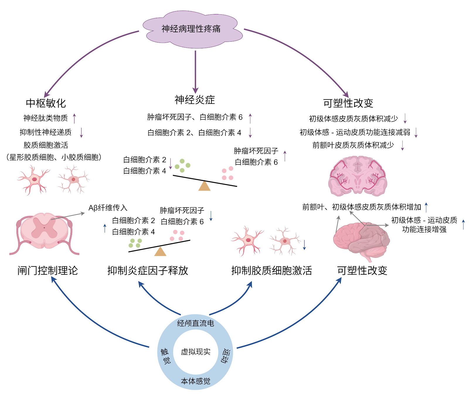
BACKGROUND: Virtual reality has demonstrated unique advantages as a novel non-pharmacological intervention in the treatment of neuropathic pain following spinal cord injury. OBJECTIVE: To systematically review the pathogenesis of neuropathic pain following spinal cord injury, summarize the specific application modes and mechanisms of virtual reality therapy for this condition, and explore key factors influencing treatment efficacy, thereby providing novel therapeutic options for patients with neuropathic pain following spinal cord injury. METHODS: A literature search was conducted in the CNKI, PubMed, Web of Science, and Cochrane Library databases up to June 2025. The Chinese and English search terms included “spinal cord injury, central sensitization, cerebral cortex, noninvasive therapy, neuropathic pain, virtual reality.” A total of 1 352 articles were retrieved, and 73 articles that met the inclusion criteria were ultimately included in the analysis. RESULTS AND CONCLUSION: The development of neuropathic pain following spinal cord injury involves complex mechanisms in both the spinal cord and brain, closely associated with central sensitization at the spinal level and abnormal plasticity changes in the brain. Virtual reality, as a novel intervention approach, can be used alone or in combination with other intervention methods, showing unique therapeutic value for the multidimensional pathogenesis of neuropathic pain after spinal cord injury. Standalone virtual reality applications primarily involve virtual walking interventions aimed at restoring abnormal brain plasticity changes, while combined virtual reality applications have shown certain effects in both inhibiting central sensitization and modulating brain plasticity. Although virtual reality intervention shows great potential in the treatment of neuropathic pain following spinal cord injury, current research and applications still have certain limitations. Future efforts should focus on addressing these issues to fully realize the therapeutic value of virtual reality in neuropathic pain following spinal cord injury.

Read Full Abstract10.12307/2026.21243
The role of exercise-regulated mitophagy in cardiovascular diseasesGraphical AbstractVerified
Chinese Journal of Tissue Engineering Research2026

The role of exercise-regulated mitophagy in cardiovascular diseases

BACKGROUND: Mitophagy plays a crucial regulatory role in the occurrence and development of cardiovascular diseases. Exercise intervention can mediate mitophagy to improve cardiovascular function, which provides new insights for the clinical prevention and treatment of cardiovascular diseases. OBJECTIVE: To summarize the regulatory role of mitophagy in cardiovascular diseases, the influence of exercise on mitophagy, and the mechanism by which exercise-mediated mitophagy improves cardiovascular diseases. METHODS: PubMed and CNKI databases were searched for relevant literature using the search terms of “mitophagy, mitochondrial function, cardiovascular disease, aerobic exercise, resistance training, combined aerobic resistance exercise, high-intensity interval training” in Chinese and English, respectively. Based on the inclusion and exclusion criteria, totally 88 documents were finally included for summary and analysis. RESULTS AND CONCLUSION: (1) Mitophagy plays a crucial role in the regulation of cardiovascular diseases such as heart failure, myocardial hypertrophy, atherosclerosis, and myocardial ischemia-reperfusion injury. Moreover, mitophagy imbalance or disorder can exacerbate the pathological process of cardiovascular diseases. (2) Various exercise modalities can activate mitophagy by regulating the expression of mitophagy-related factors. Among them, aerobic exercise can promote the formation of autophagosomes, thereby enhancing the regulation of mitophagy; resistance exercise can regulate mitochondrial biogenesis; combined aerobic and resistance exercise can further influence mitophagy by promoting lysosomal biogenesis; high-intensity interval training enhances mitophagy function by regulating the expression of mitochondrial dynamics-related proteins. (3) Exercise regulates mitophagy to alleviate myocardial fibrosis, inhibit cardiomyocyte apoptosis, regulate myocardial oxidative stress, and improve endothelial cell function, thereby playing a key role in the prevention and treatment of cardiovascular diseases, providing a new perspective for exercise promoting health and preventing cardiovascular diseases.

Read Full Abstract10.12307/2026.21247
Forkhead box transcription factor O3 affects bone metabolism and participates in the pathological processes of various bone-related diseasesGraphical AbstractVerified
Chinese Journal of Tissue Engineering Research2026

Forkhead box transcription factor O3 affects bone metabolism and participates in the pathological processes of various bone-related diseases

BACKGROUND: Bone metabolism disorders can cause the occurrence of bone-related diseases, and forkhead box transcription factor O3 (FoxO3a) can affect the processes of proliferation, differentiation and apoptosis of bone tissue cells by regulating oxidative stress and autophagy levels, and thereby regulate the bone metabolism. OBJECTIVE: To systematically analyze the relevant research literature on the regulation of bone metabolism by FoxO3a and its mechanism of action in bone diseases and to provide a reference for subsequent studies targeting FoxO3a in the treatment of bone diseases. METHODS: Literature searches were conducted using the following strategies: CNKI (China National Knowledge Infrastructure): SU=FoxO3a OR SU=Foxo3 OR SU=Forkhead box O3 OR SU=AND SU=Forkhead box transcription factor O3) AND SU=bone; WanFang Medical Database: Subject:("FoxO3a") OR Subject:("Foxo3") OR Subject:("Forkhead box O3") OR Subject:("Forkhead box transcription factor O3") AND Subject:("bone"); PubMed: ((FoxO3a) OR (Foxo3) OR (Forkhead box O3))AND ((bone) OR (Skeleton)). Outdated, repetitive, low-quality, and irrelevant studies were excluded, and 56 articles were finally included for review. RESULTS AND CONCLUSION: ①FoxO3a and bone marrow mesenchymal stem cells: FoxO3a can promote the formation of osteogenic lineage and promote early osteogenic differentiation by activating autophagy. Meanwhile, FoxO3a exhibits antioxidant properties in bone marrow mesenchymal stem cells, protecting cells from oxidative stress-induced senescence. ②FoxO3a and osteoblasts: FoxO3a can inhibit osteogenesis by interfering with the Wnt/β-catenin pathway in osteoblasts, while it can activate antioxidant enzymes to protect mature osteoblasts. FoxO3a can promote the proliferation of osteoprogenitor cells and promote osteogenic differentiation by activating autophagy. ③FoxO3a and osteoclasts: FoxO3a expression can resist oxidative stress and activate autophagy to inhibit osteoclastogenesis. ④FoxO3a and osteocytes: FoxO3a can protect osteocytes through antioxidant effects, and can also reduce bone loss by inhibiting p16 and p53 signaling pathways and inhibiting senescence-associated secretory phenotype. ⑤FoxO3a and chondrocytes: FoxO3a plays a protective role in chondrocytes in osteoarthritis, inhibiting chondrocyte breakdown or apoptosis, promoting chondrocyte extracellular matrix synthesis, and inhibiting chondrocyte hypertrophy; however, high co-expression of FoxO3a and Runt-related transcription factor 1 in chondrocytes promotes early chondrogenesis and terminal hypertrophy of chondroprogenitor cells. ⑥FoxO3a affects bone metabolism by participating in processes such as oxidative stress resistance and regulation of autophagy, and participates in the pathological processes of various bone-related diseases.

Read Full Abstract10.12307/2026.21242
Transcription factor EB improves Alzheimer’s disease via the autophagy-lysosome pathwayGraphical AbstractVerified
Chinese Journal of Tissue Engineering Research2026

Transcription factor EB improves Alzheimer’s disease via the autophagy-lysosome pathway

BACKGROUND: Studies have confirmed that transcription factor EB and its dependent autophagy-lysosome pathway play a role in the development of various neurodegenerative diseases, including Alzheimer’s disease. OBJECTIVE: To summarize the role of transcription factor EB-mediated autophagy-lysosome pathway in Alzheimer’s disease. METHODS: Electronic databases including PubMed, Web of Science, Cochrane Library, CNKI, WanFang Medical Network, and VIP were searched. The search period was from database inception to January 2025. The search terms were “Alzheimer Disease, AD, Transcription Factor EB, TFEB, Autophagy-lysosome Pathway, Autophagy, Lysosomes, Amyloid beta, Aβ, Tau, Tau protein” in English as well as “Alzheimer’s disease, transcription factor EB, autophagic lysosomes, autophagy, lysosomes, β-amyloid, Tau” in Chinese. Additionally, the references of relevant reviews and grey literature were manually supplemented. A total of 100 articles were finally included for review. RESULTS AND CONCLUSION: Amyloid-β deposition and Tau protein phosphorylation are the key pathological features of Alzheimer’s disease. Abnormalities in the autophagy-lysosome pathway promote the formation of neurotoxic protein aggregates such as amyloid-β and Tau, leading to the clinical manifestations of Alzheimer’s disease characterized by cognitive dysfunction and behavioral abnormalities. Transcription factor EB is a key regulator of the autophagy-lysosome pathway, controlling the transcription of many autophagy-related genes and lysosomal biogenesis. After entering the nucleus, transcription factor EB upregulates the expression of autophagy-lysosome pathway-related genes, significantly increasing the clearance rate of amyloid-β and Tau and significantly reducing neuronal toxicity. Therefore, increasing attention focuses on targeting transcription factor EB to influence autophagy-lysosome biological activity, thereby improving Alzheimer’s disease pathology and behavioral deficits. For example, both pharmacological and non-pharmacological interventions can activate transcription factor EB, reducing amyloid-β deposition and Tau phosphorylation, and improving cognitive function in Alzheimer’s disease. However, sustained activation of transcription factor EB may pose risks such as lysosomal storage disorders, and current intracranial delivery systems have insufficient targeting efficiency. Future development of pathological microenvironment-responsive carriers and combination therapies is needed to achieve precise intervention.

Read Full Abstract10.12307/2026.21248
Biomarkers for diabetic foot ulcers: single-cell transcriptomics bioinformatics analysis and experimental validationGraphical AbstractVerified
Chinese Journal of Tissue Engineering Research2026

Biomarkers for diabetic foot ulcers: single-cell transcriptomics bioinformatics analysis and experimental validation

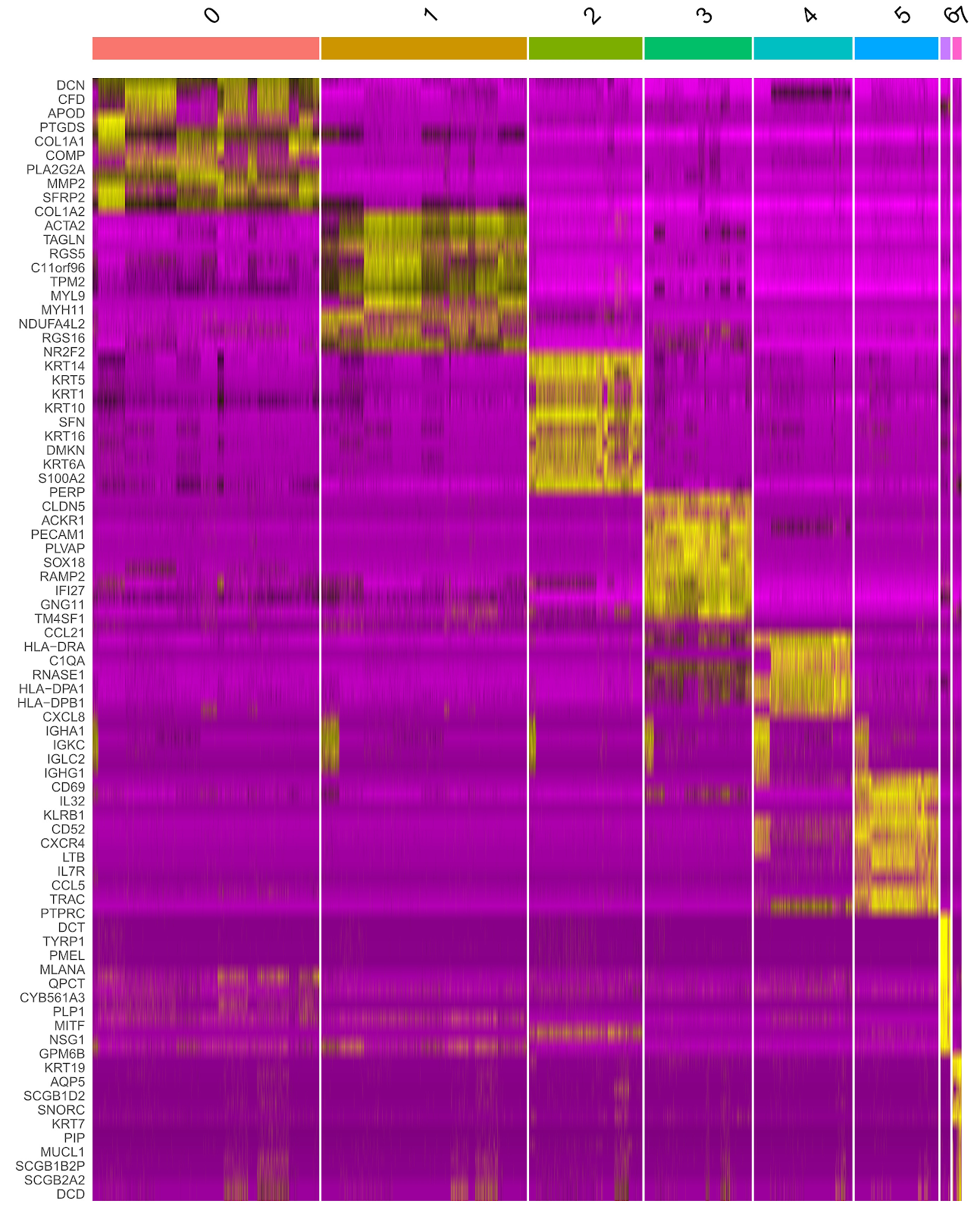
BACKGROUND: Factors such as infection, limb ischemia, and histiocyte activation are involved in diabetic foot ulcers, but the key cell subpopulations influencing diabetic foot ulcer healing remain unclear, and specific biomarkers for diabetic foot ulcers have yet to be identified. Gene Expression Omnibus (GEO) is a publicly accessible database managed by the National Center for Biotechnology Information that stores high-throughput gene expression data, allowing users to freely submit, share, query, and analyze data. Secondary analysis of published data can save research costs and uncover new research targets and ideas. OBJECTIVE: To screen biomarkers for diabetic foot ulcers using single-cell transcriptome and conventional transcriptome bioinformatics analysis, high-dimensional weighted gene co-expression network analysis (hdWGCNA), and weighted gene co-expression network analysis (WGCNA). METHODS: The single-cell transcriptome dataset GSE165816, containing non-healing ulcer tissue samples from diabetic foot ulcer patients and foot skin samples from diabetic patients, was downloaded from GEO. After data quality control, dimensionality reduction, differential analysis, cell type annotation, and pseudotime analysis, cell types spanning the entire course of diabetic foot ulcers were identified, and differentially expressed genes (DEGs) were obtained. hdWGCNA identified gene modules highly correlated with diabetic foot ulcers. Conventional transcriptome datasets GSE68183 and GSE80178, containing non-healing ulcer tissue samples from diabetic foot ulcer patients and foot skin samples from diabetic patients, were downloaded for differential analysis to screen DEGs, and WGCNA was used to identify diabetic foot ulcer-related gene modules. The DEGs from single-cell transcriptome, DEGs from conventional transcriptome samples, and module genes from WGCNA and hdWGCNA were integrated to screen biomarkers for diabetic foot ulcers. The GSE134431 dataset was downloaded as a validation conventional transcriptome dataset, and the expression levels of diabetic foot ulcer biomarkers were compared in single-cell transcriptome and validation conventional transcriptome datasets. Diabetic and diabetic foot ulcer rat models were replicated, wound tissue was collected, and immunohistochemistry and western blot were used to detect biomarker expression levels. RESULTS AND CONCLUSION: Single-cell transcriptome data analysis showed that epithelial cell differentiation spanned the entire pathological process of diabetic foot ulcers. A total of 146 DEGs were obtained from single-cell transcriptome between groups, including 59 upregulated and 87 downregulated DEGs. hdWGCNA identified 19 gene modules related to diabetic foot ulcers, containing 476 core genes. Conventional transcriptome data analysis yielded a total of 913 DEGs, including 343 upregulated and 570 downregulated DEGs. WGCNA obtained 19 diabetic foot ulcer-related gene modules, containing 887 genes. Two biomarkers for diabetic foot ulcers were screened: S100A14 and SFN. The expression levels of these two genes in diabetic foot ulcer samples were higher than those in diabetic foot skin samples in both single-cell transcriptome and validation conventional transcriptome datasets. Animal experiments showed that the expression levels of S100A14 and SFN in wound tissue of diabetic foot ulcer rats were higher than those in back skin tissue of diabetic rats. The results indicate that the pathological process of diabetic foot ulcers involves multiple cell types, among which epithelial cells are the key cell subpopulation. S100A14 and SFN are significantly upregulated in diabetic foot ulcer samples and are potential targets for the treatment of diabetic foot ulcers.

Read Full Abstract10.12307/2026.21235
The relationship between inflammatory cytokines and frozen shoulder: a large-sample analysis of the European population based on the FinnGen GWAS databaseGraphical AbstractVerified
Chinese Journal of Tissue Engineering Research2026

The relationship between inflammatory cytokines and frozen shoulder: a large-sample analysis of the European population based on the FinnGen GWAS database

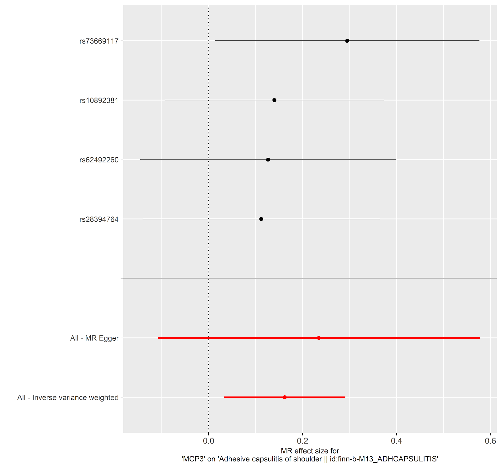
BACKGROUND: Frozen shoulder is a common disease in orthopedics, but there is no specific clinical indicator for diagnosis. There is a significant association between inflammatory cytokines and frozen shoulder, but the specific causal relationship is not yet clear. This study used summary statistical data from genome-wide association studies (GWAS) for Mendelian randomization analysis. GWAS data are based on large-sample genetic variation information, which can reduce environmental confounding factors and more reliably infer the causal relationship between inflammatory cytokines and frozen shoulder, making up for the limitation that traditional observational studies cannot determine causal associations. OBJECTIVE: To explore the causal relationship between inflammatory cytokines and the onset of frozen shoulder using bidirectional two-sample Mendelian randomization. METHODS: Using summary statistics from GWAS in the FinnGen database, we analyzed the causal relationship between 41 inflammatory cytokines and frozen shoulder. The FinnGen database, jointly initiated by the Finnish National Institute for Health and Welfare (THL), the University of Helsinki, and other Finnish research institutions, includes 2,942 cases and 167,641 European-ancestry controls, integrating genomic, clinical phenotype, and biochemical indicator data from hundreds of thousands to millions of individuals, supporting genetic association studies of diseases. This study is based on publicly available summary statistics databases and does not require ethical approval. Bidirectional Mendelian randomization analyses were performed using inverse variance weighting, weighted median, weighted model, simple model, MR-Egger regression, and sensitivity analyses (including MR-Egger, MR-PRESSO, Cochran's Q test). RESULTS AND CONCLUSION: Monocyte chemoattractant protein-3 (MCP-3) showed significant causal effects in both directions. In the forward analysis, MCP-3 was positively associated with frozen shoulder risk (OR=1.176, 95%CI: 1.034-1.338, P=0.014); in the reverse analysis, frozen shoulder was negatively associated with MCP-3 levels (OR=0.782, 95%CI: 0.625-0.979, P=0.032). Additionally, a significant association was found between tumor necrosis factor beta (TNF-β) and frozen shoulder risk (OR=1.126, 95%CI: 1.002-1.264, P=0.046); in the reverse analysis, stromal cell-derived factor 1 alpha (SDF-1α) was also significantly associated with frozen shoulder risk (OR=1.1, 95%CI: 1.011-1.196, P=0.028), indicating reliable correlations of TNF-β and SDF-1α with frozen shoulder. This bidirectional Mendelian randomization study reveals a complex interaction between MCP-3 and frozen shoulder, suggesting that MCP-3 may serve as a potential therapeutic target. Furthermore, the study indicates that TNF-β is associated with frozen shoulder risk and may be a potential risk factor; while frozen shoulder is also associated with elevated SDF-1α levels, and SDF-1α has the potential to become a diagnostic marker for frozen shoulder. However, further research is needed to elucidate the biological mechanisms underlying these causal relationships. Additionally, the analysis of international databases provides candidate molecules and causal inference paradigms for Chinese research, but it needs to be combined with local data for precise translation.

Read Full Abstract10.12307/2026.21250
Shared genetic basis and causal relationship between nutrition, nutritional status and inflammatory bowel diseaseGraphical AbstractVerified
Chinese Journal of Tissue Engineering Research2026

Shared genetic basis and causal relationship between nutrition, nutritional status and inflammatory bowel disease

BACKGROUND: Inflammatory bowel disease, encompassing ulcerative colitis and Crohn’s disease, is a chronic condition linked to malnutrition, sarcopenia, and disease severity, with limited research on their genetic associations. OBJECTIVE: To systematically explore the common genetic basis and causal relationships between nutrition, nutritional status, and inflammatory bowel disease using advanced statistical genetics. METHODS: Single nucleotide polymorphism data for nutritional markers (minerals, vitamins, albumin, hemoglobin, fatty acids) and sarcopenia traits (appendicular lean mass and hand grip strength) were obtained from the GWAS Catalog, and inflammatory bowel disease and its subtypes from the FinnGen database R10. Advanced statistical genetics methods, including linkage disequilibrium score regression, cross-phenotype association analysis, and Mendelian randomization, were used to infer associations. RESULTS AND CONCLUSION: Significant genetic correlations were found: vitamin D with inflammatory bowel disease (rg=-0.080, P=0.029) and ulcerative colitis (rg=-0.087, P=0.027); appendicular lean mass with inflammatory bowel disease (rg=-0.100, P=0.0002), ulcerative colitis (rg=-0.100, P=0.0002), and small intestine Crohn’s disease (rg=-0.081, P=0.035); hand grip strength with small intestine Crohn’s disease (rg=-0.125, P=0.035). Mendelian randomization indicated a positive causal effect of magnesium levels on inflammatory bowel disease (OR=1.41, P=0.036) and small intestine Crohn’s disease (OR=1.78, P=0.035). Cross-phenotype analysis identified shared single nucleotide polymorphisms, particularly in the human leukocyte antigen region, affecting both nutritional status and inflammatory bowel disease. These findings further explain the genetic link between nutrition, sarcopenia, and inflammatory bowel disease, suggesting that targeted nutritional management may be key to slowing disease progression. This study provides new perspectives for personalized treatment and has potential implications for prevention strategies of inflammatory bowel disease.

Read Full Abstract10.12307/2026.21251
Role of fibrosis in tissue injury repairGraphical AbstractVerified
Chinese Journal of Tissue Engineering Research2026

Role of fibrosis in tissue injury repair

BACKGROUND: Fibrosis results from dysregulated tissue healing, characterized by abnormal proliferation of fibroblasts and excessive deposition of extracellular matrix, affecting nearly all organs including liver, kidney, heart, lung, and skin. OBJECTIVE: To summarize fibrosis-related diseases such as liver, kidney, cardiac, and pulmonary fibrosis, focusing on the major abnormal cells, signaling pathways, and therapeutic approaches. METHODS: PubMed and CNKI were searched using English terms "fibrosis, fibroblasts, fibrotic organs, extracellular matrix, tissue repair, inflammatory response" and Chinese equivalents. After screening according to inclusion and exclusion criteria, 200 articles were included for review. RESULTS AND CONCLUSION: Key abnormal cells in fibrosis include immune cells (macrophages, neutrophils, lymphocytes), fibroblasts, epithelial cells, and endothelial cells, with fibroblasts playing a central role. Major abnormal pathways include TGF-β, Wnt/β-catenin, Notch, TLR4/MyD88/NF-κB, and Hippo/YAP signaling, whose dysregulation drives fibrosis. Epigenetic modifications (DNA methylation, histone modification, non-coding RNA regulation) modulate fibrosis progression. Anti-fibrotic therapies include pharmacological, cellular, and gene-based approaches, targeting signaling pathways to inhibit persistent fibroblast activation or modulating extracellular matrix deposition to alleviate fibrosis and improve organ function.

Read Full Abstract10.12307/2026.21246
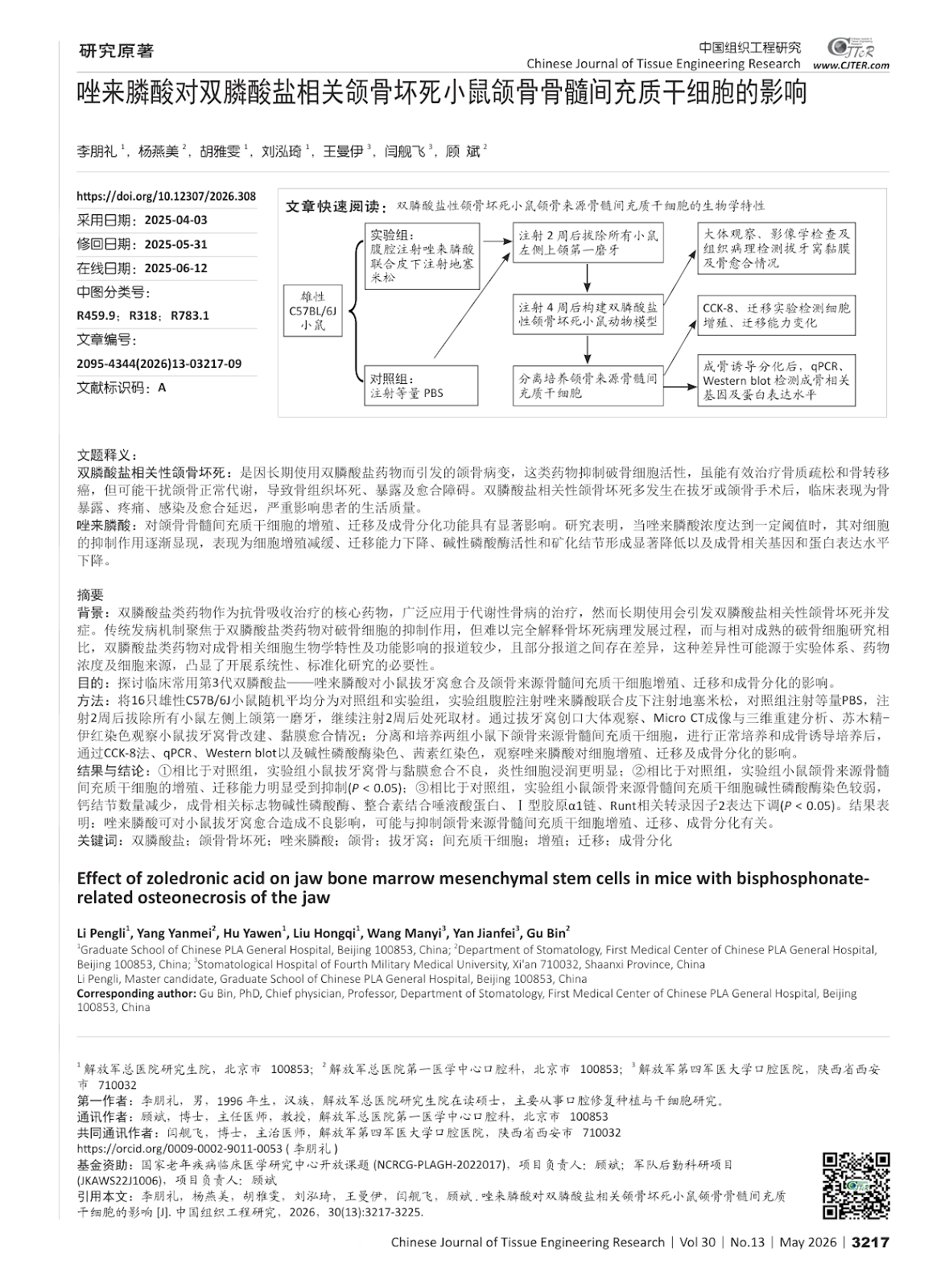
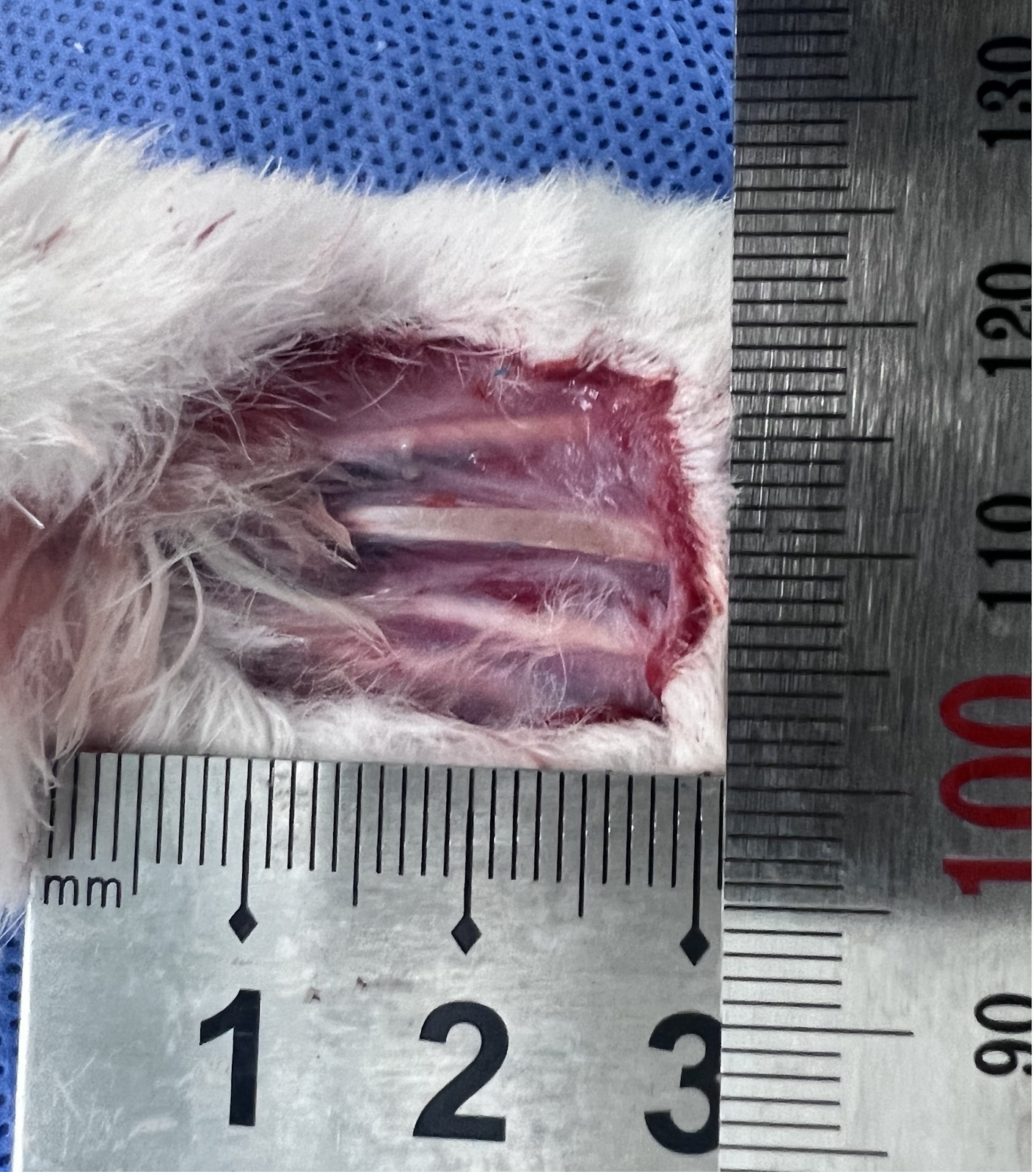
Effect of zoledronic acid on jaw bone marrow mesenchymal stem cells in mice with bisphosphonate-related osteonecrosis of the jawGraphical AbstractVerified
Chinese Journal of Tissue Engineering Research2026

Effect of zoledronic acid on jaw bone marrow mesenchymal stem cells in mice with bisphosphonate-related osteonecrosis of the jaw

BACKGROUND: Bisphosphonates, as the core drugs of anti-bone resorption therapy, are widely used in the treatment of metabolic bone diseases. However, long-term use can cause the complications of bisphosphonate related osteonecrosis of the jaw. The traditional pathogenesis focuses on the inhibitory effect of bisphosphonates on osteoclasts, but it is difficult to fully explain the pathological development of osteonecrosis. Compared with the relatively mature osteoclast research, there are fewer reports on the effects of bisphosphonates on the biological characteristics and functions of osteoblast-related cells, and there are differences between some reports. This difference may be due to the experimental system, drug concentration and cell source, highlighting the necessity of conducting systematic and standardized research. OBJECTIVE: To investigate the effect of the third-generation bisphosphonate-zoledronic acid commonly used in clinical practice on the healing of tooth extraction sockets and the proliferation, migration and osteogenic differentiation of bone marrow mesenchymal stem cells derived from the jaw in mice. METHODS: Sixteen male C57BL/6J mice were randomly divided into control and experimental groups. The experimental group received intraperitoneal injection of zoledronic acid combined with subcutaneous injection of dexamethasone, while the control group received an equal volume of PBS. After 2 weeks of injection, the left maxillary first molars of all mice were extracted, and after another 2 weeks of injection, the mice were sacrificed. The healing of extraction sockets was evaluated by gross observation, Micro CT imaging and three-dimensional reconstruction, and hematoxylin-eosin staining. Jaw bone marrow mesenchymal stem cells were isolated and cultured from both groups. After normal culture and osteogenic induction, cell proliferation, migration, and osteogenic differentiation were assessed by CCK-8 assay, qPCR, Western blot, alkaline phosphatase staining, and alizarin red staining. RESULTS AND CONCLUSION: Compared with the control group, the experimental group showed poor healing of extraction sockets with more inflammatory cell infiltration. The proliferation and migration abilities of jaw bone marrow mesenchymal stem cells were significantly inhibited in the experimental group (P < 0.05). Alkaline phosphatase staining was weaker, calcium nodule formation was reduced, and the expression of osteogenic markers (alkaline phosphatase, integrin-binding sialoprotein, collagen type I alpha 1 chain, Runt-related transcription factor 2) was downregulated in the experimental group (P < 0.05). These results indicate that zoledronic acid can adversely affect extraction socket healing, possibly by inhibiting the proliferation, migration, and osteogenic differentiation of jaw bone marrow mesenchymal stem cells.

Read Full Abstract10.12307/2026.21203
Strategies for the application of miRNA-targeted therapy in the treatment of osteoporosisGraphical AbstractVerified
Chinese Journal of Tissue Engineering Research2026

Strategies for the application of miRNA-targeted therapy in the treatment of osteoporosis

BACKGROUND: Studies have shown that miRNAs, as important post-transcriptional regulators of genes, play a key role in the onset and progression of osteoporosis. Through in-depth research on the biology of miRNA regulation of osteoporosis, its potential healing mechanisms have been revealed, and this field has become a hot focus of current research. OBJECTIVE: To explore the regulatory role of miRNAs in the development of osteoporosis and their molecular mechanisms, and to provide an overview of the key difficulties encountered in the therapeutic strategies for osteoporosis targeting miRNAs and their solutions. METHODS: We searched PubMed, Web of Science and CNKI databases for relevant literature published up to March 2025. The search terms were “miRNA, osteoporosis, angiogenesis, osteogenesis, gene therapy, drug delivery” in English and “miRNA, osteoporosis, gene therapy, ribonucleic acid drugs, delivery carrier” in Chinese. After reading the titles and abstracts for preliminary screening, we excluded the literature with poor relevance, old information, or repetitive views and lack of authority, and finally included 138 papers for review. RESULTS AND CONCLUSION: (1) miRNAs are highly efficient non-coding RNAs with a wide range of applications that can precisely regulate cellular activities, and they show significant therapeutic potential in regulating osteoblast function and bone angiogenesis. (2) Although miRNA-based targeted drugs have entered preclinical research in other disease areas, clinical translation still faces challenges of insufficient nucleic acid stability in vivo and off-target effects. (3) To address these challenges, researchers have proposed various strategies, including precise targeting of miRNA target genes to reduce off-target effects; chemical modification to improve the stability of nucleic acid drugs in vivo; reducing nucleic acid production costs to advance research; and utilizing viral vectors, exosomes, and various biomaterials to optimize nucleic acid drug delivery routes. (4) Advances in technology continue to innovate in improving the performance of nucleic acid drug carriers, and in the future, precise and efficient drug delivery and targeted therapeutic effects will be achieved.

Read Full Abstract10.12307/2026.21244
Visualization analysis of dynamic evolution of hot topics in the field of physical activity and neural plasticityGraphical AbstractVerified
Chinese Journal of Tissue Engineering Research2026

Visualization analysis of dynamic evolution of hot topics in the field of physical activity and neural plasticity

BACKGROUND: In recent years, numerous studies have shown that physical activity significantly promotes neural plasticity, contributing to improvements in cognitive abilities and neurological health. However, the current state and dynamic evolution of the research field on "physical activity and neural plasticity" have not been systematically organized and analyzed. This lack of comprehensive understanding may hinder accurate predictions regarding the future development trends in this area. OBJECTIVE: Using bibliometric methods, we analyzed the dynamic evolution trajectory of research topics in the field of physical activity and neural plasticity, organized the development trends and the evolution of the knowledge framework in this area, and provided directional references for subsequent research. METHODS: Relevant literature was retrieved from the Web of Science Core Collection database (www.webofscience.com) using the following search strategy: TS=(("physical activity" OR exercise OR "motor activity" OR "physical exercise" OR "aerobic exercise" OR "physical training") AND ("neuroplasticity" OR "brain plasticity" OR "neural plasticity" OR "cognitive plasticity" OR "brain adaptability")). A total of 2,098 eligible articles were included. Co-word analysis and visualization were performed using SciMAT software to generate keyword topic overlay maps, strategic coordinate maps, and topic evolution path maps, revealing the dynamic evolution process of research topics in this field. RESULTS AND CONCLUSION: (1) Research in the field of physical activity and neural plasticity is flourishing, with increasingly in-depth studies and ample room for future development. (2) The field comprises five main evolution directions: "physical activity", "adult rats", "stem cells", "individual differences", and "randomized controlled trials", forming 15 sub-evolution paths. (3) Topics such as "older adults", "animal models", and "disease" may become future research focuses. (4) Therefore, it is recommended that future research employ randomized controlled trials and optimized animal model designs to investigate the long-term effects and mechanisms of physical activity on neural plasticity and cognitive function, and focus on clinical validation of multimodal interventions in special populations.

Read Full Abstract10.12307/2026.21249
SinoBioData ResearchChinese Journal of Tissue Engineering Research
Chinese Journal of Tissue Engineering Research2026

Postbiotic targeting muscle aging: mechanistic insights and application prospects of urolithin A

BACKGROUND: Urolithin A is a natural active compound produced by the metabolism of dietary polyphenols, which has multiple biological effects such as promoting mitochondrial function, antioxidation and anti-inflammation. In recent years, the development of urolithin A in delaying aging-related diseases has received extensive attention. However, the specific mechanism of its action in improving muscle aging remains unclear and further systematic research is still needed. OBJECTIVE: To systematically explore the mechanism of action of urolithin A in muscle aging, providing a theoretical basis for its potential application value in delaying muscle aging. METHODS: The Web of Science, PubMed, China National Knowledge Infrastructure (CNKI) and WanFang Database were retrieved from January 2000 to April 2025. The search terms were "urolithin A, muscle aging, mitochondrial function, mitophagy, inflammation, oxidative stress, muscle function, skeletal muscle" in English and Chinese. According to the inclusion and exclusion criteria, 80 literatures were finally selected for review. RESULTS AND CONCLUSION: (1) Urolithin A is a metabolite generated by the conversion of dietary polyphenolic compounds ellagic acid and ellagic acid under the action of intestinal microorganisms, and is widely present in pomegranates, berries and nuts. It has a unique α-benzocoumarin structure, with a small molecular weight, strong lipophilicity and easy absorption. The production of urolithin A depends on an individual’s intestinal microbiota and can be classified into different metabolic types. Moreover, this ability weakens with age, reflecting changes in intestinal function and physiological state. (2) Muscle aging is a process driven by mitochondrial dysfunction, chronic inflammation, and neuromuscular degeneration. With age, mitochondrial energy metabolism declines, reactive oxygen species accumulation aggravates cellular damage; chronic low-grade inflammation accelerates protein breakdown, inhibits synthesis, and impairs muscle repair; neuromuscular junction degeneration and signal transduction disorders lead to muscle denervation and atrophy. These factors interact, leading to a continuous decline in muscle mass and function. (3) Urolithin A improves muscle aging through multi-target mechanisms, mainly including: activating PTEN-induced kinase 1/Parkin signaling pathway to clear damaged mitochondria and enhance metabolic functions such as tricarboxylic acid cycle, fatty acid oxidation, and oxidative phosphorylation; inhibiting nuclear factor κB and protein kinase B/mitogen-activated protein kinase signaling pathways, upregulating anti-inflammatory factors such as interleukin-10 and transforming growth factor β1, downregulating pro-inflammatory factors such as interleukin-1β and tumor necrosis factor α, achieving inflammatory regulation; enhancing antioxidant enzyme activities such as catalase, superoxide dismutase, and glutathione peroxidase, inhibiting reactive oxygen species generation, and alleviating oxidative stress. (4) Urolithin A promotes the proliferation and differentiation of muscle stem cells, activates anabolic pathways, enhances protein synthesis, improves muscle strength, and regulates molecular mechanisms related to endurance and anti-fatigue, thereby comprehensively improving muscle performance. (5) Urolithin A shows broad prospects in delaying muscle aging and promoting muscle health, but existing studies are limited by small sample sizes, short durations, and individual differences. Future large-scale, long-term clinical studies are urgently needed to clarify dose-response relationships, explore individualized and combined intervention strategies, and focus on the potential of urolithin A as a sports nutrition supplement in enhancing physical fitness and healthy aging.

Read Full Abstract10.12307/2026.21245
Transverse tibial bone transfer accelerates healing of foot ulcers in a rabbit model of type 2 diabetes mellitus: involvement and regulation of circular RNAGraphical AbstractVerified
Chinese Journal of Tissue Engineering Research2026

Transverse tibial bone transfer accelerates healing of foot ulcers in a rabbit model of type 2 diabetes mellitus: involvement and regulation of circular RNA

BACKGROUND: Transverse tibial bone transfer is an emerging surgical technique that enhances local blood circulation and promotes angiogenesis, thereby accelerating the healing of diabetic foot ulcers. Although this technique has demonstrated positive clinical outcomes, its specific molecular mechanisms remain unclear. Recently, the role of circular RNA in angiogenesis and wound healing has gained increasing recognition. Circular RNA may influence the healing process by regulating the expression of related genes; however, its involvement in the treatment of diabetic foot ulcers through transverse tibial bone transfer has yet to be explored. OBJECTIVE: To investigate the therapeutic effects of transverse tibial bone transfer on diabetic foot ulcers in a rabbit model and the mechanism of action. METHODS: Eighteen 3-month-old male New Zealand rabbits, weighing 2.8–3.6 kg, were included in this study. After being fed a high-sugar, high-fat diet for 1 month, type II diabetic rabbit models were induced by intravenous injection of alloxan monohydrate. After successful modeling, the right femoral artery at the mid-upper segment was ligated, and full-thickness skin on the ipsilateral foot dorsum was excised to simulate the pathological features of diabetic foot ulcers. Subsequently, the successfully modeled rabbits were randomly divided into 4 groups (4 rabbits per group): blank group (no additional treatment), dressing change group (routine iodophor disinfection after modeling), sham surgery group (installation of transverse tibial bone transfer scaffold without bone transfer), and surgery group (installation of scaffold and bone transfer). At 7 and 14 days post-surgery, the healing of foot ulcer wounds was observed. At 7, 14, and 21 days post-surgery, serum levels of vascular endothelial growth factor A (VEGF-A) and CD31 were measured by enzyme-linked immunosorbent assay. At 14 days post-surgery, ulcer tissue samples were collected for hematoxylin-eosin staining, CD31 immunofluorescence staining, and western blot analysis of VEGF-A and CD31 protein expression. At 7, 14, and 21 days post-surgery, venous blood from the surgery group was collected for whole-genome sequencing to analyze differential expression of circular RNAs. RESULTS AND CONCLUSION: At 7 and 14 days post-surgery, the surgery group showed significantly better recovery of diabetic foot ulcers compared to the other three groups, with superior promotion of epidermal repair, collagen fiber deposition, and angiogenesis. At 14 and 21 days post-surgery, serum levels of VEGF-A and CD31 in the surgery group were significantly higher than those in the other three groups (P < 0.01). Gene sequencing analysis revealed that the most significant changes in circular RNAs occurred at 21 days post-surgery, especially the expression of circular RNA PDS5B (circPDS5B) adhesion-related factor B, which gradually decreased over time, suggesting that circPDS5B may be closely related to angiogenesis and tissue repair. These results indicate that transverse tibial bone transfer can effectively promote the healing of diabetic foot ulcer wounds in rabbits. Gene sequencing results showed differential expression of circular RNAs, especially significant downregulation of circPDS5B, suggesting that transverse tibial bone transfer may promote wound repair and angiogenesis by activating related molecular pathways.

Read Full Abstract10.12307/2026.21215
Potential mechanism by which iroquois homeobox 3 regulates the browning of perivascular adipose tissue in vascular injuryGraphical AbstractVerified
Chinese Journal of Tissue Engineering Research2026

Potential mechanism by which iroquois homeobox 3 regulates the browning of perivascular adipose tissue in vascular injury

BACKGROUND: Vascular injury-related diseases have garnered significant attention in the medical field, and the browning of perivascular adipose tissue is closely linked to these diseases. However, the regulatory mechanisms of specific genes involved in this process remain unclear. OBJECTIVE: To investigate the potential mechanism by which iroquois homeobox 3 regulates the browning of perivascular adipose tissue in vascular injury. METHODS: The perivascular adipose tissue-related single-cell sequencing data matrix GSE275779 was analyzed to investigate the expression levels and functions of iroquois homeobox 3 in various cell subpopulations. In conjunction with adipocyte-related microarray and sequencing data GSE44059, GSE7032, GSE185518, and GSE168387, differentially expressed genes were identified, and the expression level of iroquois homeobox 3 during the differentiation of browning adipocytes was validated. The downstream target genes of iroquois homeobox 3 were screened using the msigdb database and the ChIP-seq database GTRD. By disrupting iroquois homeobox 3 and overexpressing retinol saturase in adipocyte precursor cells, the mRNA and protein expression levels of browning-related genes were detected by qPCR and western blot. RESULTS AND CONCLUSION: Bioinformatics analysis showed that adipocyte characteristic factors such as PR domain containing 16, cell death-inducing DFFA-like effector A, and uncoupling protein 1 were significantly downregulated in perivascular adipose tissue of diabetic patients, and these genes are involved in adipose browning. Combined with high-throughput sequencing data analysis, it was found that iroquois homeobox 3 is highly expressed in brown adipose tissue and participates in brown adipocyte differentiation. Further screening identified retinol saturase as a downstream target gene of iroquois homeobox 3, and its level was differentially expressed during brown adipocyte differentiation. In mature brown adipocytes, knockdown of iroquois homeobox 3 led to decreased expression of retinol saturase and browning-related markers (uncoupling protein 1, peroxisome proliferator-activated receptor gamma coactivator 1 alpha, PR domain containing 16). In the retinol saturase rescue experiment, overexpression of retinol saturase significantly upregulated the protein levels of browning-related markers but did not affect the expression of iroquois homeobox 3. This study preliminarily reveals the potential mechanism by which iroquois homeobox 3 regulates perivascular adipose tissue browning during vascular injury.

Read Full Abstract10.12307/2026.21228
Effects of piRNA CFAPIR in doxorubicin-induced ferroptosis models of rat and human cardiomyocytesGraphical AbstractVerified
Chinese Journal of Tissue Engineering Research2026

Effects of piRNA CFAPIR in doxorubicin-induced ferroptosis models of rat and human cardiomyocytes

BACKGROUND: Ferroptosis plays a critical role in doxorubicin-induced cardiomyopathy; however, its specific regulatory mechanisms require further elucidation. Piwi-interacting RNA 413 (piRNA413) regulates ferroptosis in doxorubicin-induced cardiomyocytes, designated as cardiac ferroptosis-associated piRNA (CFAPIR). However, the specific regulatory mechanism needs to be further elucidated. OBJECTIVE: To investigate the role and regulatory mechanism of piRNA CFAPIR in doxorubicin-induced cardiomyocyte ferroptosis and cardiomyopathy. METHODS: (1) Intraperitoneal injection of doxorubicin was used to induce cardiomyopathy in mice. The myocardium was in situ injected with CFAPIR knocking down lentivirus. The body mass and survival rate of mice were monitored and recorded; cardiac function, heart volume and mass, inflammation, and cardiac fibrosis were assessed. (2) Doxorubicin was used to induce ferroptosis in AC16 cardiomyocytes, and CFAPIR inhibitor was transfected into cells. Cell damage, ferroptosis (expression levels of ferroptosis markers, iron ion, malondialdehyde, and reduced glutathione content), and mitochondrial dysfunction were detected. The effect of CFAPIR on ABCB8 expression was also examined. RESULTS AND CONCLUSION: (1) CFAPIR levels were significantly upregulated in both doxorubicin-induced cardiomyopathy animal models (P < 0.0001) and cardiomyocyte ferroptosis models (P < 0.01). (2) In vivo, knockdown of CFAPIR significantly alleviated doxorubicin-induced cardiotoxicity, including inhibition of body weight loss (P < 0.05), improved survival rate, improved cardiac function (P < 0.01), reduced cardiac atrophy (P < 0.05), inhibited lactate dehydrogenase activity increase (P < 0.05), and reduced cardiac fibrosis (P < 0.0001). (3) In vitro, knockdown of CFAPIR significantly ameliorated doxorubicin-induced cardiomyocyte ferroptosis, manifested by increased cell viability (P < 0.05), decreased lactate dehydrogenase activity (P < 0.01), upregulated expression of ferroptosis markers xCT (P < 0.01) and glutathione peroxidase 4 (P < 0.001), downregulated mRNA level of prostaglandin-endoperoxide synthase 2 (P < 0.05), reduced iron overload (P < 0.05), decreased malondialdehyde content (P < 0.05), increased reduced glutathione content (P < 0.01), reduced reactive oxygen species accumulation (P < 0.01), and increased mitochondrial membrane potential (P < 0.05). (4) Knockdown of CFAPIR significantly attenuated the doxorubicin-induced decrease in iron transporter ABCB8 expression (P < 0.05). (5) These results indicate that CFAPIR levels are significantly upregulated in both animal models of doxorubicin-induced cardiomyopathy and cellular ferroptosis models, and knockdown of CFAPIR significantly improves doxorubicin-induced cardiotoxicity and cardiomyocyte ferroptosis, possibly by targeting mitochondrial iron transporter ABCB8.

Read Full Abstract10.12307/2026.21241