🧬 SinoBioData Academic Portal
📚 Peer-Reviewed Translated Literature

All Biomedical & Clinical Articles (Page 9)

Browse complete peer-reviewed translations from top Chinese biomedical, oncology, and genomics journals. Read verified previews and download full authentic clinical reports.

Published Research Papers

Showing 24 of 1542 peer-reviewed translated articles (Page 9 of 65)

Mechanisms by which exercise regulates gut microbiota in the prevention and treatment of non-alcoholic fatty liver diseaseGraphical AbstractVerified
Chinese Journal of Tissue Engineering Research2026

Mechanisms by which exercise regulates gut microbiota in the prevention and treatment of non-alcoholic fatty liver disease

BACKGROUND: The pathogenesis of non-alcoholic fatty liver disease is closely associated with gut microbiota dysbiosis. In recent years, accumulating evidence has indicated that exercise may exert beneficial effects on host metabolic homeostasis by modulating the composition and function of the gut microbiota, thereby playing a positive role in the prevention and treatment of non-alcoholic fatty liver disease. OBJECTIVE: To systematically summarize current research progress on the interplay between gut microbiota and non-alcoholic fatty liver disease, to further elucidate the regulatory effects of exercise on gut microbiota, and to explore in depth the potential mechanisms by which exercise intervention may prevent or ameliorate non-alcoholic fatty liver disease via the "gut–liver axis." METHODS: Search terms included "non-alcoholic fatty liver disease," "gut microbiota," "exercise," "bile acid," "short-chain fatty acid," "lipopolysaccharide," "trimethylamine oxide," and "indole" in Chinese and English, respectively. China National Knowledge Infrastructure (CNKI), and WanFang Database were searched for relevant studies published up to March 2025. A total of 85 core studies were identified based on the inclusion and exclusion criteria. RESULTS AND CONCLUSION: (1) The composition of gut microbiota in patients with non-alcoholic fatty liver disease is significantly abnormal, with increased abundance of pro-inflammatory bacteria (such as Proteobacteria, Escherichia coli, and Streptococcus) and pathogenic bacteria (such as Enterobacteriaceae), while the abundance of anti-inflammatory and homeostatic bacteria (such as Ruminococcus and Faecalibacterium) is decreased. These adverse changes in microbial composition may promote the entry of metabolites into the liver by increasing intestinal permeability, activating inflammatory pathways, and increasing endogenous ethanol production, thereby driving the pathological progression of non-alcoholic fatty liver disease. (2) Modulating gut microbiota through probiotic supplementation or fecal microbiota transplantation can effectively reduce transaminase levels and chronic inflammation in patients with non-alcoholic fatty liver disease, suggesting that gut microbiota may be an important target for the prevention and treatment of non-alcoholic fatty liver disease. (3) Exercise can regulate gut microbiota composition, increase the abundance of beneficial bacteria, reduce the abundance of pro-inflammatory bacteria, and promote the activation of key metabolic pathways, thereby improving host metabolic health. However, current research on the effects of exercise on gut microbiota in patients with non-alcoholic fatty liver disease remains relatively limited, especially the effects and mechanisms of different exercise types, intensities, and durations on gut microbiota and host metabolism are still unclear. (4) Exercise may regulate gut microbiota and increase short-chain fatty acid production to activate G protein-coupled receptors 41/43 and AMP-activated protein kinase pathways, inhibit histone deacetylase activity, thereby reducing hepatic fat accumulation, alleviating liver inflammation, and decreasing insulin resistance; exercise may regulate the gut microbiota-bile acid axis to improve bile acid metabolism, thereby mediating the farnesoid X receptor/G protein-coupled bile acid receptor 5 signaling pathway to prevent and treat non-alcoholic fatty liver disease; exercise may reshape gut microbiota to reduce the abundance of lipopolysaccharide-containing Gram-negative bacteria and improve intestinal barrier function to reduce lipopolysaccharide production and translocation, preventing and treating non-alcoholic fatty liver disease; exercise may regulate gut microbiota composition to enhance the synthesis of indole and its derivatives, while inhibiting the production of ethanol and trimethylamine oxide, thereby enhancing liver metabolic capacity, improving intestinal barrier function, and reducing liver inflammation, playing a positive role in the prevention and treatment of non-alcoholic fatty liver disease.


Read Full Abstract10.12307/2026.21301
Non-invasive brain stimulation for core symptoms in children with autism spectrum disorder: a network meta-analysisGraphical AbstractVerified
Chinese Journal of Tissue Engineering Research2026

Non-invasive brain stimulation for core symptoms in children with autism spectrum disorder: a network meta-analysis

OBJECTIVE: Non-invasive brain stimulation has been shown to improve restricted, repetitive behaviors and social deficits in children with autism; however, the efficacy of different stimulation protocols varies. This study systematically evaluated the efficacy of non-invasive brain stimulation on core symptoms in children with autism and compared the efficacy of different stimulation protocols. METHODS: Comprehensive electronic searches were conducted across CNKI, VIP, WanFang, CBM, PubMed, Embase, Cochrane Library, and Web of Science from database inception through March 2025 to identify randomized controlled trials evaluating non-invasive brain stimulation protocols targeting core symptoms in children with autism spectrum disorder. Two independent reviewers performed dual-phase screening, data extraction, and methodological quality assessment using the Cochrane Risk of Bias Tool version 2.0. Both conventional and network meta-analyses were implemented through Revman 5.4 and Stata 17.0. RESULTS: A total of 27 studies were finally included for review, involving 10 stimulation protocols of non-invasive brain stimulation and including 1 701 children with autism. (1) The results of conventional Meta-analysis showed that non-invasive brain stimulation was more effective than conventional rehabilitation in lowering the scores of Childhood Autism Rating Scale, Autism Behavior Checklist, Autism Treatment Evaluation Checklist, and Repetitive Behavior Scale-Revised. (2) The network meta-analysis showed that compared with conventional rehabilitation, high-frequency repetitive transcranial magnetic stimulation over the dorsolateral prefrontal cortex [MD=-6.00, 95%CI(-8.68, -3.33), P < 0.05, SUCRA=89.5%] was most effective in improving Childhood Autism Rating Scale scores, while high-frequency repetitive transcranial magnetic stimulation over Broca's area [MD=-15.11, 95%CI(-18.28, -11.95), P < 0.05, SUCRA=91.1%] was most effective in improving Autism Behavior Checklist scores. CONCLUSION: Current evidence indicates that high-frequency repetitive transcranial magnetic stimulation is most effective in improving core symptoms in children with autism. Due to the dual limitations of methodological heterogeneity and limited sample size in the included studies, large-sample, methodologically rigorous randomized controlled trials are urgently needed to further verify the reliability of these conclusions.

Read Full Abstract10.12307/2026.21306
SinoBioData ResearchChinese Journal of Tissue Engineering Research
Chinese Journal of Tissue Engineering Research2026

Animal models of neurogenic heterotopic ossification: key disease progression and pathogenesis

BACKGROUND: Neurogenic heterotopic ossification frequently occurs within 1 to 3 months following spinal cord injury or traumatic brain injury, characterized by abnormal bone formation in periarticular soft tissues. The precise pathogenesis remains unclear, underscoring the urgent need for systematic research to inform clinical management. OBJECTIVE: To summarize recent advances in animal models of neurogenic heterotopic ossification and elucidate its underlying mechanisms, with a particular focus on the pathological differentiation of osteogenic precursor cells, remodeling of the local tissue microenvironment, and the interplay between neural regulation and neurogenic heterotopic ossification formation. METHODS: PubMed, CNKI, and SinoMed were searched from inception to January 2025. Chinese search terms included 'neurogenic heterotopic ossification, spinal cord injury, traumatic brain injury, heterotopic ossification'; English search terms included 'Neurogenic Heterotopic Ossification, spinal cord injury, Traumatic brain injury, ossification, heterotopic, Central nervous system'. Literature related to animal models and mechanisms of neurogenic heterotopic ossification was included to summarize key pathogenic processes. RESULTS AND CONCLUSION: The recruitment and aberrant osteogenic differentiation of osteogenic precursor cells (mainly fibro-adipogenic progenitors) are regulated by local microenvironmental factors such as hypoxia, inflammation, and angiogenesis. Neurotrophic factors, calcitonin gene-related peptide, and substance P promote aberrant ossification through neuro-immune interactions. Future research should construct a systematic molecular map, explore core signaling pathways, and develop novel targeted interventions to achieve early identification and individualized treatment of neurogenic heterotopic ossification, thereby improving patient outcomes.

Read Full Abstract10.12307/2026.21299
Mechanisms of miRNAs involved in cartilage development: new strategies and targetsGraphical AbstractVerified
Chinese Journal of Tissue Engineering Research2026

Mechanisms of miRNAs involved in cartilage development: new strategies and targets

BACKGROUND: The molecular regulation of cartilage development is one of the key scientific issues in the field of orthopedics. MicroRNAs (miRNAs), as important regulators of gene expression, regulate post-transcriptional silencing or translation inhibition by binding to the 3' untranslated region (UTR) of target mRNAs, and are involved in the regulation of cell differentiation, proliferation, and metabolic homeostasis. In recent years, studies have found that miRNAs play an important role in cartilage development and various cartilage-related diseases, and their abnormal expression is closely related to diseases such as skeletal dysplasia and osteoarthritis. In-depth research on the mechanisms of miRNAs in cartilage development is of great significance for understanding the fate determination of chondrocytes and the pathogenesis of related diseases. OBJECTIVE: To comprehensively summarize the mechanisms of miRNAs in cartilage development, explore their regulatory roles in chondrocyte differentiation, proliferation, and apoptosis, and their functions in disease states such as osteoarthritis and chondrodysplasia, providing a theoretical basis for the prevention and treatment of related diseases. METHODS: PubMed, CNKI, Wanfang, and VIP databases were searched from inception to June 2025, supplemented by manual retrieval of relevant books. High-quality literature on miRNA regulation of cartilage development was screened, prioritizing studies published within the last 10 years. A total of 99 articles (95 in English, 4 in Chinese) were included for systematic analysis and summary. RESULTS AND CONCLUSION: Studies have shown that miRNAs precisely regulate key transcription factors, signaling pathways, and epigenetic modifications, thereby influencing chondrocyte differentiation, proliferation, and apoptosis. In disease states, abnormal miRNA expression is closely related to various cartilage diseases. These findings not only reveal the direct regulatory role of miRNAs in cartilage development but also provide new strategies and targets for the diagnosis, prognosis, and treatment of related diseases. With the continuous development of cutting-edge technologies such as gene editing, single-cell sequencing, and bioinformatics, the application of miRNA research in cartilage development and diseases has made significant progress, promising new means for early diagnosis, precise treatment, and prognosis assessment of skeletal diseases.

Read Full Abstract10.12307/2026.21296
Molecular dynamic characteristics of rat gastrocnemius muscle under acute and short-term exercise intervention during the subacute phase of spinal cord injuryGraphical AbstractVerified
Chinese Journal of Tissue Engineering Research2026

Molecular dynamic characteristics of rat gastrocnemius muscle under acute and short-term exercise intervention during the subacute phase of spinal cord injury

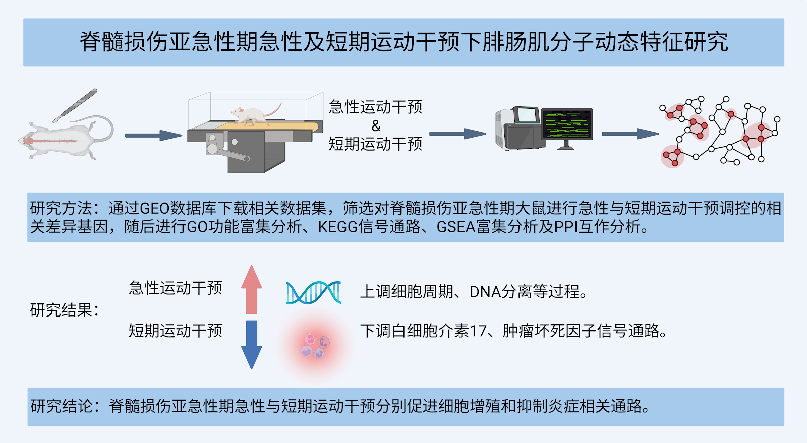
BACKGROUND: Spinal cord injury triggers a cascade of neuro-muscular system damage, involving central pattern generator dysfunction and peripheral muscle molecular network disruptions. Exercise can regulate key genes and promote the recovery of spinal cord injury. OBJECTIVE: To identify exercise-regulated key genes through bioinformatics analysis and explore the mechanisms by which exercise intervention facilitates the recovery of spinal cord injury. METHODS: The GSE45550 dataset based on the GPL1355 platform was obtained from the Gene Expression Omnibus (GEO) database. Differentially expressed genes regulated by acute and short-term exercise interventions during the subacute phase of spinal cord injury in rats were identified. Gene Ontology functional enrichment analysis, Kyoto Encyclopedia of Genes and Genomes pathway analysis, and Gene Set Enrichment Analysis were performed on these differentially expressed genes, and a protein-protein interaction network was constructed. RESULTS AND CONCLUSION: (1) After acute exercise intervention in the subacute phase of spinal cord injury, 106 genes were upregulated and 97 genes were downregulated in the gastrocnemius muscle, whereas short-term exercise intervention resulted in 138 upregulated and 105 downregulated genes. (2) Gene Ontology analysis showed that acute exercise mainly enriched genes related to chromosome segregation, while short-term exercise mainly promoted signal transduction processes. (3) KEGG analysis indicated that acute exercise was mainly associated with gastric acid secretion and upregulation of motor protein pathways, whereas short-term exercise mainly involved upregulation of neuroactive ligand-receptor interaction and downregulation of some inflammatory signaling pathways. (4) GSEA analysis revealed that acute exercise mainly upregulated cell cycle and DNA separation, while short-term exercise mainly downregulated interleukin-17 signaling and tumor necrosis factor signaling pathways. (5) Protein-protein interaction network showed that acute and short-term exercise interventions formed 2 and 3 central modules, respectively. In summary, acute exercise intervention significantly activates cell proliferation-related pathways (such as cell cycle and mitosis), upregulating pro-proliferative genes like Top2a and Sele; whereas short-term intervention exerts anti-inflammatory effects by downregulating inflammatory pathways such as interleukin-17 and tumor necrosis factor, and downregulating factors like COMP. These findings provide a reference for the molecular mechanism research of spinal cord injury rehabilitation.

Read Full Abstract10.12307/2026.21284
Correlation between cervical instability and neck muscle changes in middle-aged and young adultsGraphical AbstractVerified
Chinese Journal of Tissue Engineering Research2026

Correlation between cervical instability and neck muscle changes in middle-aged and young adults

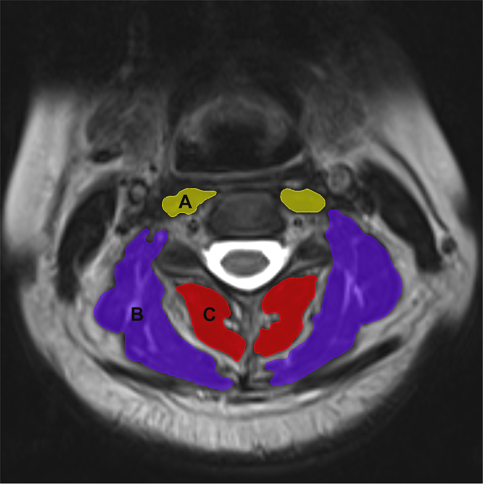
BACKGROUND: The onset of cervical instability in middle-aged and young adults often begins with neck muscle injuries. A deeper understanding of changes in neck muscles during cervical instability and their correlation can provide valuable data to support the prevention and treatment of cervical instability in this population. OBJECTIVE: To explore the correlation between cervical instability and neck muscle changes in middle-aged and young adults. METHODS: A total of 98 patients with cervical C4/5 instability and 88 healthy subjects, aged 18-45 years, were enrolled through recruitment advertisements and the Department of Spine, Wangjing Hospital, China Academy of Traditional Chinese Medicine. Cervical X-rays were collected to measure cervical curvature and C4/5 vertebral angular displacement. Cervical magnetic resonance imaging was taken to obtain data on C4/5 intervertebral disc signal intensity, as well as the relative cross-sectional area and fat ratio of neck muscles, including prevertebral muscles, deep posterior cervical muscles, and superficial muscles. A univariate intergroup comparison of X-ray and magnetic resonance imaging data was conducted between cervical instability subjects and healthy controls, along with Spearman correlation analysis between C4/5 angular displacement and disc signal intensity, relative cross-sectional area of neck muscles and fat percentage at the C4/5 level in cervical instability patients. RESULTS AND CONCLUSION: The cervical instability group had significantly greater age, C4/5 horizontal displacement, C4/5 angular displacement, and fat ratio of deep posterior cervical muscles than the healthy group (P < 0.05), while cervical curvature and relative cross-sectional area of deep posterior cervical muscles were significantly smaller (P < 0.05). Spearman correlation analysis showed a negative correlation between C4/5 angular displacement and relative cross-sectional area of deep posterior cervical muscles (P < 0.05). These findings suggest that changes in deep posterior cervical muscles may be closely related to the occurrence of cervical instability in middle-aged and young adults.

Read Full Abstract10.12307/2026.21287
Stroboscopic visual interference combined with balance training improves the balance ability of older adultsGraphical AbstractVerified
Chinese Journal of Tissue Engineering Research2026

Stroboscopic visual interference combined with balance training improves the balance ability of older adults

BACKGROUND: Numerous studies have indicated that stroboscopic visual interference combined with balance training can promote postural stability by reducing visual compensation in the central nervous system and increasing residual proprioceptive and vestibular input during training. OBJECTIVE: To clarify the effects of stroboscopic visual interference combined with balance training on improving balance ability of older adults by comparing the effects of balance training under different visual conditions. METHODS: Forty-three older adults were recruited and randomized into a normal balance training group (n=23) and a strobe vision training group (n=20). Among them, the balance training content was the same, and the training was conducted 3 times per week for 8 weeks. For the strobe vision training group, strobe glasses were worn during the balance training, and the strobe difficulty level (levels 1-8) was adjusted adaptively. Indicator tests were conducted at weeks 0, 4, 8, and 10 to assess the dynamic and static postural stability and the scores on the Berg Balance Scale of all subjects. RESULTS AND CONCLUSION: (1) Static postural stability: In the flat-surface eyes-closed single-leg stance test, both groups showed a significant time main effect (P < 0.001), but the group effect (P=0.530) and group×time interaction effect (P=0.780) were not significant. In the foam-surface eyes-closed single-leg stance test, both groups showed significant time (P < 0.001) and group (P=0.024) effects, but no group×time interaction effect (P=0.063). Compared with before training, both groups showed significant improvements in static postural stability after 8 weeks (P=0.034, P < 0.001) and 10 weeks (P=0.003, P < 0.001). (2) Dynamic postural stability: There was a significant group×time interaction effect (P < 0.001), while the main effects of group (Timed Up and Go and 3-m heel-to-toe walk: P=0.461, P=0.926) and time (P=0.120, P=0.937) were not statistically significant. Compared with before training, both groups showed significant improvements in dynamic postural stability after 4, 8, and 10 weeks of training (all P < 0.05). (3) Berg Balance Scale scores: There was a significant time effect (P < 0.001); group (P=0.420) and group×time (P=0.239) factors had no statistical significance on Berg Balance Scale scores. Compared with before training, both groups had higher Berg Balance Scale scores after 4 weeks (P=0.025), 8 weeks (P < 0.001), and 10 weeks (P=0.003). (4) It is suggested that both traditional balance training and strobe training can significantly improve the dynamic and static stability of older adults. Compared with traditional balance training, balance training based on stroboscopic visual interference can significantly improve the dynamic balance ability of older adults.

Read Full Abstract10.12307/2026.21292
Differences in angiogenesis and osteogenic effects between autogenous bone and mixed bone in guided bone regenerationGraphical AbstractVerified
Chinese Journal of Tissue Engineering Research2026

Differences in angiogenesis and osteogenic effects between autogenous bone and mixed bone in guided bone regeneration

BACKGROUND: Angiogenesis is a key factor in the success of guided bone regeneration, but the impact of different bone graft materials on angiogenesis and osteogenic effects remains unclear. OBJECTIVE: To compare the angiogenesis and osteogenic effects of autologous bone powder and the mixture of autologous bone powder and artificial bone powder during guided bone regeneration. METHODS: Preoperative scanning of the jawbone data of New Zealand white rabbits using cone beam CT was performed to extract a rabbit jawbone model, which was then 3D printed to create a bone cutting guide plate. Twenty-one New Zealand white rabbits were used to construct a rabbit mandibular bone defect model. Each rabbit had two bone defect areas on both sides, one side was the autologous bone powder + artificial bone powder group, implanted with a mixture of autologous bone powder and artificial bone powder at a volume ratio of 1:1; the other side was the autologous bone powder group, implanted with autologous bone powder only. Angiogenesis and osteogenesis in the bone defect area were detected at 2, 5, 7, 14 days and 4, 8, 12 weeks after modeling. RESULTS AND CONCLUSION: (1) At 12 weeks postoperatively, the tissue morphology and angiogenesis of the autologous bone powder group and the autologous bone powder + artificial bone powder group were similar; (2) The number of vascular cross-sections in the autologous bone powder group showed a more significant upward trend at 2-7 days, while the trends of the two groups were similar in other periods; (3) At 8 weeks postoperatively, the distribution range of new bone in the autologous bone powder + artificial bone powder group was not as large as that in the autologous bone powder group. At 12 weeks, the continuity and density of bone tissue in both groups were better, close to normal bone tissue morphology; (4) At 8 weeks, the bone volume fraction of the autologous bone powder group was significantly higher than that of the autologous bone powder + artificial bone powder group (P < 0.05). At 12 weeks, there was no significant difference in bone volume parameters between the two groups (P > 0.05); (5) The results indicate that early angiogenesis and bone regeneration were more obvious with autologous bone powder implantation, but by 12 weeks postoperatively, the effect of mixed implantation of autologous bone powder and artificial bone powder was similar to that of autologous bone powder alone in repairing rabbit mandibular defects, indicating that with the progress of bone remodeling, the comprehensive effects of the two materials tend to be consistent.

Read Full Abstract10.12307/2026.21285
Serum remnant cholesterol reduces bone quality in obese miceGraphical AbstractVerified
Chinese Journal of Tissue Engineering Research2026

Serum remnant cholesterol reduces bone quality in obese mice

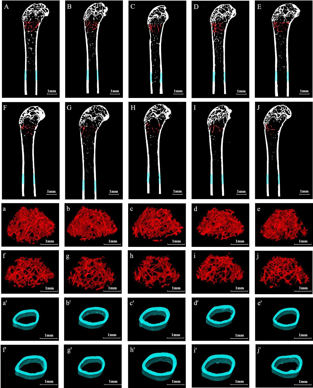
BACKGROUND: High cumulative remnant cholesterol levels are associated with the risk of various metabolic diseases, but their impact on bone quality remains to be explored. OBJECTIVE: To investigate the effects of high cumulative remnant cholesterol levels on bone mass, microstructure, and biomechanical properties in high-fat diet treated mice. METHODS: Ten healthy male SPF-grade C57BL6 mice were randomly allocated into normal control group and high-fat diet group. The normal control group was fed a normal diet for 20 weeks, while the high-fat diet group was fed a high-fat, high-cholesterol diet for 20 weeks. Mouse body mass was detected every week. After 20 weeks of feeding, serum levels of total cholesterol, remnant cholesterol, low-density lipoprotein cholesterol, high-density lipoprotein cholesterol, type I collagen carboxy-terminal cross-linked telopeptide, and type I procollagen amino-terminal propeptide were measured. Micro-CT was used to assess the microstructure of femoral cancellous and cortical bone. Three-point bending test was performed to measure the elastic modulus and maximum stress of the femur. RT-qPCR was used to detect the mRNA expression of RUNT-related transcription factor 2, type I collagen, osteocalcin, alkaline phosphatase, osteoprotegerin, receptor activator of nuclear factor-κB ligand, nuclear factor of activated T cells 1, and cathepsin K in the tibia. Pearson correlation analysis was used to analyze the correlation between remnant cholesterol, total cholesterol, high-density lipoprotein cholesterol, and low-density lipoprotein cholesterol levels and bone mineral density, as well as the correlation between remnant cholesterol levels and bone volume fraction, trabecular number, structure model index, and trabecular separation. RESULTS AND CONCLUSION: (1) From the 6th week of feeding, the body mass of mice in the high-fat diet group was higher than that in the control group (P < 0.05). The serum levels of total cholesterol, remnant cholesterol, and type I collagen carboxy-terminal cross-linked telopeptide in the high-fat diet group were higher than those in the control group (P < 0.05). (2) Micro-CT detection showed that compared with the control group, the bone microstructure of mice in the high-fat diet group underwent obvious degeneration, specifically manifested as significantly decreased bone mineral density, bone volume fraction, trabecular connectivity density, and trabecular number in cancellous bone, and significantly increased trabecular separation, structure model index, and trabecular pattern factor (P < 0.05). There were no significant changes in cortical bone thickness, volume, and area (P > 0.05). There was no significant difference in elastic modulus and maximum stress of the femur between the two groups (P > 0.05). (3) RT-qPCR detection showed that the mRNA expression of RUNT-related transcription factor 2, type I collagen, osteocalcin, alkaline phosphatase, and osteoprotegerin in the high-fat diet group was lower than that in the control group (P < 0.05), while the mRNA expression of nuclear factor of activated T cells 1 and cathepsin K was higher than that in the control group (P < 0.05). (4) Pearson analysis showed that remnant cholesterol and total cholesterol levels were significantly negatively correlated with bone mineral density (P < 0.05), and remnant cholesterol was significantly negatively correlated with bone volume fraction and trabecular number (P < 0.05), and significantly positively correlated with structure model index and trabecular separation (P < 0.05). These results indicate that remnant cholesterol may reduce bone quality by affecting the balance of bone turnover.

Read Full Abstract10.12307/2026.21283
Methylation alterations of Fbln1 gene in the hippocampus of PSEN1/PSEN2 double knockout and APP/PS1 transgenic miceGraphical AbstractVerified
Chinese Journal of Tissue Engineering Research2026

Methylation alterations of Fbln1 gene in the hippocampus of PSEN1/PSEN2 double knockout and APP/PS1 transgenic mice

BACKGROUND: Alzheimer's disease (AD) is a progressive neurodegenerative disorder characterized by abnormal deposition of β-amyloid (Aβ) and neurofibrillary tangles of tau protein. Current medications only alleviate some symptoms, and despite extensive efforts to develop new therapies, such as anti-Aβ immunotherapy and β-secretase inhibitors, clinical trials have not been successful. OBJECTIVE: To investigate the methylation changes of the Fbln1 gene in the hippocampus of PSEN1/PSEN2 double knockout (dKO) mice, which lack Aβ deposition, and APP/PS1 double transgenic (DTG) mice, which exhibit Aβ deposition, to explore non-Aβ-related mechanisms and potential targets in AD. METHODS: Hippocampal tissues were collected from female dKO mice at 7 months (early AD) and 12 months (mid-stage AD) of age, with age-matched wild-type (WT) mice as controls. Epigenetic reduced representation bisulfite sequencing (RRBS) was used to screen for aberrantly methylated genes, identifying Fbln1. Bisulfite sequencing PCR (BSP) was performed to validate the methylation status of Fbln1 in mid-stage dKO mice. RT-PCR and western blot were used to measure Fbln1 mRNA and protein expression in early and mid-stage dKO mice, as well as in 12-month-old DTG mice. Finally, the expression levels of Fbln1 and Aβ were compared between dKO and DTG mice, with age-matched WT mice as controls. RESULTS AND CONCLUSION: RRBS showed that Fbln1 was hypomethylated in the hippocampus of mid-stage dKO mice (P < 0.05), while early-stage dKO mice showed a trend of hypomethylation but without statistical significance (P > 0.05). BSP confirmed the abnormal hypomethylation of Fbln1 in mid-stage dKO mice. In early-stage dKO mice, Fbln1 mRNA and protein levels were not significantly different from WT (P > 0.05). In mid-stage dKO mice, Fbln1 mRNA and protein levels were significantly higher than in WT (t=5.336, P < 0.01; t=8.985, P < 0.01). Similarly, mid-stage DTG mice showed significantly higher Fbln1 mRNA and protein levels than WT (t=4.151, P < 0.01; t=8.392, P < 0.01), but there was no significant difference between the two AD models (P > 0.05). In mid-stage dKO mice, there was no significant difference between Fbln1 and Aβ protein levels (P > 0.05), whereas in DTG mice, the difference was significant (t=6.348, P < 0.01), indicating that Fbln1 plays a role in both Aβ-dependent and Aβ-independent mechanisms. These findings suggest that Fbln1 methylation changes may contribute to age-dependent neurodegeneration in dKO mice and may be involved in both Aβ and non-Aβ pathways in AD, providing new insights and potential targets for non-Aβ-related mechanisms. Fbln1, as an aging-related factor, holds promise as a novel target.

Read Full Abstract10.12307/2026.21290
Roles of pregnane X receptor in sodium arsenite-induced oxidative stress and inflammatory injury in human normal hepatocytesGraphical AbstractVerified
Chinese Journal of Tissue Engineering Research2026

Roles of pregnane X receptor in sodium arsenite-induced oxidative stress and inflammatory injury in human normal hepatocytes

BACKGROUND: As the primary organ for arsenic metabolism in the body, the liver has become a focal point for research on the mechanisms of arsenic toxicity. OBJECTIVE: To investigate the role of pregnane X receptor in sodium arsenite-induced oxidative stress and inflammatory injury in human normal hepatocytes. METHODS: Human normal hepatocyte MIHA cells were exposed to 0 (control), 10, 20, 30 μmol/L sodium arsenite for 48 hours. Changes in cell morphology were observed. Cell viability was measured via the cell counting kit-8 assay. Intracellular reactive oxygen species levels were detected using fluorescence probe staining combined with a microplate reader. Malondialdehyde levels were measured by thiobarbituric acid method. Glutathione reductase activity was detected by NADPH method. Total superoxide dismutase activity was measured by WST-8 method. Levels of interleukin-6, interleukin-1β, and tumor necrosis factor-α in cell supernatant were detected by ELISA. mRNA expression of pregnane X receptor and cytochrome P450 3A4 enzyme was detected by qRT-PCR. Protein expression of pregnane X receptor, cytochrome P450 3A4, nuclear factor-κB p65, nuclear factor-κB p-p65, proliferating cell nuclear antigen, interleukin-6, interleukin-1β, tumor necrosis factor-α, nuclear factor-κB inhibitor protein α, cyclooxygenase-2, p-nuclear factor-κB inhibitor protein α, nuclear factor erythroid 2-related factor 2, Keap1, and p-nuclear factor erythroid 2-related factor 2 was detected by western blot. RESULTS AND CONCLUSION: Compared with the control group, cells in all sodium arsenite groups showed unclear cell membrane boundaries, reduced cytoplasm, decreased cell fusion rate, and widened intercellular spaces. Compared with the control group, intracellular reactive oxygen species and malondialdehyde levels were increased (P < 0.05), levels of interleukin-6, interleukin-1β, and tumor necrosis factor-α in cell supernatant were increased (P < 0.05), protein expression of p-nuclear factor-κB inhibitor protein α, nuclear factor-κB p-p65, nuclear factor-κB p65, tumor necrosis factor-α, and interleukin-1β were increased (P < 0.05), cell viability was decreased (P < 0.05), protein expression of proliferating cell nuclear antigen, nuclear factor erythroid 2-related factor 2, p-nuclear factor erythroid 2-related factor 2 and total superoxide dismutase activity were decreased (P < 0.05), and mRNA and protein expression of pregnane X receptor and cytochrome P450 3A4 enzyme were decreased (P < 0.05). Compared with the control group, glutathione reductase activity was decreased in 20 and 30 μmol/L sodium arsenite groups (P < 0.05), and protein expression of Keap1, interleukin-6, and cyclooxygenase-2 was increased (P < 0.05). These results indicate that sodium arsenite may induce oxidative stress and inflammatory injury in hepatocytes by downregulating pregnane X receptor expression, inhibiting the nuclear factor erythroid 2-related factor 2 antioxidant pathway, and activating the nuclear factor-κB inflammatory pathway, while also inhibiting the expression of the drug-metabolizing enzyme cytochrome P450 3A4.

Read Full Abstract10.12307/2026.21293
Mechanical loading of the patellofemoral joint and its correlation with various body mass indices in older adults practicing Tai Chi exercisesGraphical AbstractVerified
Chinese Journal of Tissue Engineering Research2026

Mechanical loading of the patellofemoral joint and its correlation with various body mass indices in older adults practicing Tai Chi exercises

BACKGROUND: As the aging population grows in China, overweight and obesity are increasingly leading to lower limb joint injuries in older adults. OBJECTIVE: To compare the biomechanical data of the lower limb patellofemoral joint during Tai Chi movements and normal walking among older adults with different body mass indices, analyze the correlation between body mass index and biomechanical data of the lower limb patellofemoral joint, and investigate the impact of different body mass indices on the mechanical loading of the patellofemoral joint in older adults. METHODS: The older adults with obesity (n=17), overweight (n=17), and normal body mass index (n=19) who had been practicing Tai Chi for an extended period were recruited based on body mass index classification. The VCION 3D infrared motion capture system and Kistler force measurement system were used to collect kinematic and dynamic data during typical Tai Chi movements and normal walking. A biomechanical model of the patellofemoral joint was established to calculate biomechanical parameters of the patellofemoral joint. The effects of body mass index on the mechanical characteristics of the patellofemoral joint during walking and Tai Chi in the older adults were quantified using analysis of variance and correlation analysis. RESULTS AND CONCLUSION: (1) Compared with the normal group, patellofemoral joint force and contact area peak during normal walking were significantly lower in the overweight and obese groups (P < 0.05); during Tai Chi movements, knee flexion moment, patellofemoral joint force, and quadriceps muscle force were significantly lower (P < 0.05). (2) Body mass index was significantly negatively correlated with the maximum knee flexion angle, maximum and minimum patellofemoral joint contact area during the Tai Chi movement 'Reversing the Wind' (P < 0.05); and with the maximum knee extension moment, maximum and minimum patellofemoral joint reaction force, maximum quadriceps muscle force, maximum knee angle, and maximum and minimum patellofemoral joint contact area during the 'Brush Knee and Twist Step' movement (P < 0.05). These results indicate that as body mass index increases, the dynamic load on the patellofemoral joint during Tai Chi movements decreases; compared with normal-weight individuals, overweight and obese older adults show significantly lower maximum patellofemoral joint force during the 'Brush Knee and Twist Step' movement, indicating lower dynamic load. Therefore, Tai Chi is recommended as a fitness exercise for overweight and obese older adults to avoid sports injuries caused by high joint loads.

Read Full Abstract10.12307/2026.21282
Role and mechanism of ABL1 in myocardial necroptosis and cardiac ischemia/reperfusion injuryGraphical AbstractVerified
Chinese Journal of Tissue Engineering Research2026

Role and mechanism of ABL1 in myocardial necroptosis and cardiac ischemia/reperfusion injury

BACKGROUND: ABL1 is involved in the regulation of multiple cellular processes, yet its functions within the cardiovascular system remains largely unexplored. In particular, its role in cardiac ischemia/reperfusion injury and necroptosis has not been reported. OBJECTIVE: To investigate the role of ABL1 in cardiac ischemia/reperfusion injury and myocardial necroptosis, as well as the underlying molecular mechanisms. METHODS: (1) Animal experiment: C57BL/6J mice were randomly divided into four groups: sham surgery group, ischemia/reperfusion group, ABL1 knockdown + ischemia/reperfusion group, and ABL1 negative control + ischemia/reperfusion group. Lentiviral vectors targeting ABL1 were injected in situ into the myocardium. One week later, ischemia/reperfusion injury was induced by ligation of the left anterior descending coronary artery followed by reperfusion. ABL1 protein expression, cardiac function, myocardial fibrosis, and cardiomyocyte surface area were assessed. (2) Cell experiment: H9c2 cells were divided into four groups: negative control cell line + PBS, ABL1 knockdown cell line + PBS, negative control cell line + H2O2 500 µmol/L, and ABL1 knockdown cell line + H2O2 500 µmol/L. Additionally, H9c2 cells were divided into five groups: negative control cell line + PBS, negative control cell line + H2O2 500 µmol/L, ABL1 knockdown cell line + H2O2 500 µmol/L, ABL1 knockdown cell line + Parkin overexpression adenovirus + H2O2 500 µmol/L, and ABL1 knockdown cell line + Parkin negative control adenovirus + H2O2 500 µmol/L. Cell viability, necroptosis, reactive oxygen species levels, and mitochondrial membrane potential were measured. Expression of ABL1, Parkin, and cyclophilin D was detected, and the interaction between ABL1 and Parkin was examined. RESULTS AND CONCLUSION: (1) ABL1 protein expression was significantly downregulated in the mouse cardiac ischemia/reperfusion model. (2) Knockdown of ABL1 exacerbated ischemia/reperfusion-induced cardiac dysfunction, as evidenced by decreased left ventricular ejection fraction and fractional shortening, and increased left ventricular end-systolic and end-diastolic diameters. (3) Knockdown of ABL1 promoted ischemia/reperfusion-induced myocardial fibrosis and aggravated ventricular remodeling. (4) ABL1 protein expression was significantly downregulated in the cardiomyocyte oxidative stress model. (5) Knockdown of ABL1 exacerbated oxidative stress-induced cell viability loss, necroptosis, and reactive oxygen species accumulation. (6) ABL1 regulated mitochondrial membrane permeability, modulated the expression of Parkin and cyclophilin D, and regulated cellular oxidative stress levels by targeting Parkin. (7) These results indicate that ABL1 expression is significantly downregulated in both in vivo ischemia/reperfusion and in vitro oxidative stress models, and knockdown of ABL1 aggravates cardiac ischemia/reperfusion injury and cardiomyocyte oxidative stress injury, acting through the Parkin-CypD pathway.

Read Full Abstract10.12307/2026.21291
Role of chondrocyte ferroptosis in the pathogenesis of osteoarthritisGraphical AbstractVerified
Chinese Journal of Tissue Engineering Research2026

Role of chondrocyte ferroptosis in the pathogenesis of osteoarthritis

BACKGROUND: As a programmed cell death, ferroptosis relies on lipid peroxidation triggered by iron overload, leading to the death of chondrocytes and the exacerbation of joint degeneration. OBJECTIVE: To summarize the molecular mechanism of chondrocyte ferroptosis in the progression of osteoarthritis, and intervention strategies for this mechanism. METHODS: A literature search was conducted in CNKI, PubMed, and Web of Science databases using Chinese and English search terms including 'cartilage, ferroptosis, Nrf2, NF-E2-related factor 2, SIRT, Sirtuin, PINK1, PTEN induced putative kinase 1, stromal cell-derived factor 1, SDF1, nanoparticle, mitophagy, hydrogel' etc. According to inclusion criteria, 69 articles were finally included for review. RESULTS AND CONCLUSION: The mechanism of ferroptosis involves iron deposition, lipid peroxidation, and abnormal amino acid metabolism, among which reactive oxygen species accumulation and lipid peroxidation are necessary conditions for generating ferroptosis signals. In osteoarthritis, chondrocyte ferroptosis is one of the key pathological mechanisms leading to cartilage degeneration. Current research on the mechanism of chondrocyte ferroptosis and related materials mainly focuses on antioxidant pathways. Multiple studies have attempted to construct materials with antioxidant properties or regulate metal ion distribution to alleviate cartilage damage caused by osteoarthritis and improve disease progression. In addition, developing materials that interfere with ferroptosis through other pathways is also a focus of current research on osteoarthritis treatment. However, these research results have only produced effective therapeutic effects in cell and animal experiments, and clinical studies have not yet been conducted. Future research needs to further explore ferroptosis-related signaling networks and the interaction between materials and biological interfaces to develop more efficient and safe treatment strategies.

Read Full Abstract10.12307/2026.21295
Mechanical differences between medial collateral ligament and lateral collateral ligament and influence of elastin degradationGraphical AbstractVerified
Chinese Journal of Tissue Engineering Research2026

Mechanical differences between medial collateral ligament and lateral collateral ligament and influence of elastin degradation

BACKGROUND: As crucial stabilizers of the knee joint, the medial collateral ligament and lateral collateral ligament play essential roles in restricting valgus and varus movements, respectively. However, the mechanical differences between the medial collateral ligament and lateral collateral ligament, the microstructure characteristics, and the effect of elastin degradation on their mechanical properties remain poorly understood. OBJECTIVE: To compare the mechanical differences between the medial collateral ligament and lateral collateral ligament, quantify the structural characteristics of the collagen fiber alignment, and investigate the effect of elastin degradation on the mechanical properties of both ligaments. METHODS: Left medial collateral ligaments and lateral collateral ligaments were harvested from adult pigs, frozen, and thawed. Quasi-static uniaxial tensile tests were performed to measure the mechanical properties of the medial collateral ligament and lateral collateral ligament, and the effects of repeated stretching on their mechanical properties were compared. Second harmonic generation imaging using a two-photon microscope was used to quantify the collagen fiber structure of the medial collateral ligament and lateral collateral ligament. After repeated stretching, the medial collateral ligament and lateral collateral ligament were incubated in elastase solution for 12 hours, followed by uniaxial tensile tests to determine the effect of elastin treatment on ligament mechanical properties. RESULTS AND CONCLUSION: (1) Quasi-static uniaxial tensile tests showed that the high-tension elastic modulus of the medial collateral ligament was higher than that of the lateral collateral ligament (P < 0.05), while there was no significant difference in the low-tension elastic modulus between the two groups (P > 0.05). Repeated stretching significantly reduced the low-tension elastic modulus of both the medial collateral ligament and lateral collateral ligament. (2) Elastase treatment significantly reduced the low-tension and high-tension elastic moduli of both the medial collateral ligament and lateral collateral ligament, and the decrease in the high-tension elastic modulus of the lateral collateral ligament was greater than that of the medial collateral ligament. After elastase treatment, both the low-tension and high-tension elastic moduli of the medial collateral ligament were higher than those of the lateral collateral ligament (P < 0.05). (3) Two-photon imaging showed that the collagen fibers of the medial collateral ligament maintained a crimped structure, and its fiber waviness was significantly higher than that of the lateral collateral ligament. (4) These results indicate that the medial collateral ligament has stronger elastic properties than the lateral collateral ligament, and elastase treatment has a greater effect on the mechanical properties of the lateral collateral ligament. These mechanical results may be related to the more crimped collagen fiber arrangement in the medial collateral ligament.

Read Full Abstract10.12307/2026.21288
Constructing an in vitro model of ulcerative colitis in mice based on organoid technologyGraphical AbstractVerified
Chinese Journal of Tissue Engineering Research2026

Constructing an in vitro model of ulcerative colitis in mice based on organoid technology

BACKGROUND: The pathogenesis of ulcerative colitis is highly complex, necessitating the development of models that more closely mimic human physiological and pathological responses to study the mechanisms underlying its onset and progression. OBJECTIVE: To establish a mouse ulcerative colitis organoid model. METHODS: Colon organoids of C57BL/6J mice were extracted, cultured and passaged in vitro. Colon organoids from mice after three generations of passage were taken and incubated in lipopolysaccharide at varying concentrations [0 (control), 150, 175, 200, 225, 250, 275, 300, 325, and 350 μg/mL] to induce inflammation for 24 hours. The morphology of mouse colon organoids was observed under a microscope, and changes in proliferation viability were assessed using the cell counting kit-8 assay. After 24 hours of incubation with 0, 225, 250, 275 μg/mL lipopolysaccharide, the levels of tumor necrosis factor α, interleukin-6, interleukin-9, and interleukin-10 were measured by ELISA. After 24 hours of incubation with 0 and 275 μg/mL lipopolysaccharide, the expression of occludin and zonula occludens-1 was detected by immunofluorescence staining, and the mRNA expression of tumor necrosis factor α, interleukin-6, interleukin-9, occludin, and zonula occludens-1 was detected by q-PCR. RESULTS AND CONCLUSION: (1) Under the microscope, colon organoids in the 150-275 μg/mL lipopolysaccharide group showed varying degrees of swelling, while those in the 300-350 μg/mL lipopolysaccharide group had inhibited growth and swelling. CCK-8 assay showed that 150-350 μg/mL lipopolysaccharide reduced the proliferation viability of mouse colon organoids, with 225-350 μg/mL having a more pronounced effect. Based on cell morphology and proliferation viability results, 225, 250, and 275 μg/mL lipopolysaccharide were selected for ELISA. (2) Compared with the control group, the levels of interleukin-6 and tumor necrosis factor α were increased in the 225, 250, and 275 μg/mL lipopolysaccharide groups (P < 0.05), and the level of interleukin-9 was increased in the 275 μg/mL lipopolysaccharide group (P < 0.05). (3) Immunofluorescence staining showed that compared with the control group, the expression of occludin and zonula occludens-1 was decreased in the 275 μg/mL lipopolysaccharide group. q-PCR detection showed that compared with the control group, the mRNA expression of interleukin-6 and tumor necrosis factor α was increased (P < 0.05), occludin mRNA expression was decreased (P < 0.05), and there was no significant difference in the expression of interleukin-9 and zonula occludens-1 (P > 0.05). (4) These results indicate that an in vitro mouse ulcerative colitis model based on organoids was successfully constructed, providing a powerful tool for studying the mechanisms of ulcerative colitis and screening effective drugs.

Read Full Abstract10.12307/2026.21289
Mechanism of cuproptosis in the diagnosis and treatment of orthopedic-related diseasesGraphical AbstractVerified
Chinese Journal of Tissue Engineering Research2026

Mechanism of cuproptosis in the diagnosis and treatment of orthopedic-related diseases

BACKGROUND: Studies have shown that cuproptosis plays a critical role in the pathogenesis and treatment of orthopedic diseases. However, the regulatory roles and mechanisms of cuproptosis in orthopedic-related diseases remain unclear. OBJECTIVE: To review the roles and mechanisms of cuproptosis in orthopedic-related diseases. METHODS: A literature search was conducted in the PubMed database using the following English keywords: "cuproptosis," "copper steady state," "osteoarthritis," "osteoporosis," "rheumatoid arthritis," "osteosarcoma," and "oxidative stress." The search included publications up to March 2025. According to the inclusion criteria, 55 articles were finally included for review. RESULTS AND CONCLUSION: In osteoarthritis, excessive copper ions induce the expression of metal-regulatory transcription factor 1, indirectly activating matrix metalloproteinases and leading to cartilage degradation. Cuproptosis disrupts the tricarboxylic acid cycle and inhibits glutamine metabolism, triggering oxidative stress and accelerating chondrocyte death. In osteoporosis, cuproptosis suppresses glutamine metabolism and mineralization in osteoblasts while promoting osteoclast differentiation, thereby disrupting the balance between bone formation and resorption. In rheumatoid arthritis, copper ions activate the phosphatidylinositol 3-kinase/protein kinase B/mitogen-activated protein kinase signaling pathway, promoting synovial cell abnormal activation and inflammatory factor release, exacerbating joint destruction. In osteosarcoma, high concentrations of copper ions selectively kill tumor cells by targeting ferredoxin 1, inducing mitochondrial dysfunction and proteotoxic stress. The mechanisms of cuproptosis in orthopedic diseases are complex and diverse, involving multiple cell types and signaling pathways. Targeting cuproptosis or its related pathways may provide new strategies for the treatment of orthopedic diseases, such as the application of copper chelators or copper ion carriers, which hold significant clinical potential. Future research should further explore the specific regulatory mechanisms of cuproptosis and its therapeutic value in disease treatment.

Read Full Abstract10.12307/2026.21294
X-ray imaging and finite element analysis of the L6-S1 intervertebral disc in rats under abnormal forward-flexed postureGraphical AbstractVerified
Chinese Journal of Tissue Engineering Research2026

X-ray imaging and finite element analysis of the L6-S1 intervertebral disc in rats under abnormal forward-flexed posture

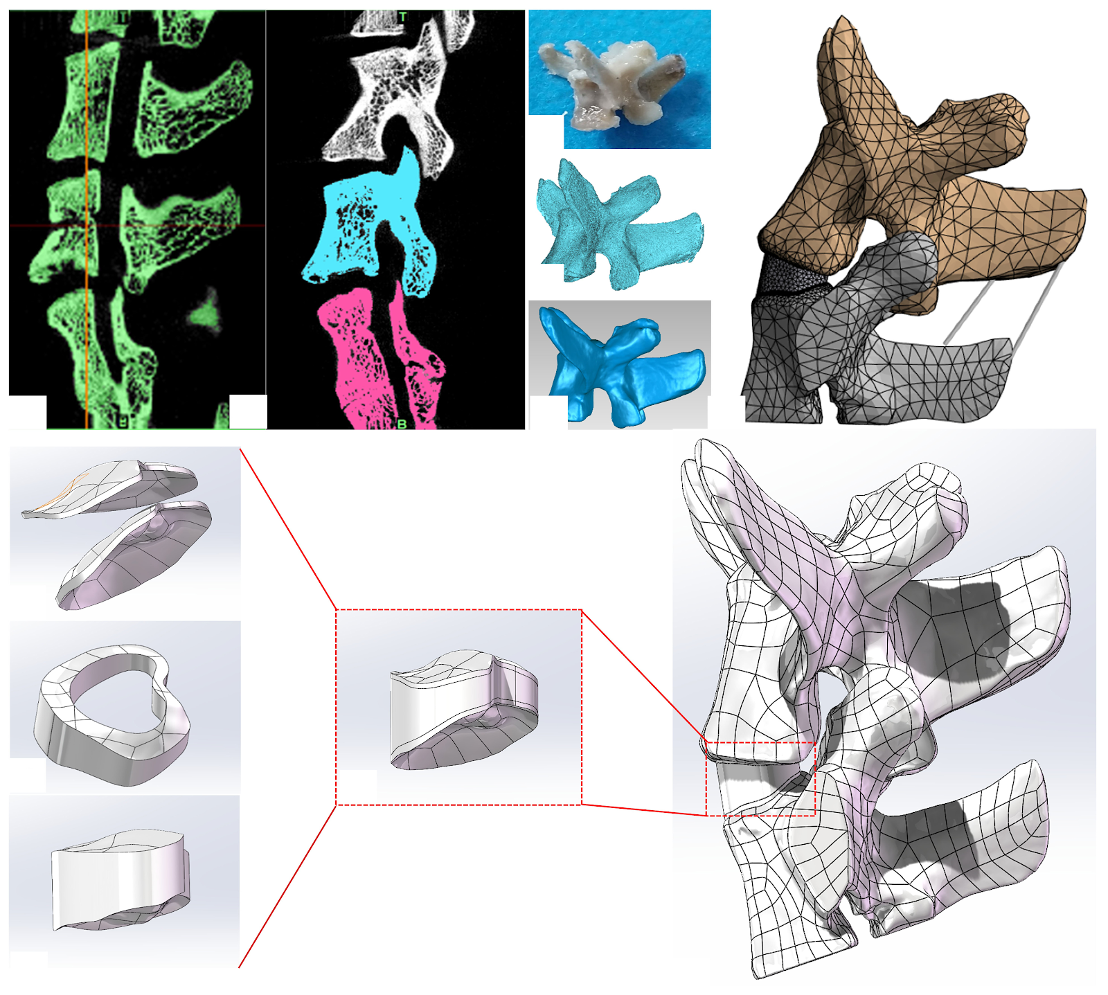
BACKGROUND: Our group developed a rat lumbar spine model inducing L6-S1 segmental degeneration by prolonged fixation in an abnormal forward-bending posture through a specific device. However, biomechanical evaluation of this model remains lacking. OBJECTIVE: To evaluate the biomechanical properties of L6-S1 motion segment in rats with abnormal forward bending posture through X-ray verification and finite element analysis. METHODS: This study utilized a previously established SD rat model of abnormal forward-flexed posture. Lateral X-ray images of three healthy female SD rats were taken in both restrained (unanesthetized) and relaxed (anesthetized) states to measure the L6-S1 disc angle and analyze its changes under different postures. Micro-CT data from one healthy female SD rat were used to reconstruct a 3D L6-S1 model with Mimics, Geomagic Wrap, and SolidWorks. The model was then meshed, assigned material properties, and subjected to forward flexion loading simulation in ANSYS Workbench to calculate stress distribution in L6-S1 disc structures. RESULTS AND CONCLUSION: (1) The mean L6-S1 intervertebral disc angle was (12.16±0.57)° in relaxed posture and (1.26±0.26)° in restrained posture. (2) Under 10° forward flexion, the maximum von Mises stresses in the upper endplate, lower endplate, annulus fibrosus, and nucleus pulposus were 10.398, 19.928, 6.819, and 0.104 MPa, respectively, with endplates showing significantly higher stresses. (3) The forward-flexed posture reduced the L6-S1 disc angle, altering disc morphology and load distribution. The finite element model simulated the biomechanical environment under abnormal posture, indicating that endplates may be the earliest structures to undergo degenerative changes.

Read Full Abstract10.12307/2026.21415
Bushen Tongshi Pills improves osteogenic disorders in alcoholic femoral head necrosis ratsGraphical AbstractVerified
Chinese Journal of Tissue Engineering Research2026

Bushen Tongshi Pills improves osteogenic disorders in alcoholic femoral head necrosis rats

BACKGROUND: Bushen Tongshi Pills has been proven to delay the progression of collapse in alcoholic femoral head necrosis, but the mechanism is still unclear. OBJECTIVE: To explore the mechanism by which Bushen Tongshi Pills improve osteogenic disorders of alcoholic femoral head necrosis. METHODS: Fifty male Sprague-Dawley rats were randomly divided into a control group, a model group, and low, medium, and high dose groups of Bushen Tongshi Pills, with 10 rats in each group. Except for the control group, other groups were fed with ethanol-containing Lieber-DeCarli liquid diet for 8 weeks to construct alcoholic femoral head necrosis models, while the low, medium, and high dose groups were given 1.05, 2.1, 4.2 g/kg Bushen Tongshi Pills by gavage daily. After 8 weeks, Micro-CT was used to observe the gross morphology of the femoral head, hematoxylin-eosin staining was used to observe the pathological morphology of the femoral head, ELISA was used to detect serum levels of interleukin-1β and interleukin-18, immunohistochemical staining and western blot were used to detect the expression of NOD-like receptor protein 3, Caspase-1, gasdermin D, Runt-related transcription factor 2, osteocalcin, and type I collagen in rat femoral head tissues, and RT-qPCR was used to detect the mRNA expression of these genes. RESULTS AND CONCLUSION: (1) Micro-CT and hematoxylin-eosin staining results showed that the medium and high dose groups of Bushen Tongshi Pills significantly improved bone loss and pathological morphological development in the femoral head of alcoholic femoral head necrosis model rats. (2) ELISA results showed that compared with the control group, serum levels of interleukin-1β and interleukin-18 in the model group were significantly increased (P < 0.05); compared with the model group, serum levels of interleukin-1β and interleukin-18 in all Bushen Tongshi Pills dose groups were significantly decreased (P < 0.05), in a dose-dependent manner, with the most significant decrease in the high dose group (P < 0.05). (3) Immunohistochemistry, western blot, and RT-qPCR results showed that compared with the control group, the protein and mRNA expression of NOD-like receptor protein 3, Caspase-1, and gasdermin D in the femoral head of model rats were significantly upregulated (P < 0.05), while the protein and mRNA expression of osteogenic factors Runt-related transcription factor 2, osteocalcin, and type I collagen were significantly downregulated (P < 0.05); compared with the model group, the protein and mRNA expression of NOD-like receptor protein 3, Caspase-1, and gasdermin D in the femoral head of rats in all Bushen Tongshi Pills dose groups were downregulated in a dose-dependent manner, while the protein and mRNA expression of Runt-related transcription factor 2, osteocalcin, and type I collagen were upregulated in a dose-dependent manner, with the most significant upregulation in the high dose group (P < 0.05). (4) Western blot results showed that compared with the control group, the expression of cleaved-Caspase-1, gasdermin D-N protein, cleaved-Caspase-1/Caspase-1 ratio, and gasdermin D-N/gasdermin D ratio in the femoral head of model rats were significantly upregulated (P < 0.05); compared with the model group, the expression of cleaved-Caspase-1, gasdermin D-N protein, and gasdermin D-N/gasdermin D ratio in all Bushen Tongshi Pills dose groups were significantly downregulated (P < 0.05), and the cleaved-Caspase-1/Caspase-1 ratio in the medium and high dose groups was significantly downregulated (P < 0.05). (5) These results indicate that Bushen Tongshi Pills may inhibit the release of inflammatory factors in alcoholic femoral head necrosis model rats through the NOD-like receptor protein 3/Caspase-1/gasdermin D pyroptosis pathway, promote osteogenic differentiation, and repair necrotic bone tissue.

Read Full Abstract10.12307/2026.21280
Morphological measurement of anterior cervical pedicle screw placement assisted by Mimics three-dimensional CT reconstructionGraphical AbstractVerified
Chinese Journal of Tissue Engineering Research2026

Morphological measurement of anterior cervical pedicle screw placement assisted by Mimics three-dimensional CT reconstruction

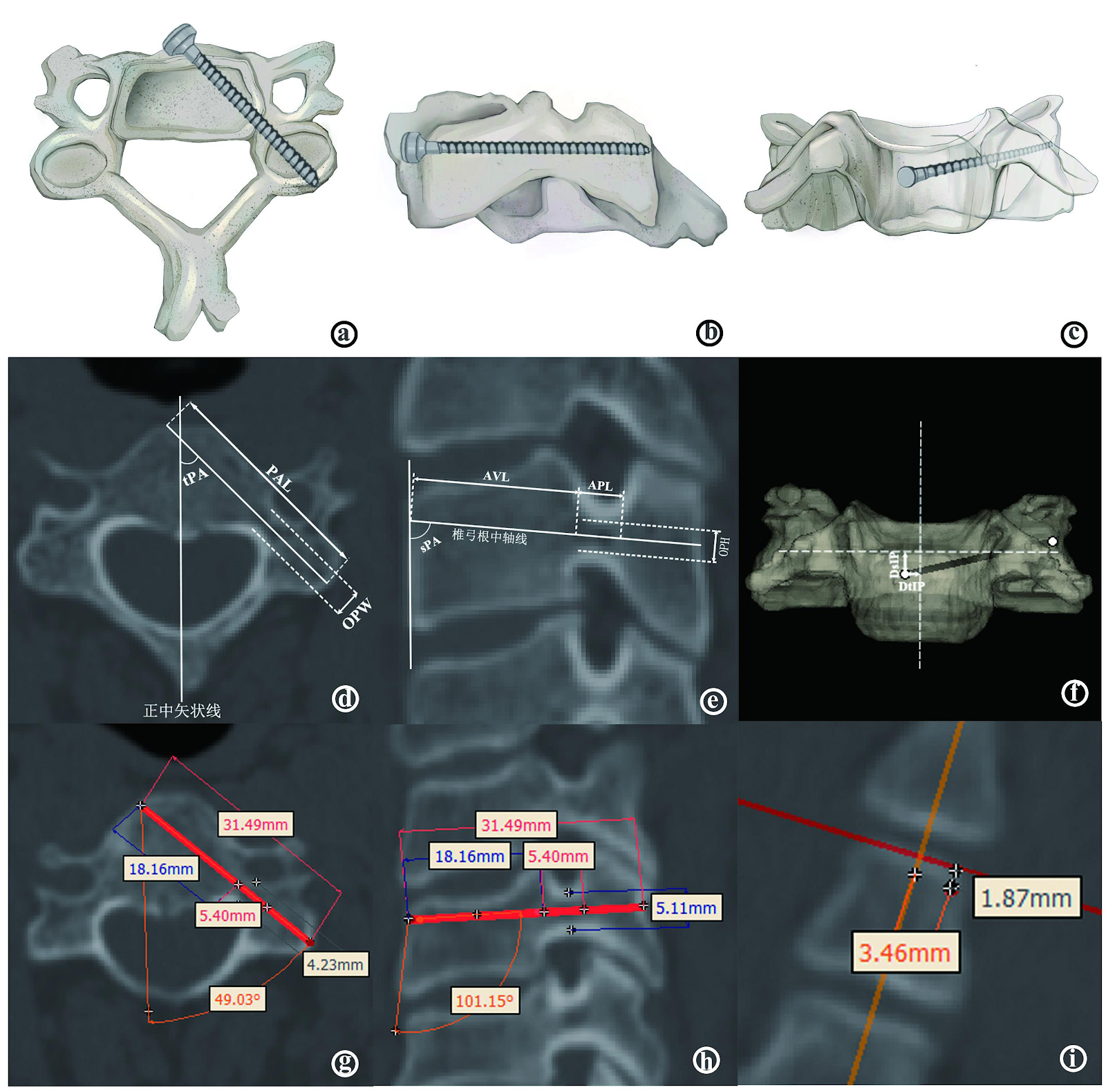
BACKGROUND: The anterior transpedicular screw fixation system can achieve adequate decompression and strong fixation in a single anterior surgery. However, due to its unique anatomical structure, the risk of screw placement is relatively high. Previous domestic and international scholars have confirmed the feasibility of this approach through anatomical measurements, but there are some drawbacks such as relatively small sample sizes, difficulties in locating the pedicle axis, and limitations in measurement methods. OBJECTIVE: To perform morphological measurements of the adult cervical spine based on imaging to provide anatomical guidance for anterior transpedicular pedicle screw fixation. METHODS: 3D CT scan data of 50 adult cervical vertebrae were imported into the Mimics system for 3D reconstruction. Morphological data were measured, including pedicle axis distance, pedicle width, pedicle height, pedicle horizontal axial angle, pedicle sagittal angle, distance from the entry point in the transverse plane, distance to the entry point in the sagittal plane, axial vertebral length and axial pedicle length. RESULTS AND CONCLUSION: (1) Positioning of nail entry points: C3 and C4 were located on the opposite side of the median sagittal plane of the vertebral body, with distances from the median sagittal line of approximately 2.060 mm and 2.310 mm. C5 could be located on the same side of the median sagittal line as or opposite to the median sagittal line, with an average value of approximately 1.224 mm. C6-C7 were located on the same side of the body, with distances of 1.132 mm and 2.538 mm from the midline; the distance from the upper endplate increased gradually from C3 to C7, with average values ranging from 2.362 to 7.350 mm. (2) Direction of nail entry: the transverse angle increased gradually from C3 to C4 (46.32°-47.36°) and decreased from C5 to C7 (44.03° to 37.80°); the sagittal angle required caudal deviation for C3-C4 (95.75° and 100.93°) and cephalad deviation for C5-C7 (104.38°, 110.34°, and 104.86°). (3) There were no significant differences in entry point location and direction between genders or sides (P > 0.05). For screw selection, except for individual patients with cervical developmental abnormalities, for most subaxial cervical pedicle screws, it is safe and reliable to choose screws at least 30 mm long and 4.0 mm in diameter for males, and at least 28 mm long and 3.5 mm in diameter for females. (4) Morphological measurements confirmed that anterior transpedicular screw fixation of the subaxial cervical spine is feasible, but individualized principles should be followed to formulate personalized fixation plans.

Read Full Abstract10.12307/2026.21417
Perioperative hidden blood loss and risk factors in transforaminal lumbar interbody fusion calculated by a new methodGraphical AbstractVerified
Chinese Journal of Tissue Engineering Research2026

Perioperative hidden blood loss and risk factors in transforaminal lumbar interbody fusion calculated by a new method

BACKGROUND: Transforaminal lumbar interbody fusion is one of the main surgical methods for treating degenerative lumbar diseases such as lumbar disc herniation, lumbar spinal stenosis, and lumbar spondylolisthesis. Hidden blood loss refers to the concealed loss of blood volume in patients, which is often overlooked by people. OBJECTIVE: To evaluate the perioperative blood loss during transforaminal lumbar interbody fusion using a new method, calculate the hidden blood loss based on the new method, and analyze its risk factors. METHODS: The medical records of 93 patients with lumbar degenerative diseases (lumbar spinal stenosis, lumbar disc herniation, and lumbar spondylolisthesis) who were hospitalized in the Department of Spine Surgery of Second Affiliated Hospital of Soochow University from October 2023 to October 2024 were retrospectively analyzed. The general data of patients were collected, such as age, gender, height, body mass, body mass index, and whether they had hypertension and diabetes; surgical data, such as the number of surgical segments, operation time, and American Society of Anesthesiologists anesthesia grade; laboratory tests, such as prothrombin time, activated partial thromboplastin time, international normalized ratio, platelet count, fibrinogen, and D-dimer level. Pearson or Spearman correlation analysis was used to explore the correlation between patient characteristics and postoperative hidden blood loss, and multivariate linear regression analysis was utilized to determine the independent risk factors for postoperative hidden blood loss. RESULTS AND CONCLUSION: (1) The average hidden blood loss calculated by the new method for transforaminal lumbar interbody fusion was (284.24±352.76) mL, accounting for 58.6% of total blood loss; while the traditional method calculated an average hidden blood loss of (165.77±339.89) mL, accounting for 34.15% of total blood loss, with a significant difference between the two (P < 0.05). (2) In univariate analysis, hidden blood loss was positively correlated with the number of segments (r=0.213, P=0.040) and operation time (r=0.210, P=0.043), and negatively correlated with platelet count (r=-0.324, P=0.018). (3) In multivariate linear regression analysis, decreased platelet count was an independent risk factor for hidden blood loss (P=0.016). (4) These findings suggest that the new method provides a more accurate estimation of hidden blood loss, and hidden blood loss is an important component of perioperative total blood loss; increased number of segments, prolonged operation time, and decreased platelet count are risk factors for hidden blood loss in transforaminal lumbar interbody fusion, with decreased platelet count being an independent risk factor.

Read Full Abstract10.12307/2026.21414
Meta-analysis of application effect of 3D-printed artificial vertebral bodies in anterior cervical corpectomy and fusionGraphical AbstractVerified
Chinese Journal of Tissue Engineering Research2026

Meta-analysis of application effect of 3D-printed artificial vertebral bodies in anterior cervical corpectomy and fusion

OBJECTIVE: In recent years, many scholars have applied 3D-printed artificial vertebrae to anterior cervical vertebral subtotal vertebral resection and bone grafting fusion, but whether it is more effective than traditional titanium cages remains controversial. This study aims to systematically evaluate the effectiveness and safety of 3D-printed artificial vertebrae compared with traditional titanium cages as implants for anterior cervical corpectomy and fusion in the treatment of spondylosis. METHODS: Databases such as CNKI, WangFang, CBM, VIP, PubMed, EMBASE, and The Cochrane Library were searched to collect the clinical research on the application of 3D-printed artificial vertebrae in anterior cervical corpectomy and fusion from the establishment of each database to February 2025. After screening the literature, extracting the data and evaluating the methodological quality of the included studies, the meta-analysis was performed using Rev Man 5.4 software. RESULTS: A total of 10 studies were included, comprising 2 prospective randomized controlled studies, 6 retrospective cohort studies, and 2 prospective cohort studies, all of high quality. The included studies involved 534 patients, with 273 in the 3D-printed group and 261 in the control group. Meta-analysis results showed that the 3D-printed group was superior to the control group in terms of operation time [SMD=-1.13, 95%CI(-1.87, -0.39), P=0.003], loss of intervertebral disc height at last follow-up [SMD=-3.01, 95%CI(-5.74, -0.29), P=0.03], neck disability index at 3 months postoperatively [SMD=-0.34, 95%CI(-0.66, -0.03), P=0.03], prosthesis subsidence rate [OR=0.19, 95%CI(0.11, 0.32), P < 0.000 01], and postoperative dysphagia incidence [OR=0.43, 95%CI(0.21, 0.90), P=0.03], with significant differences. There were no significant differences in blood loss, hospital stay, postoperative Japanese Orthopaedic Association score, postoperative visual analogue scale score, postoperative neck disability index (at 6 months and last follow-up), and fusion rate between the two groups (P > 0.05). CONCLUSION: Compared with traditional titanium cages, 3D-printed artificial vertebral bodies have significant advantages in improving surgical efficiency, maintaining postoperative intervertebral disc height, reducing postoperative dysphagia incidence, and reducing prosthesis subsidence rate.

Read Full Abstract10.12307/2026.21428
Characteristics of lower limb muscle motor activation in patients with unilateral knee osteoarthritisGraphical AbstractVerified
Chinese Journal of Tissue Engineering Research2026

Characteristics of lower limb muscle motor activation in patients with unilateral knee osteoarthritis

BACKGROUND: There are mechanical changes in the lower limb muscles in patients with knee osteoarthritis. These changes can be objectively reflected through surface electromyography. Currently, there is limited research on abnormal electrophysiological signals of lower limb muscles, especially of calf muscles, during walking in patients with unilateral knee osteoarthritis. OBJECTIVE: To investigate the motor activation status and activation mode of the medial thigh muscle, lateral thigh muscle, tibialis anterior muscle, and gastrocnemius muscle during daily walking in patients with unilateral knee osteoarthritis using surface electromyography. METHODS: Fifty subjects with unilateral knee osteoarthritis who met the inclusion criteria underwent surface electromyography walking tests. The target muscles were the medial thigh muscle, lateral thigh muscle, anterior tibialis muscle, and gastrocnemius muscle. The walking time was 60 seconds. Surface electromyography signals were collected from both healthy and affected sides. The main observation indicators were root mean square value, integrated electromyography value, median frequency, and muscle contraction ratio. Whether there is an abnormal activation status and its characteristics of lower limb muscles in unilateral knee osteoarthritis patients were investigated by surface electromyography. The abnormal movement activation of lower limb muscles in unilateral knee osteoarthritis patients was explored from an electrophysiological perspective. RESULTS AND CONCLUSION: (1) The integrated electromyography value and root mean square value of the medial and lateral thigh muscles on the affected side were significantly lower than those on the healthy side (P < 0.001); the median frequency values of the medial and lateral thigh muscles on the affected side were significantly lower than those on the healthy side (P < 0.001), with a more significant shift to low frequency, indicating a higher fatigue risk. (2) The integrated electromyography value and root mean square value of the tibialis anterior muscle on the affected side were significantly higher than those on the healthy side (P < 0.001); the median frequency value of the tibialis anterior muscle on the affected side was significantly lower than that on the healthy side (P < 0.001), with a more significant shift to low frequency, indicating a significantly increased fatigue risk. (3) The integrated electromyography value and root mean square value of the gastrocnemius muscle on the affected side were significantly higher than those on the healthy side (P < 0.001); the median frequency value of the gastrocnemius muscle on the affected side did not show a significant excessive shift compared with the healthy side (P > 0.05), indicating similar fatigue levels. (4) The muscle activation ratio on the affected side was significantly lower than that on the healthy side (P < 0.001), and the activation of the medial and lateral thigh muscles on the affected side was significantly unbalanced compared with the healthy side. These results indicate that during daily walking, patients with unilateral knee osteoarthritis exhibit insufficient activation and reduced activation efficiency of the medial and lateral thigh muscles, with high fatigue levels and risk, and unbalanced activation of these muscles, which is detrimental to knee joint stability and balance. The tibialis anterior and gastrocnemius muscles on the affected side show compensatory activation and abnormal co-activation levels, which are important factors aggravating joint stiffness, with the most obvious compensation in the tibialis anterior muscle, and there is a risk of fatigue-induced muscle atrophy.

Read Full Abstract10.12307/2026.21281
AI Algorithm Analysis for CT Three-Dimensional Diagnosis and Accurate Assessment of Posterior Cruciate Ligament Tibial Avulsion FracturesGraphical AbstractVerified
Chinese Journal of Tissue Engineering Research2026

AI Algorithm Analysis for CT Three-Dimensional Diagnosis and Accurate Assessment of Posterior Cruciate Ligament Tibial Avulsion Fractures

BACKGROUND: Surgical decision-making for posterior cruciate ligament avulsion fractures is highly dependent on imaging evaluation. Traditional methods rely on subjective interpretation of CT images, which suffer from limitations such as difficulties in quantifying three-dimensional spatial displacement parameters and insufficient precision in assessing rotational angles. Given the advancements in artificial intelligence (AI) technology, there is a need to develop automated, intelligent image recognition software based on AI algorithms. OBJECTIVE: To investigate the intelligent diagnostic capabilities of AI algorithms for posterior cruciate ligament tibial avulsion fractures in 3D CT images and their effectiveness in accurately assessing 3D parameters of fracture fragments. METHODS: Knee CT data from 24 patients with posterior cruciate ligament tibial avulsion fractures who were treated at the Wangjing Hospital of the China Academy of Chinese Medical Sciences between December 1, 2022, and August 30, 2024, were retrospectively collected. Three-dimensional reconstruction, intelligent fracture point recognition, and simulated reduction were performed using self-developed AI image recognition software. Translational and rotational parameters of the fracture fragments along the X, Y, and Z axes were obtained. These measurements were compared with those from traditional radiology reading software (PACS system) using rank-sum tests, Bland-Altman analysis, and linear regression models to assess consistency, and coefficients of variation were calculated to verify software stability. RESULTS AND CONCLUSION: ①There were no significant differences between AI software and traditional methods in measuring fracture fragment displacement (X/Y/Z axis translation and rotation) (P > 0.05). ②Bland-Altman analysis showed good consistency between the two methods, with no significant differences (P > 0.05). ③Linear regression models for X, Y, Z axis displacement and angles showed R² values > 0.99. ④The coefficients of variation for three repeated fracture point identifications by the AI software showed that for total fracture identification, 21 cases had coefficients of variation < 20%, and for articular surface fracture points, 18 cases had coefficients of variation < 20%. ⑤These findings indicate that the AI image recognition software can accurately quantify three-dimensional parameters of posterior cruciate ligament avulsion fracture fragments, with measurement results consistent with traditional methods and good stability. It can assist doctors in judging the degree of displacement and provide precise data support for preoperative planning. The software has good application prospects in avulsion fractures, and future studies should expand the sample size and further verify its impact on surgical outcomes.

Read Full Abstract10.12307/2026.21420