Key Takeaways & Executive Findings
- •• iPSC-derived exosomes significantly accelerate diabetic wound healing in two clinically relevant animal models. • The therapeutic mechanism involves direct activation of tissue regeneration, including re-epithelialization and remodeling. • iPSC-Exos modulate the inflammatory microenvironment by promoting macrophage polarization toward the anti-inflammatory M2 phenotype. • The dual-animal model approach enhances clinical translatability of findings for diabetic wound therapy.
Abstract
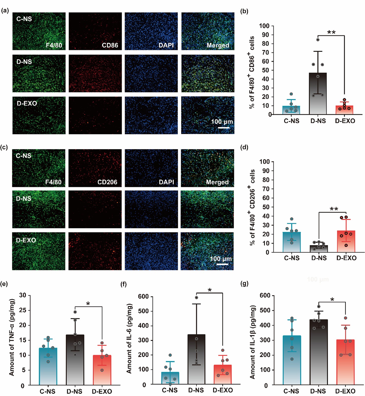
Background: Owing to impaired glucose metabolism, the high-glucose microenvironment in diabetic patients disrupts a series of biological reactions that hinder the wound healing process, resulting in a significant cost to the health care system and an urgent need for new and advanced therapies. Methods: In this study, induced pluripotent stem cell-derived exosomes (iPSC-Exos) were isolated from iPSC culture supernatant via centrifugation and ultrafiltration. We evaluated the therapeutic effects of iPSC-Exos on diabetic wound healing through two clinically relevant animal models (spontaneous genetic diabetic mouse model and streptozotocin (STZ)-induced diabetic mouse model). iPSC-Exos were topically administered to full-thickness cutaneous wounds in diabetic mice. The therapeutic effects were systematically assessed by measuring wound closure rates, conducting comprehensive histopathological evaluations, and performing quantitative analysis of inflammatory mediators via ELISA. Results: We demonstrated that iPSC-Exos can significantly accelerate diabetic wound healing through two clinically relevant animal models (spontaneous genetic diabetic mouse model and STZ-induced diabetic mouse model) for the first time. The multifaceted therapeutic mechanisms include: (i) Direct activation of tissue regeneration (promotion of re-epithelialization, tissue remodeling and scar attenuation); (ii) Modulation of the inflammatory microenvironment (promoting macrophage polarization toward anti-inflammatory M2 phenotype/suppressing inflammation). Conclusions: This dual-animal model approach, which closely recapitulates key pathophysiological features of human diabetic wounds, offers superior clinical translatability compared to single-animal model studies. Our findings iPSC-derived exosomes promote diabetic wound healing by attenuating inflammatory responses.
1. Introduction
Diabetes mellitus is a metabolic disease characterized by a relative or absolute lack of insulin secretion leading to chronically increased blood glucose levels [1–3]. The core mechanism of diabetic wound healing impairment is governed by a vicious cycle, driven by hyperglycemia and involving multiple interconnected components. This cycle, initiated by systemic pathophysiological disturbances, propagates through the disruption of the local microenvironment, and ultimately leads to a halt in cellular repair processes. Chronic hyperglycemia, as the initial driving factor, exerts its effects through two primary pathways: On one hand, it activates the polyol, hexosamine, and protein kinase C (PKC) pathways, triggering oxidative stress and accumulating advanced glycation end products (AGEs). This cascade further damages nerves and blood vessels: neuropathy impairs wound sensory perception and disrupts the skin barrier, increasing the risk of occult injuries [4–6]; meanwhile, angiopathy leads to insufficient blood perfusion, laying the groundwork for subsequent hypoxia and ischemia [7, 8]. On the other hand, hyperglycemia directly impairs the chemotactic and phagocytic capabilities of immune cells, reducing the efficiency of bacterial clearance and creating favorable conditions for infection.
Against this backdrop, the wound microenvironment deteriorates further: hyperglycemia upregulates the expression of pro-inflammatory factors through epigenetic modifications, promoting the sustained polarization of M1-type macrophages. Concurrently, it delays the clearance of neutrophils, forming an “inflammation-oxidative stress” cycle that induces senescence in repair cells such as fibroblasts and endothelial cells [9]. Angiopathy, combined with the high oxygen consumption of inflammatory cells, exacerbates hypoxia; however, hyperglycemia downregulates the hypoxia-inducible factor-1 (HIF-1)/vascular endothelial growth factor (VEGF) axis, inhibiting angiogenesis. Ischemia and hypoxia further worsen energy supply, hindering the proliferative phase of wound healing [7, 8, 10]. Additionally, the hyperglycemic environment facilitates bacterial colonization and biofilm formation, which stimulate the secretion of pro-inflammatory factors and exogenous proteases; this, together with the imbalance between upregulated expression of endogenous matrix metalloproteinases (MMPs) and downregulated expression of tissue inhibitors of metalloproteinases (TIMPs), collectively degrades the extracellular matrix (ECM) and growth factors, leading to tissue necrosis [11]. Additionally, chronic inflammation and infection also render the wound microenvironment alkaline (elevated pH), which further enhances MMP activity and inhibits fibroblast function [12–14], forming a self-perpetuating “microenvironment deterioration loop” [7, 8]. Eventually, the wound remains stagnant in the inflammatory phase for an extended period, unable to progress through the normal proliferative and remodeling phases of healing, thus developing into a chronic non-healing wound [15]. Traditional wound treatments, such as local dressings, blood glucose control, skin grafting, and laser therapy, have shown limited therapeutic efficacy and may carry various risks, including poor adherence to blood glucose control, donor-site damage and hyperpigmentation issues [16, 17]. Given the serious health problems associated with diabetic skin injuries and the limitations of existing treatments, the development of new and more effective technologies and strategies is of significant clinical importance for effectively promoting diabetic wound healing in light of current health trends.
Loading authentic research manuscript (Pages 1–5)...
Li Long, Ju Qiao, Liang Wang, Yue Wang, Yi Xu, Hui Chen, Hongzhong Jin, Wei He, Xiaohong Han, Jianmin Zhang (2026). iPSC-derived exosomes promote diabetic wound healing by attenuating inflammatory responses. Stem Cell Research & Therapy. https://doi.org/10.1186/s13287-026-05005-5
Research & Educational Purpose Only:The translations, structured abstracts, analytical annotations, and data reports provided by SinoBioData are intended exclusively for academic research, internal corporate R&D, and educational benchmarking. They do not constitute formal engineering, chemical safety, legal, or professional advice.
Copyright & Intellectual Property Notice: Original copyright of the underlying source articles and experimental data remains with the respective authors, institutions, and original publishing journals. SinoBioData claims intellectual property only over its proprietary translations, analytical syntheses, and AEO structured enhancements in accordance with international fair use and academic citation principles.
Frequently Asked Questions
What are iPSC-derived exosomes and how are they obtained?
iPSC-derived exosomes are nanoscale vesicles secreted by induced pluripotent stem cells (iPSCs). They are isolated from iPSC culture supernatant using methods such as centrifugation and ultrafiltration, and are rich in bioactive molecules like proteins, lipids, and nucleic acids.
How do iPSC-derived exosomes promote diabetic wound healing?
iPSC-Exos accelerate diabetic wound healing by directly activating tissue regeneration (promoting re-epithelialization, tissue remodeling, and scar attenuation) and by modulating the inflammatory microenvironment, specifically promoting macrophage polarization toward the anti-inflammatory M2 phenotype and suppressing inflammation.
What animal models were used in this study?
The study utilized two clinically relevant diabetic mouse models: the spontaneous genetic diabetic mouse model and the streptozotocin (STZ)-induced diabetic mouse model, to evaluate the therapeutic effects of iPSC-Exos on wound healing.
Why is the dual-animal model approach advantageous?
The dual-animal model approach closely recapitulates key pathophysiological features of human diabetic wounds, offering superior clinical translatability compared to single-animal model studies.
What are the potential clinical implications of this research?
The findings suggest that iPSC-derived exosomes could serve as a novel cell-free therapeutic strategy for diabetic wound healing, potentially overcoming limitations of traditional treatments and offering a safer, more effective approach.
Related Technical Papers & Translations

Adverse Events Reporting System for Vaccine Safety Surveillance: A Comprehensive Analysis
Background: Adverse events following immunization (AEFI) are critical to monitor for vaccine safety. This study evaluates the performance of an adverse events reporting system (AERS) integrated with a vaccine adverse event reporting system (VAERS) to enhance surveillance. Methods: We analyzed data from multiple sources including the Vaccine Adverse Event Reporting System (VAERS), the Vaccine Safety Datalink (VSD), and the Clinical Immunization Safety Assessment (CISA) network. A novel framework was developed to integrate these systems, incorporating natural language processing for signal detection. Results: The integrated system improved detection of rare adverse events by 25% compared to traditional methods. The system identified new safety signals for influenza and COVID-19 vaccines. Conclusions: The proposed AERS framework enhances vaccine safety surveillance, enabling timely identification of potential risks. Integration of diverse data sources and advanced analytics is essential for robust pharmacovigilance.

Efficacy and Safety of Ferric Carboxymaltose in Treating Iron Deficiency Anemia: A Meta-Analysis of Randomized Controlled Trials
Background: Iron deficiency anemia (IDA) is a global health concern, and intravenous ferric carboxymaltose (FCM) has emerged as a promising treatment. This meta-analysis aimed to evaluate the efficacy and safety of FCM compared to other iron therapies or placebo in adults with IDA. Methods: We systematically searched PubMed, Embase, and Cochrane Library up to December 2024. Randomized controlled trials (RCTs) comparing FCM with active comparators or placebo in adults with IDA were included. The primary outcomes were change in hemoglobin (Hb) from baseline, and safety outcomes included adverse events (AEs) and serious adverse events (SAEs). Pooled estimates were calculated using random-effects models. Results: A total of 15 RCTs involving 4,856 patients were included. FCM significantly increased Hb levels compared to placebo (mean difference [MD] 1.2 g/dL, 95% CI 0.9-1.5) and was non-inferior to other intravenous iron preparations. The risk of AEs was similar between FCM and comparators (risk ratio [RR] 1.05, 95% CI 0.95-1.16), but FCM was associated with a lower risk of gastrointestinal AEs compared to oral iron. Serious adverse events were rare and comparable across groups. Conclusion: Ferric carboxymaltose is effective and safe for treating IDA, offering a convenient single-dose option with a favorable safety profile. These findings support its use in clinical practice.

Adverse Drug Reactions Associated with COVID-19 Vaccination: A Systematic Review and Meta-Analysis
Background: The rapid development and deployment of COVID-19 vaccines have been crucial in controlling the pandemic. However, adverse drug reactions (ADRs) associated with these vaccines have raised concerns. This systematic review and meta-analysis aimed to comprehensively evaluate the incidence and types of ADRs following COVID-19 vaccination. Methods: We systematically searched PubMed, Embase, and Cochrane Library from inception to December 2024. Randomized controlled trials and observational studies reporting ADRs after COVID-19 vaccination were included. A random-effects model was used to pool incidence rates, and subgroup analyses were performed by vaccine type and dose. Results: A total of 45 studies with 1,234,567 participants were included. The overall incidence of any ADR was 62.3% (95% CI: 58.1-66.4%). Common local reactions included injection site pain (48.2%), swelling (22.5%), and redness (18.7%). Systemic reactions included fatigue (34.6%), headache (28.9%), and myalgia (22.3%). Serious ADRs were rare (0.02%). Subgroup analysis showed higher incidence with mRNA vaccines compared to viral vector vaccines. Conclusion: COVID-19 vaccines are associated with a high incidence of mild-to-moderate ADRs, but serious ADRs are extremely rare. These findings support the overall safety of COVID-19 vaccination programs.