🧬 SinoBioData Academic Portal
📚 Peer-Reviewed Translated Literature

All Biomedical & Clinical Articles (Page 10)

Browse complete peer-reviewed translations from top Chinese biomedical, oncology, and genomics journals. Read verified previews and download full authentic clinical reports.

Published Research Papers

Showing 24 of 1542 peer-reviewed translated articles (Page 10 of 65)

Degree of paraspinal muscle fat infiltration predicts non-infectious poor wound healing following lumbar surgeryGraphical AbstractVerified
Chinese Journal of Tissue Engineering Research2026

Degree of paraspinal muscle fat infiltration predicts non-infectious poor wound healing following lumbar surgery

BACKGROUND: Non-infectious poor wound healing following lumbar surgery is a significant clinical complication that prolongs hospitalization and increases the risk of reoperation. However, its predictive indicators remain unclear. Based on the hypothesis that paraspinal muscle degeneration may impede tissue repair by altering the local microenvironment, this study aimed to investigate the predictive value of preoperative paraspinal muscle fatty infiltration for non-infectious poor wound healing and its association with osteoporosis. OBJECTIVE: To quantify the degree of paraspinal muscle fatty infiltration using preoperative MRI and evaluate its predictive value for non-infectious poor wound healing after lumbar surgery. METHODS: A retrospective analysis was conducted on medical records of 4,368 patients who underwent traditional open posterior lumbar surgery at Third Affiliated Hospital of Guangzhou University of Chinese Medicine between 2019 and 2024. We screened 190 patients with a postoperative hospital stay of 15 days or longer. Based on postoperative wound healing and infection indicators, 41 patients with non-infectious poor healing were selected as the poor healing group. From the remaining 4,178 patients, 40 patients with good healing were selected as the good healing group. The poor healing group was further subdivided into osteoporosis and non-osteoporosis subgroups. Preoperative lumbar MRI images were collected, and Image J software was used to measure the cross-sectional area of the psoas major muscle and the percentage of fat infiltration in the erector spinae and multifidus muscles. RESULTS AND CONCLUSION: (1) There were no significant differences in gender, age, or diabetes between the poor healing and good healing groups (P > 0.05). (2) The functional cross-sectional area and fat infiltration percentage of the psoas major, erector spinae, and multifidus muscles were significantly different between the two groups (P < 0.05). (3) Logistic regression analysis showed that fat infiltration percentage was an independent risk factor for poor wound healing. (4) Receiver operating characteristic curve analysis showed that fat infiltration percentage had high predictive value for poor wound healing (area under the curve > 0.7). (5) One-way ANOVA indicated that osteoporosis was a risk factor for fat infiltration in the L4 multifidus muscle (P < 0.05). (6) The results indicate that paraspinal muscle fat infiltration percentage is an important predictor of non-infectious poor wound healing after traditional open posterior lumbar surgery, providing clinical reference. Osteoporosis was also confirmed as a risk factor for L4 multifidus fat infiltration, but due to the small subgroup sample size, whether osteoporosis affects non-infectious poor healing after lumbar surgery requires further clinical trials.


Read Full Abstract10.12307/2026.21416
Impact of disease duration on lumbar curvature correction in patients with rigid post-traumatic thoracolumbar kyphosisGraphical AbstractVerified
Chinese Journal of Tissue Engineering Research2026

Impact of disease duration on lumbar curvature correction in patients with rigid post-traumatic thoracolumbar kyphosis

BACKGROUND: Currently, most studies on rigid post-traumatic thoracolumbar kyphosis focus on overall sagittal balance and surgical intervention, while the effect of the disease duration on the change of lumbar compensatory curvature and degeneration is still unclear. OBJECTIVE: To explore the effect of the disease duration on lumbar degeneration and the potential mechanism of rigid post-traumatic thoracolumbar kyphosis in patients with rigid post-traumatic thoracolumbar kyphosis, and provide a basis for optimizing treatment strategies. METHODS: Clinical and imaging data from 79 rigid post-traumatic thoracolumbar kyphosis patients were retrospectively analyzed. The patients were divided into two groups according to the disease duration: Patients with a disease duration of ≤ 5 years were categorized as group A (n=40), and those with > 5 years as group B (n=39). X-ray images were used to measure the local kyphosis angle of the injured vertebra, the height of the posterior walls of the injured vertebra and adjacent vertebrae, lumbar lordosis, the intervertebral space angle for each lumbar segment, and sacral slope. The Weishaupt-CT classification system was employed to assess lumbar facet joint degeneration. Pfirrmann-MRI grading was used to evaluate intervertebral disc degeneration. Clinical outcomes including visual analog scale for back pain, Oswestry Disability Index, SRS-22 score, and American Spinal Injury Association impairment scale were compared between groups. The influence of disease duration on clinical symptoms and imaging features was analyzed. RESULTS AND CONCLUSION: (1) There were no significant differences in age, sex, visual analog scale score, fracture site, fracture morphology, or American Spinal Injury Association grade between the two groups (P > 0.05). The SRS-22 subscore was significantly higher in group A than in group B (P < 0.05), while the Oswestry Disability Index was significantly higher in group B than in group A (P < 0.05). (2) The local kyphosis angle, lumbar lordosis, and L4/5 intervertebral space angle were significantly greater in group B than in group A (P < 0.05). (3) There were no significant differences in L1/2, L2/3, L3/4, L5/S1 intervertebral space angles and sacral slope between groups (P > 0.05). (4) The degree of facet joint degeneration at L3/4, L4/5, and L5/S1 was significantly more severe in group B than in group A (P < 0.05). The degree of intervertebral disc degeneration at L2/3, L3/4, L4/5, and L5/S1 was significantly more severe in group B than in group A (P < 0.05). (5) Pearson correlation analysis showed that within group B, disease duration was positively correlated with local kyphosis angle and lumbar lordosis (r=0.335, 0.418, P < 0.05). (6) In patients with rigid post-traumatic thoracolumbar kyphosis, long-term compensation leads to increased lumbar lordosis and accelerated lumbar degeneration. The L4/5 segment is the main compensatory segment in lumbar curvature compensation, and special attention should be paid to the correction of lower lumbar curvature during surgical correction.

Read Full Abstract10.12307/2026.21412
Comparison of stability of percutaneous minimally invasive pedicle screw insertion in thoracolumbar fractures through and across the injured vertebra under navigationGraphical AbstractVerified
Chinese Journal of Tissue Engineering Research2026

Comparison of stability of percutaneous minimally invasive pedicle screw insertion in thoracolumbar fractures through and across the injured vertebra under navigation

BACKGROUND: Recently, percutaneous minimally invasive pedicle screw fixation systems have become a popular treatment option for thoracolumbar fractures due to ongoing innovation in orthopedic internal fixation devices. Currently, while many studies compare the effectiveness of open pedicle screw fixation through or across the fractured vertebra, fewer studies compare these two methods when using navigation-assisted percutaneous minimally invasive techniques. OBJECTIVE: To compare the clinical efficacy of percutaneous minimally invasive pedicle screw fixation in the treatment of thoracolumbar fracture through and across the injured vertebra. METHODS: A retrospective analysis was performed on 67 patients with single level thoracolumbar fracture without spinal cord nerve injury who were treated in the Department of Spine Surgery, Affiliated Hospital of Yan'an University from October 2021 to June 2023. All of them were treated with percutaneous pedicle screw fixation with the assistance of computer navigation, and were followed up to 6 months after surgery. The injured vertebrae were divided into transinjured vertebrae group (n=35) and cross-injured vertebrae group (n=32). RESULTS AND CONCLUSION: ① There were no significant differences in preoperative general data between the two groups (P > 0.05), indicating comparability. ② Intragroup comparison: In both groups, the anterior vertebral height ratio, Cobb angle, visual analog scale score, and Oswestry disability index at 7 days and 6 months after surgery were significantly improved compared with preoperative values (P < 0.05). ③ Intergroup comparison: The operation time, intraoperative blood loss, incision length, and intraoperative fluoroscopy times in the cross-injured vertebrae group were significantly less than those in the trans-injured vertebrae group (P < 0.05), while there was no significant difference in hospital stay between the two groups (P > 0.05). At 7 days and 6 months after surgery, there were no significant differences in anterior vertebral height ratio, Cobb angle, visual analog scale score, and Oswestry disability index between the two groups (P > 0.05). ④ It is suggested that for patients with single-level thoracolumbar fractures without spinal cord nerve injury, compared with trans-injured vertebra fixation, percutaneous minimally invasive pedicle screw fixation across the injured vertebra has advantages of shorter operation time, less bleeding, smaller surgical incision, and fewer fluoroscopy times. In the medium-term follow-up period, both trans-injured and cross-injured percutaneous minimally invasive fixation can effectively restore and maintain the height of the injured vertebra, correct kyphotic deformity, rebuild spinal stability, and relieve pain, achieving excellent clinical results. For patients with AO type A thoracolumbar fractures with normal body mass index, without spinal cord nerve injury and posterior ligamentous complex injury, the less traumatic cross-injured vertebra percutaneous pedicle screw fixation is recommended.

Read Full Abstract10.12307/2026.21413
Anatomical risk factor analysis of posterior cruciate ligament tibial avulsion fracture in adultsGraphical AbstractVerified
Chinese Journal of Tissue Engineering Research2026

Anatomical risk factor analysis of posterior cruciate ligament tibial avulsion fracture in adults

BACKGROUND: The anatomical morphology of the distal femur and proximal tibia has an important influence on the movement of the tibiofemoral joint, and its role in anterior cruciate ligament injury has been well described. However, the research on the anatomical risk factors of posterior cruciate ligament tibial avulsion fracture is still limited, and no unified consensus has been formed. OBJECTIVE: To investigate the anatomical risk factors for posterior cruciate ligament tibial avulsion fracture. METHODS: The medical records of 53 patients who underwent surgical treatment for posterior cruciate ligament tibial avulsion fracture from March 2021 to September 2024 were retrospectively collected as the posterior cruciate ligament avulsion group (32 males and 21 females), and the data of 53 subjects without posterior cruciate ligament injury in the same period were included as the posterior cruciate ligament normal group (24 males and 29 females). The intercondylar notch width, femoral condyle width, intercondylar notch width index, intercondylar notch height, intercondylar notch shape index, intercondylar notch angle, Blumensaat's line inclination angle, medial tibial posterior slope, and lateral tibial posterior slope were measured and calculated in Magnetic Resonance Imaging to analyze the differences in the anatomical data of the two groups. Binary logistic regression analysis was used to determine the independent risk factors and to establish a risk factor model by receiver operating characteristic curve. RESULTS AND CONCLUSION: (1) The intercondylar notch width index, intercondylar notch shape index, intercondylar notch angle, and medial tibial posterior slope in the posterior cruciate ligament avulsion group were significantly smaller than those in the posterior cruciate ligament normal group (P < 0.05); there were no significant differences in intercondylar notch width, femoral condyle width, intercondylar notch height, Blumensaat's line inclination angle, and lateral tibial posterior slope between the two groups (P > 0.05). (2) Binary logistic regression found that intercondylar notch width index was associated with posterior cruciate ligament tibial avulsion fracture. (3) It is suggested that compared with those with normal posterior cruciate ligament, patients with posterior cruciate ligament tibial avulsion fracture have smaller intercondylar notch width index, intercondylar notch shape index, intercondylar notch angle, and medial tibial posterior slope; intercondylar notch width index is an independent risk factor for posterior cruciate ligament tibial avulsion fracture.

Read Full Abstract10.12307/2026.21410
Diagnostic value of diffusion-weighted imaging with different diffusion sensitivity coefficients for spinal tuberculosis and Brucellar spondylitisGraphical AbstractVerified
Chinese Journal of Tissue Engineering Research2026

Diagnostic value of diffusion-weighted imaging with different diffusion sensitivity coefficients for spinal tuberculosis and Brucellar spondylitis

BACKGROUND: Although the clinical manifestations of spinal tuberculosis and Brucella spondylitis are similar, the treatment options are different, and traditional imaging examinations are difficult to accurately distinguish them. Diffusion-weighted imaging can quantify the diffusion differences of water molecules in tissues through apparent diffusion coefficient, which may provide a new way for early identification. However, the correlation between apparent diffusion coefficient and inflammatory markers under different diffusion sensitivity coefficients is not clear. OBJECTIVE: To explore the clinical value of diffusion-weighted imaging with different diffusion sensitivity coefficients in the diagnosis of spinal tuberculosis and Brucella spondylitis, and to analyze the correlation between apparent diffusion coefficient and inflammatory indicators. METHODS: Sixty patients with spinal tuberculosis and 60 patients with Brucella spondylitis who were admitted and diagnosed in Shengjing Hospital Affiliated to China Medical University from June 2021 to June 2024 were randomly selected as the research objects. Among them, there were 35 males and 25 females in the spinal tuberculosis group, with an average age of (53.36±5.45) years; and 38 males and 22 females in the Brucella spondylitis group, with an average age of (55.47±6.43) years. The area under the curve was used to evaluate the diagnostic value of diffusion-weighted imaging-apparent diffusion coefficient at different diffusion sensitivity coefficients in differentiating spinal tuberculosis and Brucella spondylitis. Pearson correlation analysis was used to test the relationships between variables. Paired t-test was used to compare the apparent diffusion coefficients between operator A and B. Intraclass correlation coefficient was used to analyze the consistency of measurements between different operators and the same operator. RESULTS AND CONCLUSION: (1) There were significant differences in erythrocyte sedimentation rate, C-reactive protein, white blood cell level, and distribution of lumbar and thoracic involvement between the two groups (P < 0.05). Spinal tuberculosis most commonly involved the lumbar and thoracic spine, while Brucella spondylitis most commonly involved the lumbar spine. (2) The apparent diffusion coefficients of the affected vertebral bodies in the spinal tuberculosis group were significantly higher than those in the Brucella spondylitis group at all diffusion sensitivity coefficients (P < 0.05). Within the same group, there were significant differences in apparent diffusion coefficients between the affected vertebral body and paravertebral abscess at different diffusion sensitivity coefficients (P < 0.05). (3) The diagnostic efficiency of apparent diffusion coefficient was the highest at a diffusion sensitivity coefficient of 400 s/mm2, with a sensitivity of 95.00% and specificity of 96.67%. (4) The intraclass correlation coefficients of diffusion-weighted imaging-apparent diffusion coefficient between different operators for both spinal tuberculosis and Brucella spondylitis were greater than 0.75. (5) Pearson correlation analysis showed that the apparent diffusion coefficients of the affected vertebral bodies at different diffusion sensitivity coefficients (1.03×10⁻³-1.49×10⁻³ mm2/s) were significantly correlated with erythrocyte sedimentation rate, C-reactive protein, and white blood cell level (P < 0.001). (6) These findings indicate that diffusion-weighted imaging is a simple and stable method for detecting spinal tuberculosis and Brucella spondylitis. The diffusion-weighted imaging-apparent diffusion coefficient at a diffusion sensitivity coefficient of 400 s/mm2 has the greatest diagnostic efficacy in differentiating spinal tuberculosis from Brucella spondylitis, and the apparent diffusion coefficients of the affected vertebral bodies at different diffusion sensitivity coefficients are closely related to inflammatory status.

Read Full Abstract10.12307/2026.21421
Dynamic evolution of evaluation standards for effectiveness and safety after anterior cruciate ligament reconstruction in the kneeGraphical AbstractVerified
Chinese Journal of Tissue Engineering Research2026

Dynamic evolution of evaluation standards for effectiveness and safety after anterior cruciate ligament reconstruction in the knee

BACKGROUND: A combination of subjective and objective evaluation criteria is often required to more accurately and comprehensively assess knee function in patients undergoing anterior cruciate ligament reconstruction. OBJECTIVE: To review the evolving trends in effectiveness and safety evaluation criteria after anterior cruciate ligament reconstruction and analyze the dynamic shift in the use of subjective and objective assessment tools. METHODS: A systematic search of PubMed and Embase was conducted up to August 22, 2023 to identify studies assessing knee function after anterior cruciate ligament reconstruction. A total of 136 eligible studies meeting the inclusion criteria were included. The frequency of each evaluation standard was extracted and analyzed over time using Origin 2025 software. RESULTS AND CONCLUSION: (1) Between 1990 and 2005, objective measures were widely applied. Since 2005, subjective scoring systems, particularly patient-reported outcome measures, have increased sharply, surpassing objective standards in frequency from 2009 onward. (2) Early use was dominated by the Lysholm scale and Tegner activity score, while the International Knee Documentation Committee-Subjective Knee Form, Knee Injury and Osteoarthritis Outcome Score, and anterior cruciate ligament–return to sport after injury gradually emerged as the main tools in later years. (3) In contrast, objective assessments such as the KT1000/2000 arthrometer, Lachman test, and hop test remained relatively stable but showed an overall declining trend. (4) These findings indicate a paradigm shift from objective knee stability to patient-centered subjective experience in evaluating ACL reconstruction outcomes. (5) This study is the first to quantitatively reveal the dynamic evolution of mainstream evaluation tools, highlighting the current emphasis on combining subjective and objective criteria. The recommended combination is the International Knee Documentation Committee-Subjective Knee Form or Knee Injury and Osteoarthritis Outcome Score plus anterior cruciate ligament–return to sport after injury plus KT1000/2000 or hop test, to comprehensively reflect knee function recovery and patient perception, providing an evidence base for future comprehensive assessment approaches.

Read Full Abstract10.12307/2026.21424
Micromorphological characteristics of human scaphoid bone based on Micro CT imaging technologyGraphical AbstractVerified
Chinese Journal of Tissue Engineering Research2026

Micromorphological characteristics of human scaphoid bone based on Micro CT imaging technology

BACKGROUND: Clinically, due to the special anatomical characteristics and internal trabecular bone distribution of the scaphoid, the treatment effect of fractures is generally poor, often leading to nonunion and ischemic necrosis, which in turn causes wrist arthritis and loss of function. OBJECTIVE: To scan scaphoid specimens using Micro CT technology, analyze their internal microstructure characteristics, measure the trabecular bone microstructure parameters in each region, and discover regional differences in scaphoid trabecular bone, aiming to provide a scientific basis for the prevention, treatment, and fracture mechanism research of scaphoid fractures. METHODS: Bilateral scaphoid bones (10 cases) from 5 adult cadaver specimens were scanned by Micro CT. By selecting and reconstructing trabecular bone in three regions of interest (tubercle, waist, and body), the internal micromorphological characteristics of the scaphoid were observed in detail, and the differences in trabecular bone microstructure parameters among regions were measured and compared. RESULTS AND CONCLUSION: (1) Micro CT images showed that the cortical bone on the surface of the scaphoid was relatively thin, and the interior was filled with complex trabecular bone microstructure; the lamellar trabecular bone near the cortical bone was relatively dense, extending inward into rod-like trabecular bone. From sagittal, coronal, and transverse sections, the trabecular bone distribution in the waist was relatively sparse, while that in the body and tubercle was denser. (2) There were significant differences in bone volume fraction, bone surface area, bone surface area to tissue volume ratio, trabecular separation, trabecular number, trabecular connectivity, trabecular connection density, fractal dimension, bone mineral density, and bone mineral content of the scaphoid tubercle between left and right sides (P < 0.05). There were no significant differences in the trabecular bone microstructure parameters of the waist and body between left and right sides (P > 0.05). (3) There were significant differences in bone volume, bone volume fraction, bone surface area, bone surface area to tissue volume ratio, bone surface area to bone volume ratio, bone mineral density, and bone mineral content between the body and the tubercle/waist (P < 0.05). There was a significant difference in trabecular thickness between the body and the tubercle (P < 0.05). There were significant differences in trabecular separation and fractal dimension among the body, tubercle, and waist (P < 0.05). There were significant differences in trabecular number, trabecular connectivity, and trabecular connection density between the waist and the tubercle/body (P < 0.05). There were no significant differences in tissue volume and degree of anisotropy among the body, tubercle, and waist (P > 0.05). (4) The results showed that the trabecular bone microstructure parameters of the scaphoid had regional differences, among which the waist had lower bone density and strength, making it the most prone to fracture. This finding provides a theoretical basis for understanding the fracture mechanism of the scaphoid from the perspective of trabecular bone microstructure. At the same time, the trabecular bone structure characteristics of different parts of the scaphoid revealed in this study also provide a theoretical basis for designing targeted internal fixation instruments.

Read Full Abstract10.12307/2026.21418
Bone grafting for repairing scaphoid nonunionGraphical AbstractVerified
Chinese Journal of Tissue Engineering Research2026

Bone grafting for repairing scaphoid nonunion

BACKGROUND: Due to the relatively special symptoms and anatomical structure of scaphoid fractures, untreated fractures or those with delayed treatment often lead to non-union of the fracture, carpal joint collapse, or loss of function, subsequently resulting in persistent or intermittent pain, swelling, and limited mobility in the carpal joint. Currently, although there are a wide variety of bone grafting techniques and the choice of treatment methods is complex, there is still no consensus on which bone grafting surgical method is superior. OBJECTIVE: To review the research status of scaphoid nonunion, summarize different bone grafting surgeries for the treatment of nonunion of fractures at home and abroad in recent years, explore the clinical efficacy, advantages and disadvantages of various bone grafting techniques, and provide guidance for clinical diagnosis and treatment. METHODS: A computer was used to search for relevant articles published in the PubMed, MEDLINE, EMBASE, CNKI, China Medical Library, VIP, and WanFang databases from 1980 to 2024. The Chinese and English search terms were “scaphoid nonunion, surgery, bone graft, bone flap, scaphoid proximal pole.” A total of 687 articles were retrieved, and 54 articles were selected for review through inclusion and exclusion criteria. RESULTS AND CONCLUSION: (1) Non-vascularized graft is still the mainstream treatment for scaphoid nonunion. Among them, simple cancellous bone transplantation has fast bone formation (healing rate 85%-100%) and is suitable for cases without deformity; cortical cancellous bone has strong support (healing rate 88%-93%), which is more conducive to restoring the morphology of the scaphoid. (2) There is no significant difference in the healing rate between iliac bone and distal radial donors, but the distal radial donor site has fewer complications (5% vs. 18%). (3) Vascularized grafts can significantly improve the efficacy of complex cases: radial styloid bone flap healing rate 81%-92%, pronator quadratus pedicled flap healing rate 93.3%, free medial femoral condyle flap has outstanding effect on joint surface reconstruction (healing rate 95%), free iliac bone flap success rate 91.7%-100%, but high technical requirements. (4) The treatment of scaphoid nonunion needs to follow the principle of individualization: non-vascularized grafts are suitable for simple, well-vascularized fractures, while vascularized grafts are aimed at ischemic necrosis or complex deformities. (5) In the future, standardized imaging evaluation (such as MRI grading), biological enhancement techniques (bone morphogenetic protein 2, 3D printed scaffolds), and the popularization of microsurgery are needed to improve efficacy, while combining patient age and occupational needs to formulate the optimal plan, balancing functional recovery and complication prevention.

Read Full Abstract10.12307/2026.21427
Arthroscopy-assisted loop titanium plate versus clavicular hook plate for acute acromioclavicular joint dislocation: clinical efficacy and cost-effectivenessGraphical AbstractVerified
Chinese Journal of Tissue Engineering Research2026

Arthroscopy-assisted loop titanium plate versus clavicular hook plate for acute acromioclavicular joint dislocation: clinical efficacy and cost-effectiveness

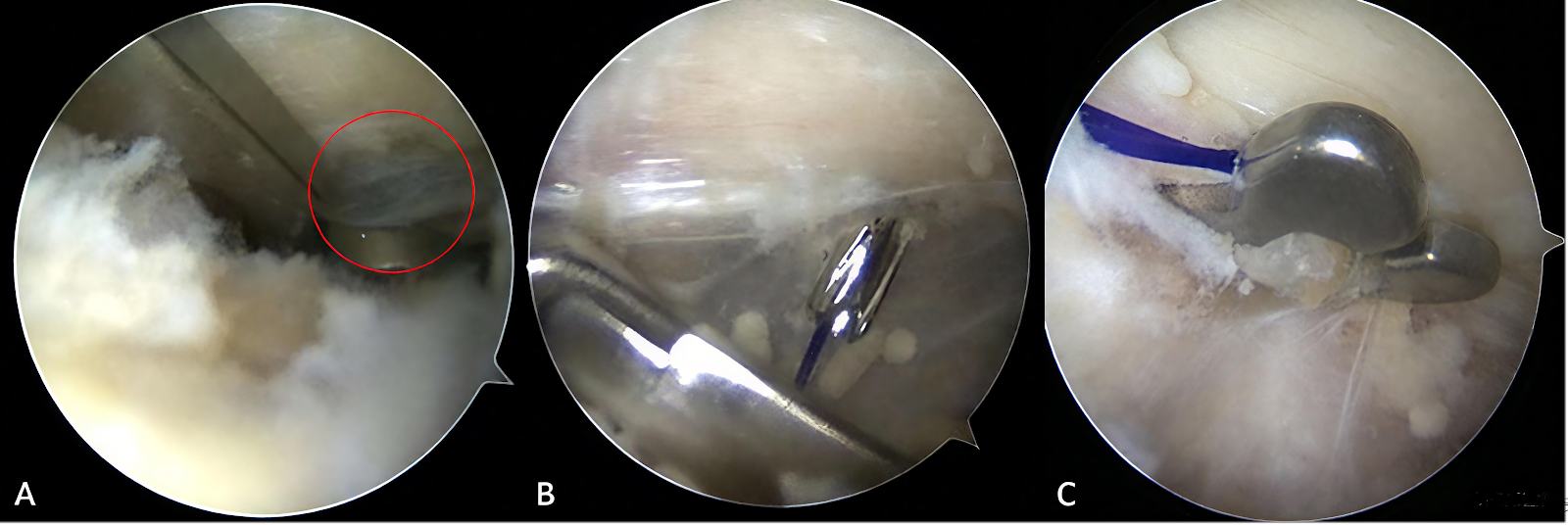
BACKGROUND: Surgical management of acute Rockwood type III-V acromioclavicular joint dislocations remains controversial. While clavicular hook plate fixation is widely adopted, it carries risks of postoperative complications. The emerging arthroscopy-assisted loop titanium plate technique, despite its minimally invasive advantages, requires further validation regarding long-term efficacy and cost-effectiveness. OBJECTIVE: To compare the clinical efficacy and cost-effectiveness of modified arthroscopic loop titanium plate fixation versus clavicular hook plate fixation for the treatment of acute Rockwood type III-V acromioclavicular joint dislocations. METHODS: Eighty eligible patients with acute Rockwood type III-V acromioclavicular joint dislocations admitted at Zhongshan Hospital of Traditional Chinese Medicine Affiliated to Guangzhou University of Chinese Medicine between January 2021 and July 2024 were enrolled and randomly assigned to either the loop titanium plate group (n=40) or the hook plate group (n=40). Ultimately, 31 and 33 patients in each group completed follow-up, respectively. The loop titanium plate group underwent fixation using the FixButtonTM suspension system with 2.4 mm bone tunnels; the hook plate group received open reduction and internal fixation with a clavicular hook plate. Outcome measures included operative time, incision length, blood loss, hospitalization costs, visual analog scale (VAS) for pain, Constant-Murley score, coracoclavicular distance, acromioclavicular distance, and complication rates, with a follow-up of 6 months. RESULTS AND CONCLUSION: The loop titanium plate group had longer operative time, shorter incisions, less blood loss, but higher hospitalization costs (P < 0.05). At 6 months postoperatively, the excellent-to-good rate in the loop titanium plate group (94%) was significantly higher than that in the hook plate group (82%) (P < 0.05); VAS scores were lower and Constant-Murley scores were higher (P < 0.05); coracoclavicular and acromioclavicular distances were better restored (P < 0.05). The total complication rate in the loop titanium plate group (6%) was lower than that in the hook plate group (12%) (P < 0.05). These findings suggest that although the modified arthroscopic loop titanium plate technique requires longer operative time and higher costs, it significantly reduces trauma, alleviates pain, improves shoulder function, and lowers complication risk, offering superior short-term clinical efficacy. Despite higher upfront costs, the potential reduction in secondary surgeries may confer long-term economic benefits. This technique is more suitable for patients with high demands for cosmetic incisions and functional recovery. Future studies with larger sample sizes and longer follow-up are needed to further validate these conclusions.

Read Full Abstract10.12307/2026.21411
Application of correction leverage technique in primary failure of distal locking screw during antegrade femoral intramedullary nailingGraphical AbstractVerified
Chinese Journal of Tissue Engineering Research2026

Application of correction leverage technique in primary failure of distal locking screw during antegrade femoral intramedullary nailing

BACKGROUND: Interlocking intramedullary nail fixation is the "gold standard" for the treatment of femoral shaft fractures, and the difficulty of distal locking nail implantation has always been a difficult problem to solve. OBJECTIVE: By comparing the clinical effects of correction leverage technique and free-hand locking nail technique, it is further explained whether the correction leverage technique can be fast and accurate. The distal locking screw of femoral intramedullary nail was placed without direct X-ray radiation exposure, thereby solving the problem of difficult distal locking screw placement. METHODS: A total of 52 patients with femoral shaft fractures who had difficulty in distal locking screw placement during interlocking intramedullary nail fixation were enrolled from the Department of Orthopedics and Traumatology, Affiliated Traditional Chinese Medicine Hospital of Southwest Medical University from July 2022 to September 2024. They were divided into two groups according to the placement protocol: correction leverage group (26 cases) used the correction leverage technique for distal locking screw placement, and free-hand group (26 cases) used the traditional free-hand technique. The distal locking screw placement time, number of X-ray exposures, and first-attempt accuracy were compared between the two groups. RESULTS AND CONCLUSION: (1) The distal locking screw placement time in the correction leverage group was significantly shorter than that in the free-hand group (t=-4.136, P < 0.001). (2) The number of X-ray exposures in the correction leverage group was less than that in the free-hand group (t=-19.696, P < 0.001). (3) The first-attempt accuracy in the correction leverage group (100%) was higher than that in the free-hand group (71%), with a significant difference (χ2=5.253, P < 0.05). (4) These results indicate that compared with simple free-hand screw placement, the correction leverage technique has the advantages of faster locking, higher accuracy, and lower X-ray radiation. This technique does not require auxiliary equipment and has strong operability, and is worthy of further clinical validation and promotion for interlocking intramedullary nail fixation of femoral shaft fractures.

Read Full Abstract10.12307/2026.21408
Relationship between gait parameter characteristics and joint function recovery after arthroscopic minimally invasive surgery in patients with knee osteoarthritisGraphical AbstractVerified
Chinese Journal of Tissue Engineering Research2026

Relationship between gait parameter characteristics and joint function recovery after arthroscopic minimally invasive surgery in patients with knee osteoarthritis

BACKGROUND: There are individual differences in the effectiveness of arthroscopic surgery in improving knee osteoarthritis, and subjective scoring scales may be biased in evaluating the clinical efficacy of arthroscopic treatment for knee osteoarthritis. OBJECTIVE: To explore the correlation between gait parameters and joint function recovery in patients with knee osteoarthritis after arthroscopic surgery. METHODS: A total of 98 patients with knee osteoarthritis admitted to Chongqing Armed Police Corps Hospital from October 2023 to October 2024 were selected as the research subjects. According to Lysholm knee function score after 6 months of follow-up, they were divided into the excellent group (n=63) and the fair group (n=35). Clinical data including gender, age, body mass index, disease duration, respiration, heart rate, Kellgren-Lawrence grade, smoking history, drinking history, hypertension history, location of onset, and postoperative complications were collected. The intraoperative and postoperative indicators, as well as the knee joint function scores and gait parameters at different times before and after surgery were compared between the two groups. Multivariate Logistic regression was used to analyze the independent influencing factors of knee joint function recovery. Stratified regression analysis was conducted to explore the impact of different clinical and pathological characteristics after treatment on gait parameters. Generalized estimating equations were used to analyze the differences in gait parameters among patients with different knee joint functions after treatment. Generalized additive models were used to analyze the impact of gait parameters on Lysholm score after treatment. Receiver operating characteristic curves were drawn to analyze the value of gait parameters in judging the recovery of knee joint function after treatment. RESULTS AND CONCLUSION: (1) There were significant differences in age, disease duration, Kellgren-Lawrence grade, and postoperative complications between the excellent and fair groups (P < 0.05). (2) The fair group had longer operation time, more intraoperative blood loss, longer postoperative swelling regression time, and longer rehabilitation time than the excellent group (P < 0.05). (3) After surgery, the fair group had higher Western Ontario and McMaster Universities Osteoarthritis Index and visual analog scale scores, and lower Lysholm score, step frequency, and step speed than the excellent group (P < 0.05). (4) Logistic regression analysis showed that age, Kellgren-Lawrence grade, postoperative complications, and postoperative swelling regression time were independent risk factors affecting knee joint function recovery (P < 0.05). (5) Stratified regression analysis showed that age, Kellgren-Lawrence grade, postoperative swelling regression time, and postoperative complications all had negative effects on step frequency and step speed (β < 0, P < 0.05). (6) Generalized estimating equation analysis showed that the degree of knee joint function recovery was associated with gait characteristics (β > 0, P < 0.05). (7) Generalized additive model analysis showed that the effects of step frequency and step speed on Lysholm score after treatment were linear. (8) Receiver operating characteristic curve analysis showed that the combined detection of step frequency and step speed had higher predictive efficacy (area under the curve > 0.85, P < 0.05). (9) These findings suggest that arthroscopic surgery can improve knee joint function in patients with knee osteoarthritis, and dynamic tracking of postoperative gait parameter changes combined with functional scoring scales reveals the association between step frequency, step speed, and knee joint function outcome, further clarifying its clinical predictive value and providing a new quantitative tool for clinical functional assessment to achieve more precise postoperative rehabilitation guidance.

Read Full Abstract10.12307/2026.21402
Application of a modified rectangular locking device in the treatment of femoral shaft fractures with intramedullary nailingGraphical AbstractVerified
Chinese Journal of Tissue Engineering Research2026

Application of a modified rectangular locking device in the treatment of femoral shaft fractures with intramedullary nailing

BACKGROUND: Conventional rectangular targeting devices are divided into two modules, one left and one right, which hinders flexible intraoperative use and is prone to deformation and error. Even after connection, repeated fluoroscopic confirmation is still required for screw drilling and placement. OBJECTIVE: To investigate the key technology development and clinical application of a modified rectangular locking device for precise distal locking screw placement in intramedullary nailing of femoral shaft fractures. METHODS: Medical records of patients with femoral shaft fractures admitted to Affiliated Suqian Hospital of Xuzhou Medical University and Suyu District People's Hospital from 2021 to 2023 were collected. Patients aged 18-65 years, diagnosed with femoral shaft fractures (AO classification: A, B, or C) by radiographic examination, who underwent closed reduction and intramedullary nailing with distal locking screw placement using the modified rectangular locking device, and whose clinical data (including medical history, radiographic findings, surgical records, and follow-up records) were selected. Forty-one cases met the criteria, including 30 males and 11 females, aged 20-62 years, with an average age of (41.17±8.14) years. Intraoperative fluoroscopy times, locking success rate, time for successful distal locking screw placement, American Knee Society Score at 1 month postoperatively and after fracture healing, and fracture healing time were collected. RESULTS AND CONCLUSION: (1) All 41 patients underwent distal locking screw placement using the modified rectangular locking device. Intraoperative fluoroscopy times ranged from 0 to 2 times, with an average of (1.1±0.5) times; the locking success rate was 98%; the time for successful distal locking screw placement ranged from 6 to 10 minutes, with an average of (7.0±1.5) minutes; the American Knee Society Score at 1 month postoperatively ranged from 140 to 190, with an average of (150±15) points. (2) Thirty-eight patients were followed up completely for 12-24 months. Fracture healing time ranged from 9 to 14 months, with an average of (10.5±2.5) months. After fracture healing, the American Knee Society Score ranged from 150 to 190, with an average of (185±8) points, with 35 excellent and 3 good results. (3) The results indicate that compared with freehand locking, oblique fluoroscopic placement, arthroscopic-assisted placement, and electromagnetic navigation locking of distal locking screws, the modified rectangular locking device offers advantages including no dependence on arthroscopic or electromagnetic navigation equipment, no requirement for extensive surgical experience, simple steps, accurate locking, high repeatability, and reduced radiation exposure.

Read Full Abstract10.12307/2026.21409
Morphometric analysis of principal stress trabeculae in the proximal femurGraphical AbstractVerified
Chinese Journal of Tissue Engineering Research2026

Morphometric analysis of principal stress trabeculae in the proximal femur

BACKGROUND: The principal compressive and tensile trabecular bones in the proximal femur are essential structures responsible for transmitting forces through the hip joint. Given their complex and heterogeneous composition, exploring their microstructural variations will help improve our understanding of principal stress trabeculae. OBJECTIVE: To extract trabecular microstructural information from various regions of femoral head specimens and study their morphometric patterns, elucidate the microstructural variations of principal stress trabeculae, and provide a theoretical basis for the design of future bionic hip prostheses. METHODS: Totally 11 fresh femoral heads were obtained from patients with femoral neck fractures. They were scanned by Micro-CT and reconstructed by image analysis software (InveonTM Acquisition Workplace). The principal compressive and tensile trabecular bones were segmented into five regions respectively. After selecting the volumes of interest for each region, morphometric analyses were subsequently performed on bone volume fraction, bone surface area fraction, trabecular thickness, trabecular number, trabecular spacing, trabecular pattern factor, and grayscale value. RESULTS AND CONCLUSION: (1) Among the principal compressive trabeculae, Region 5 exhibited the highest bone volume fraction, trabecular thickness, and grayscale value, followed by Region 4 and 1, with statistically significant differences between Region 5 and 1 (all P < 0.05). Moving from Region 3 to Region 2, 1, and Regions 4, 5, there was a decreasing trend in bone surface area fraction and trabecular number, accompanied by an increasing trend in trabecular thickness. (2) Among the principal tensile trabeculae, Region 3 displayed the highest values for bone volume fraction, trabecular thickness, trabecular number, and grayscale value, while Region 2 showed opposite trends, with statistically significant differences between the two regions, (excluding grayscale value; all P < 0.001). (3) This study proposes a reliable method for extracting the volume of interest of principal stress trabeculae, and for the first time proposes the theory that principal compressive trabeculae are a functionally graded porous structure, providing new insights into the microstructural anatomy of principal compressive trabeculae and potentially proving useful for the design of novel bionic hip prostheses.

Read Full Abstract10.12307/2026.21404
Determination of vancomycin blood concentration and its relationship with safety in patients with orthopedic infection by ultra-performance liquid chromatography-tandem mass spectrometryGraphical AbstractVerified
Chinese Journal of Tissue Engineering Research2026

Determination of vancomycin blood concentration and its relationship with safety in patients with orthopedic infection by ultra-performance liquid chromatography-tandem mass spectrometry

BACKGROUND: Vancomycin is widely used as a first-line drug for treating methicillin-resistant Staphylococcus aureus infections in orthopedic perioperative infection prevention and treatment. However, its narrow therapeutic window and large individual differences make blood drug concentration monitoring crucial for ensuring efficacy and safety. OBJECTIVE: To establish the ultra-performance liquid chromatography-tandem mass spectrometry method to determine the concentration of vancomycin in plasma, and explore the correlation of blood drug concentration changes with clinical efficacy and acute renal impairment. METHODS: Totally 200 orthopedic patients with bone infection who were hospitalized in Beijing Jishuitan Hospital Guizhou Hospital from January 2020 to May 2022 were selected. Blood drug concentration was measured by ultra-performance liquid chromatography-tandem mass spectrometry. Renal function indexes (blood creatinine and urea nitrogen) were monitored, and the clinical efficacy and acute renal impairment were analyzed in patients with different vancomycin blood concentration levels. RESULTS AND CONCLUSION: (1) The established UPLC-MS/MS method had a linear range of 0.5-120.0 μg/mL (R²=0.997), intra-day relative standard deviation of 4.82%-6.57%, inter-day relative standard deviation of 10.2%-12.3%, and accuracy of 97.3%-106%. (2) The clinical effective rate in the 10-20 μg/mL group (82%) was significantly higher than that in the <10 μg/mL group (67%) (P < 0.05). (3) The incidence of renal impairment in the >20 μg/mL group (35.0%) was significantly higher than that in the 10-20 μg/mL group (16.0%) and <10 μg/mL group (6.7%) (P < 0.05). (4) The established UPLC-MS/MS method is sensitive and accurate, and can be used for clinical monitoring of vancomycin blood concentration. Maintaining vancomycin blood concentration within 10-20 μg/mL can achieve the best clinical efficacy and reduce the risk of nephrotoxicity. Blood concentration monitoring is of great significance for guiding individualized administration.

Read Full Abstract10.12307/2026.21405
Biomechanical evaluation of oblique pulling manipulation on C5/6 intervertebral discs with different levels of degenerationGraphical AbstractVerified
Chinese Journal of Tissue Engineering Research2026

Biomechanical evaluation of oblique pulling manipulation on C5/6 intervertebral discs with different levels of degeneration

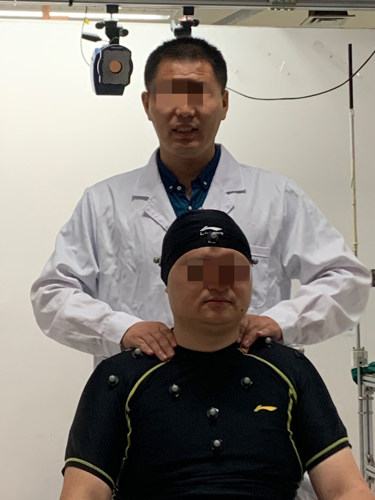
BACKGROUND: The oblique pulling manipulation has good therapeutic effects on cervical spondylosis, but its biomechanical mechanism of action on intervertebral discs with different degrees of degeneration is not clear. OBJECTIVE: To explore the biomechanical mechanism of the oblique pulling manipulation on the discs with different degrees of degeneration through three-dimensional finite element model. METHODS: The motion capture system was used to measure the kinematic parameters of the key steps during the oblique pulling manipulation, and a three-dimensional finite element model of the whole cervical spine of the C5/6 mildly, moderately, and severely degenerated intervertebral discs was established. The kinematic parameters were converted into moments and loaded onto the whole cervical spine of the mildly, moderately, and severely degenerated intervertebral discs in a step-by-step manner, so as to obtain the biomechanical parameters of the stress-strain of each structure during the simulated oblique pulling manipulation. RESULTS AND CONCLUSION: (1) In the simulation of the oblique pulling manipulation to the right side, with the increase of disc degeneration, the Von-Mise stress of the annulus fibrosus gradually increased, and the stress was concentrated on the right lateral side of the annulus fibrosus; the Von-Mise stress of the nucleus pulposus decreased, and the stress was concentrated at the junction of the nucleus pulposus and annulus fibrosus; the overall displacement of the intervertebral disc decreased, the stress of the left facet joint decreased, and the Von-Mise stress of the spinal cord increased, with the stress concentrated in the upper cervical spine. (2) In the simulation of the oblique pulling manipulation to the right side, with the increase of disc degeneration, the overall strain of the C5/6 intervertebral disc, the intradiscal pressure of the nucleus pulposus, and the stress of the left nerve root decreased, while the shear force of the intervertebral disc increased, the relative distance between the left nerve root and the intervertebral disc increased, and the relative distance between the right nerve root and the intervertebral disc did not change significantly. (3) The results indicate that the cervical oblique pulling manipulation improves the biomechanical imbalance of mildly and moderately degenerated intervertebral discs by adjusting the stress of the facet joints and increasing the distance between nerve roots, but for severely degenerated intervertebral discs, it should be used with caution due to the sharp increase of annulus fibrosus stress and the risk of spinal cord compression.

Read Full Abstract10.12307/2026.21388
Screening biomarkers for premature ovarian insufficiency based on cellular senescence and endoplasmic reticulum stress with experimental validationGraphical AbstractVerified
Chinese Journal of Tissue Engineering Research2026

Screening biomarkers for premature ovarian insufficiency based on cellular senescence and endoplasmic reticulum stress with experimental validation

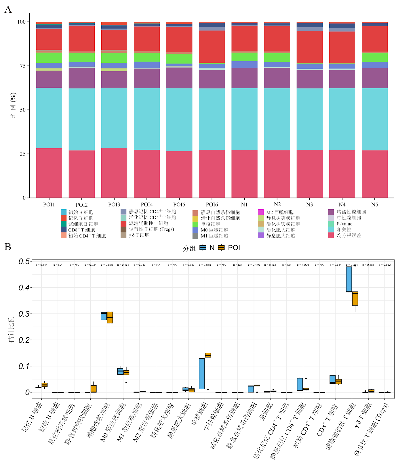
BACKGROUND: Ovarian granulosa cell senescence and endoplasmic reticulum stress are closely related to the development and progression of premature ovarian insufficiency; however, the underlying regulatory mechanisms remain unelucidated. OBJECTIVE: To identify potential biomarkers associated with cellular senescence and endoplasmic reticulum stress in granulosa cells in premature ovarian insufficiency using bioinformatic analysis and machine learning algorithms, with subsequent validation in animal experiments. METHODS: The premature ovarian insufficiency dataset GSE201276 was downloaded from the GEO database. Differentially expressed genes were screened, and weighted gene co-expression network analysis was performed to identify module genes. Gene sets related to cellular senescence and endoplasmic reticulum stress were obtained from the GeneCards database, and intersected with differentially expressed genes and module genes. Consensus clustering analysis was then performed to identify subtype-specific differentially expressed genes, followed by Gene Ontology and Kyoto Encyclopedia of Genes and Genomes enrichment analyses and immune infiltration analysis. Two machine learning algorithms were applied to screen key genes associated with cellular senescence and endoplasmic reticulum stress in granulosa cells, and a diagnostic model was constructed and validated. Finally, a premature ovarian insufficiency mouse model was established in C57BL/6J mice, and the model was verified by estrous cycle monitoring, hematoxylin-eosin staining, and serum ELISA. The expression of key genes was validated by real-time quantitative PCR and western blot. RESULTS AND CONCLUSION: Consensus clustering identified 911 subtype-specific differentially expressed genes associated with cellular senescence and endoplasmic reticulum stress. Gene Ontology enrichment analysis showed that these genes were mainly involved in biological processes such as negative regulation of cell cycle, meiosis, and female gonad development. Kyoto Encyclopedia of Genes and Genomes analysis revealed enrichment in pathways such as oocyte meiosis, progesterone-mediated oocyte maturation, and transforming growth factor beta signaling. Immune infiltration analysis showed significantly higher infiltration levels of M1 macrophages and resting dendritic cells in the premature ovarian insufficiency group (P < 0.05). Machine learning algorithms identified four key genes, and the diagnostic model and calibration curves showed that aurora kinase A and actin binding protein exhibited good predictive performance. Animal experiments showed that compared with the control group, the model group exhibited disrupted estrous cycles, reduced numbers of primary, secondary, and antral follicles, and increased numbers of atretic follicles (P < 0.01). Serum follicle-stimulating hormone levels were elevated, while anti-Müllerian hormone levels were decreased, with significant differences (P < 0.01). Compared with the control group, the mRNA and protein expression levels of aurora kinase A and actin binding protein in ovarian tissues of the model group were significantly decreased (P < 0.05). These results indicate that aurora kinase A and actin binding protein may participate in the development of premature ovarian insufficiency by regulating granulosa cell senescence and endoplasmic reticulum stress, and their specific regulatory roles and molecular mechanisms require further experimental validation.

Read Full Abstract10.12307/2026.21371
Unicompartmental knee arthroplasty for severe medial compartment osteoarthritis with moderate lateral involvement: clinical outcomesGraphical AbstractVerified
Chinese Journal of Tissue Engineering Research2026

Unicompartmental knee arthroplasty for severe medial compartment osteoarthritis with moderate lateral involvement: clinical outcomes

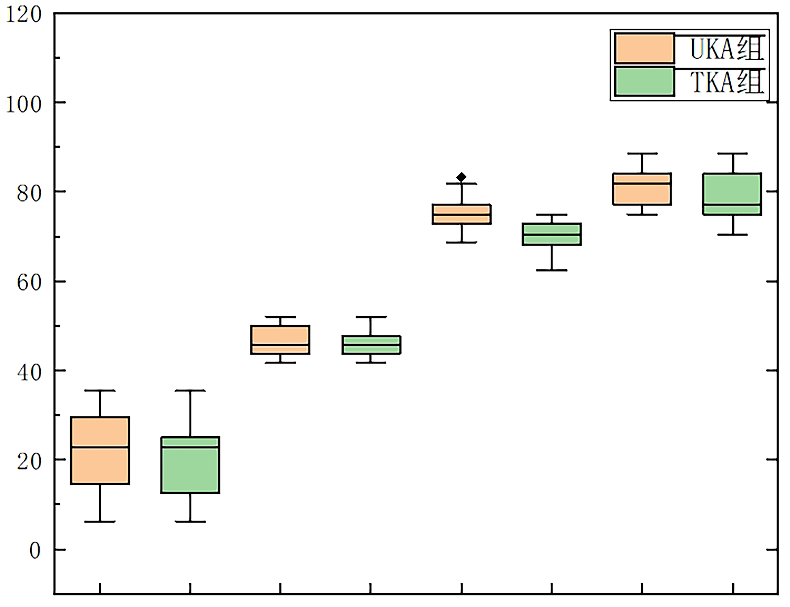
BACKGROUND: In patients with severe medial compartment osteoarthritis of the knee combined with moderate lateral compartment osteoarthritis, it is controversial whether medial monondylar replacement alone can achieve satisfactory clinical benefit. OBJECTIVE: To compare and analyze the difference in clinical outcomes of unicompartmental knee arthroplasty and total knee arthroplasty in patients with heterogeneous degenerative changes of the knee (medial compartment Kellgren-Lawrence grade III-IV combined with lateral compartment Kellgren-Lawrence grade II). METHODS: Knee arthroplasty patients with severe medial ventricular osteoarthritis combined with mild and moderate lateral ventricular osteoarthritis shown by preoperative knee X-ray were selected and divided into unicompartmental knee arthroplasty group and total knee arthroplasty group according to different operation methods, and 50 patients were included in each group according to 1:1 pairing. Western Ontario and McMaster University Osteoarthritis Index (WOMAC), American Knee Association Score, joint amnesia score, postoperative gait parameters, and complications were collected at 3, 6 months, 1 and 2 years after surgery, and the relevant data were statistically analyzed to compare the differences in postoperative efficacy between the two groups. RESULTS AND CONCLUSION: (1) The American Knee Association Score and WOMAC scores of the unicompartmental knee arthroplasty group were better than those of the total knee arthroplasty group at 3, 6 months and 1 year after surgery (P < 0.05). Two years after surgery, American Knee Association Score and WOMAC score were not significantly different between two groups (P > 0.05). (2) There was no significant difference in joint amnesia score between the two groups at 3 months postoperatively (P > 0.05), but at 6 months, 1 year, and 2 years postoperatively, the unicompartmental knee arthroplasty group was higher than the total knee arthroplasty group (P < 0.05). (3) At 1 year postoperatively, the unicompartmental knee arthroplasty group had better gait speed and stride length than the total knee arthroplasty group (P < 0.05), while there was no significant difference in cadence between the two groups (P > 0.05). (4) No complications occurred in either group within 2 years postoperatively. (5) Follow-up confirmed that in patients with severe medial compartment osteoarthritis (Kellgren-Lawrence grade III-IV) combined with moderate lateral compartment degeneration (Kellgren-Lawrence grade II), unicompartmental knee arthroplasty had equivalent clinical efficacy to total knee arthroplasty at 2 years postoperatively, and the biomechanical advantages of unicompartmental knee arthroplasty were significant within 1 year postoperatively. During the follow-up period, no obvious progression of lateral compartment osteoarthritis (increase in Kellgren-Lawrence grade) was found, but long-term observation is needed. Before making a decision, clinicians should establish a multidimensional evaluation framework (including patient age, exercise load expectation, surgeon's unicompartmental knee arthroplasty volume, etc.), and fully inform patients of the risk of lateral compartment progression, and comprehensively evaluate whether to perform unicompartmental knee arthroplasty.

Read Full Abstract10.12307/2026.21397
Mechanical analysis of a bone cement-augmented cortical bone trajectory screw versus a new variable-diameter all-cortical bone screwGraphical AbstractVerified
Chinese Journal of Tissue Engineering Research2026

Mechanical analysis of a bone cement-augmented cortical bone trajectory screw versus a new variable-diameter all-cortical bone screw

BACKGROUND: The scarcity of bone trabecular structures caused by osteoporosis is not enough to maintain screw holding power, which often leads to the failure of internal fixation surgery. The screw holding power is often increased by increasing the diameter and length of screws, changing the surface coating of screws, and reinforcing the nail track with bone cement. The cement reinforced cortical bone track nailing technique and the modified cortical bone track nailing technique using a new type of variable diameter screw have been proven to have good fixation effects, and now the related mechanical properties of the two need to be analyzed and compared. OBJECTIVE: Finite element analysis was used to compare the mechanical properties of bone cement-strengthened cortical bone trajectory nailing technology, cortical bone trajectory nailing technology, and modified cortical bone trajectory nailing technology using a new variable-diameter total cortical bone thread screw in lumbar spine internal fixation surgery. METHODS: Based on the CT scan data processing of three osteoporotic vertebrae, the L4 lumbar spine model was constructed, and the innovative variable-diameter all-cortical bone screw was applied in the modified cortical bone nailing technique, with the screw having a total length of 45 mm and a diameter varying from 5.5 to 4.3 mm. This was compared with the un-augmented cortical bone trajectory group (diameter 5.5 mm, length 40 mm) and the bone cement-augmented cortical bone trajectory group (diameter 5.5 mm, length 40 mm, central hole diameter 1 mm). The fixation strength of each group was compared by measuring axial pullout force, screw stability (load-displacement ratio in superior, inferior, left, and right directions), and vertebral range of motion. RESULTS AND CONCLUSION: (1) Axial pullout force: augmented cortical bone trajectory group > modified cortical bone trajectory screw group (P=0.0246), and both augmented and modified groups were greater than the un-augmented cortical bone trajectory group (P=0.0001 and P=0.00264, respectively). (2) Screw stability: when load was applied inferiorly, the load-displacement ratios were augmented cortical bone trajectory group > un-augmented and modified groups (all P < 0.05), and modified group > un-augmented group (P < 0.05). (3) Vertebral range of motion: under five loading conditions, the augmented cortical bone trajectory group showed less motion than the modified group, but the differences were not statistically significant (P > 0.05), and both augmented and modified groups showed less motion than the un-augmented group. (4) Compared with the modified group, the augmented group showed improved mechanical properties in screw load-displacement ratio and lumbar range of motion, but the differences were not statistically significant (P > 0.05). (5) These findings suggest that the bone cement-augmented cortical bone trajectory technique has better biomechanical properties than the new variable-diameter all-cortical bone screw, and may be a more suitable screw placement option for internal fixation in patients with osteoporosis.

Read Full Abstract10.12307/2026.21389
Finite element analysis of biomechanics of two internal fixation methods for Pauwels type III fractures based on fatigue life calculationGraphical AbstractVerified
Chinese Journal of Tissue Engineering Research2026

Finite element analysis of biomechanics of two internal fixation methods for Pauwels type III fractures based on fatigue life calculation

BACKGROUND: In patients with Pauwels type III femoral neck fracture who are unable to undergo closed reduction, the traditional cannulated compression screw internal fixation method cannot effectively counteract the large shear force. To solve this problem, this study personalized an internally supported plate and used it in conjunction with the cannulated compression screw internal fixation protocol to improve treatment outcomes. OBJECTIVE: To compare the biomechanical performance of two internal fixation methods for Pauwels type III femoral neck fractures under gait loading: an "inverted triangle" arrangement of three screws and an "inverted triangle" arrangement of three screws combined with a medial support plate by finite element calculation. METHODS: Based on the CT scan data, inverse modeling was first performed using Mimics software to generate a point cloud model of the femur. Subsequently, the model was refined using Geomagic software to optimize its geometry and ensure the accuracy of the model. Finally, the processed model was imported into NX software to establish a femoral neck fracture model with a Pauwels angle of 70°. The mechanical and fatigue life results of the 3-nail [fully threaded (model 1), unthreaded (model 2)] model, 3-nail + personalized internal support plate [fully threaded (model 3), unthreaded (model 4)] model were computed based on the Ansys software for the gait loading. RESULTS AND CONCLUSION: (1) Under gait loading, the introduction of an internal support plate reduced the mean femoral stress compared with the 3-nail fixation approach, including a decrease in fracture and stump stresses of 6.6 MPa and 11.0 MPa, respectively; a decrease in displacement of 0.24 mm and 0.12 mm, respectively, and a reduction in relative displacement of the fracture surface. (2) The internal fixation method reduced the fatigue life of the bone system, and the addition of the internal support plate further reduced fatigue life. (3) The finite element analysis was sensitive to thread parameters, so the thread type and characteristics of screws should be considered in model construction. Compared with the 3-screw fixation alone, the addition of an internal support plate reduced stress and deformation levels, providing a more stable mechanical environment for bone healing. From the perspective of fatigue life, internal fixation reduced the life of the femoral system, and the more implants, the lower the life; the fixation scheme with the internal support plate had the lowest fatigue life. (4) This indicates that in clinical design of fixation schemes, the impact of implants on long-term healing outcomes should be fully considered, and the number of implants and fixation method should be comprehensively evaluated.

Read Full Abstract10.12307/2026.21391
InterTAN versus proximal femoral nail anti-rotation for intertrochanteric fractures in the elderly: a comparison of joint function and stabilityGraphical AbstractVerified
Chinese Journal of Tissue Engineering Research2026

InterTAN versus proximal femoral nail anti-rotation for intertrochanteric fractures in the elderly: a comparison of joint function and stability

BACKGROUND: Elderly patients with intertrochanteric fractures often have multiple medical comorbidities and significant osteoporosis, posing considerable challenges for treatment. Proximal femoral nail anti-rotation (PFNA) and InterTAN are both commonly used intramedullary fixation systems. However, there remains insufficient evidence regarding the optimal selection of fixation systems for patients with different medical conditions and fracture types. OBJECTIVE: To compare the clinical outcomes and complication differences between InterTAN and PFNA in the treatment of elderly patients with intertrochanteric fractures. METHODS: A prospective randomized controlled trial was conducted. A total of 118 elderly patients with intertrochanteric fractures who met the inclusion criteria were enrolled. Patients were randomly divided into two groups: PFNA group (59 cases) underwent internal fixation with PFNA, and InterTAN group (59 cases) underwent internal fixation with InterTAN. Perioperative parameters, including operation time, intraoperative blood loss, fluoroscopy frequency, and hospital stay, were recorded. Follow-up assessments were conducted at 1, 3, 6, and 12 months postoperatively. The fracture healing rate, internal fixation stability, and complications were evaluated using Harris hip scores and imaging examinations. RESULTS AND CONCLUSION: (1) The PFNA group had significantly shorter operation time, less intraoperative blood loss, and fewer fluoroscopy exposures than the InterTAN group (P < 0.001), while hospital stay showed no significant difference (P > 0.05). (2) At 12 months postoperatively, the fracture healing rate was similar between the two groups (P > 0.05). However, the InterTAN group had a significantly lower rate of internal fixation instability (P < 0.05) and better Harris pain, function, and range of motion scores (P < 0.05). The overall complication rate did not differ significantly between groups (P > 0.05). (3) These findings suggest that PFNA offers advantages in perioperative trauma, while InterTAN shows potential benefits in fixation stability and functional recovery at 1-year follow-up. The choice of internal fixation system should be tailored to the patient's condition and fracture type.

Read Full Abstract10.12307/2026.21401
Single-dose liposomal bupivacaine versus continuous ropivacaine infusion for adductor canal block analgesia after total knee arthroplastyGraphical AbstractVerified
Chinese Journal of Tissue Engineering Research2026

Single-dose liposomal bupivacaine versus continuous ropivacaine infusion for adductor canal block analgesia after total knee arthroplasty

BACKGROUND: Effective analgesia after total knee arthroplasty is crucial for rehabilitation, and adductor canal block is a common method. Traditional single injection of local anesthetic has limited analgesic duration, while continuous catheter infusion is complex and costly. OBJECTIVE: To compare the efficacy and safety of single-dose liposomal bupivacaine versus continuous catheter infusion of ropivacaine for adductor canal block analgesia after total knee arthroplasty. METHODS: Eighty patients undergoing primary unilateral knee arthroplasty at Gaoyou People's Hospital from March 2024 to February 2025 were randomly divided into liposomal bupivacaine group and continuous catheter infusion group. The liposomal bupivacaine group received a single adductor canal block with 133 mg (10 mL) liposomal bupivacaine mixed with 5 mL of 0.75% ropivacaine. The continuous catheter infusion group received an adductor canal catheter with 0.25% ropivacaine via a pump (load 10 mL, infusion rate 6 mL/h). Pain scores at different time points, pain-free time, morphine rescue dose, and walking distance were compared to assess analgesic efficacy; complications were observed for safety. Operation time, cost, patient satisfaction, and hospital stay were also compared. RESULTS AND CONCLUSION: (1) At 6, 12, 24, 48, and 72 h postoperatively, there were no significant differences in resting and movement pain scores between groups (P > 0.05). Pain-free time was 19 h in the liposomal bupivacaine group and 22 h in the continuous infusion group (P > 0.05). (2) At 72 h, morphine rescue dose was 78.6 mg morphine equivalents in the liposomal bupivacaine group and 80.5 mg in the continuous infusion group (P > 0.05). (3) Operation time was significantly shorter in the liposomal bupivacaine group (6.1±1.4 min vs. 20.3±1.2 min, P < 0.05). (4) Patient satisfaction, hospital stay, and walking ability showed no significant differences (P > 0.05). (5) One case of transient femoral nerve palsy occurred in the continuous infusion group at 6 h postoperatively, resolving spontaneously. Each patient in the liposomal bupivacaine group saved 132 RMB in analgesic costs. (6) Within 72 h after total knee arthroplasty, single-dose liposomal bupivacaine and continuous ropivacaine infusion showed no differences in analgesic scores, pain-free time, morphine rescue dose, hospital stay, satisfaction, or walking ability, but single-dose liposomal bupivacaine was more convenient, saving time and cost.

Read Full Abstract10.12307/2026.21392
Finite element analysis of core decompression with ceramic rod implantation in osteonecrosis of the femoral head during the peri-collapse stageGraphical AbstractVerified
Chinese Journal of Tissue Engineering Research2026

Finite element analysis of core decompression with ceramic rod implantation in osteonecrosis of the femoral head during the peri-collapse stage

BACKGROUND: The elastic modulus of β-tricalcium phosphate bioceramic rods is close to that of normal bone tissue, and it exhibits excellent biocompatibility and mechanical properties. It can be used as a supporting material inside the femoral head after core decompression. However, there are few biomechanical studies on osteonecrosis of the femoral head and the changes in stress and displacement of the femoral head after ceramic rod implantation. OBJECTIVE: To explore the biomechanical effects of core decompression with ceramic rod implantation in the treatment of osteonecrosis of the femoral head during the peri-collapse stage. METHODS: A total of 21 hips were selected from 19 patients with osteonecrosis of the femoral head implanted with ceramic rods at the peri-collapse stage. Preoperative and postoperative imaging data were obtained, and relevant CT images were loaded in Mimics 21.0 software to construct a three-dimensional model of the femoral head. A global model of the proximal femur that includes cortical and cancellous bone, as well as a model of the proximal cancellous bone of the femur were created. The preoperative MRI image data of the patients were imported, and the necrotic lesion model was made by using the graphic matching technology, which was saved in .stl format. They were transferred to Geomagic 2012 software for smoothing and precise surface processing. Subsequently, the ceramic rod was designed and modeled in SolidWorks 2021 software, and the relevant models were imported for assembly and Boolean operations. After ensuring no interference, ANSYS 2021 software was used to calculate and observe the stress and displacement of the weight-bearing area and necrotic area of the femoral head during single-leg stance and the push-off phase of walking. RESULTS AND CONCLUSION: (1) The area of maximum stress on the femoral head was located in the anterolateral superior part of the necrotic area. During single-leg stance, the stress values in the weight-bearing area and necrotic area were significantly lower postoperatively than preoperatively (P < 0.05), and the femoral head collapse value (displacement of the weight-bearing area) was lower than preoperatively (P < 0.05). (2) During the push-off phase of walking, with the increase in load, the stress values in the weight-bearing area and necrotic area and the femoral head collapse value (displacement of the weight-bearing area) increased, but they were still lower than preoperatively (P < 0.05). (3) It is suggested that core decompression combined with ceramic rod implantation helps to reduce the load on the weight-bearing area of the femoral head, effectively disperse the stress in the weight-bearing area, partially transfer the load to the femoral calcar, improve the local stress concentration, and effectively support the femoral head to prevent further collapse.

Read Full Abstract10.12307/2026.21393
Preoperative Planning Assisted Sleeve+ Extension Rod Combined with MBT Prosthesis in Revision for Non-infectious Knee Prosthesis LooseningGraphical AbstractVerified
Chinese Journal of Tissue Engineering Research2026

Preoperative Planning Assisted Sleeve+ Extension Rod Combined with MBT Prosthesis in Revision for Non-infectious Knee Prosthesis Loosening

BACKGROUND: With the widespread application of total knee arthroplasty in China, non-infectious prosthesis loosening has become one of the main reasons for postoperative revision. For complex loosening cases, traditional revision techniques are relatively complex and difficult. The application of artificial intelligence-assisted preoperative planning combined with Sleeve extension rods and mobile bearing tray prostheses provides a new solution for precisely reconstructing joint stability and mechanical alignment, which is expected to improve the long-term outcomes of revision surgeries. OBJECTIVE: To explore the mid-and early-term clinical efficacy of revision surgery for non-infectious total knee prosthesis loosening using Sleeve extension rods combined with mobile bearing tray prostheses under the assistance of artificial intelligence-assisted preoperative planning. METHODS: A retrospective analysis was conducted on 17 patients with non-infectious prosthesis loosening after total knee arthroplasty in Department of Orthopedics and Traumatology, Jiangsu Provincial Hospital of Traditional Chinese Medicine from January 2021 to September 2024. There were 6 males and 11 females, aged 59-81 years (mean 72.06±6.10 years). The affected side was left in 8 cases and right in 9 cases. The duration of prosthesis use ranged from 2 to 22 years (mean 10.53±4.60 years). All cases were revisions after primary arthroplasty. Revision reasons included periprosthetic osteolysis with liner wear in 15 cases, femoral condyle old fracture causing loosening in 1 case, and tibial plateau prosthesis fracture in 1 case. According to AORI classification, there were 13 cases of type IIB and 4 cases of type IIA. The artificial intelligence-designed prosthesis sizes were recorded and compared with the actual intraoperative sizes. Visual analog scale (VAS) score, American Knee Society knee score, hip-knee-ankle angle, and knee range of motion were compared preoperatively, at 1 week, 6 months, and 12 months postoperatively to evaluate surgical efficacy. RESULTS AND CONCLUSION: (1) Except for one patient with poor incision healing at 1 month postoperatively, all other patients recovered well without adverse events such as deep vein thrombosis, infection, periprosthetic fracture, or prosthesis loosening. (2) The follow-up period ranged from 6 to 41 months (mean 23.63±12.50 months). At the last follow-up, 2 patients had slight soreness and discomfort after activity, and 1 patient had obvious pain during activity. (3) At the last follow-up after revision, resting pain, exercise pain VAS scores, affected side range of motion, hip-knee-ankle angle, and American Knee Society knee score were significantly improved compared with preoperative values (P < 0.01). (4) The matching rate of artificial intelligence preoperative design for femoral condyle and tibial plateau prostheses was 85%, and the matching rate for other components was 62%. (5) The use of Sleeve+ extension rod combined with MBT prosthesis for non-infectious knee revision can effectively correct joint alignment, fill bone defects, improve pain and knee range of motion, and enhance patients' quality of life, with good early and mid-term efficacy. Artificial intelligence preoperative planning generally helps improve surgical accuracy, reduce revision difficulty, minimize risks, and promote postoperative recovery.

Read Full Abstract10.12307/2026.21395
Establishment and validation of a high-fidelity finite element model of the wrist jointGraphical AbstractVerified
Chinese Journal of Tissue Engineering Research2026

Establishment and validation of a high-fidelity finite element model of the wrist joint

BACKGROUND: Current finite element models of the wrist joint predominantly focus on osseous and ligamentous structures, with insufficient incorporation of musculotendinous components, thereby limiting their fidelity and accuracy. OBJECTIVE: To establish a high-fidelity finite element model of the wrist joint, providing a reference for in-depth biomechanical investigations. METHODS: Upper limb CT and MRI data from a 33-year-old healthy male volunteer were imported into Mimics 20.0. Threshold-based selection, region growing, and image segmentation techniques were employed to reconstruct wrist-related bones and soft tissues (including muscles). The model underwent surface optimization, patch generation, and meshing in SolidWorks 2020 and HyperMesh 14.0. Material property assignment and ligament-cartilage contact interfaces were implemented in ABAQUS 6.13 to construct a three-dimensional finite element model of the wrist joint. Stress distribution across wrist structures under axial compression was analyzed. RESULTS AND CONCLUSION: (1) A three-dimensional finite element model encompassing the ulna, radius, distal humerus, carpal bones, metacarpals, pronator teres, pronator quadratus, supinator, lateral muscle group, volar muscle group, dorsal muscle group, interosseous membrane, major ligaments, and cartilage structures was successfully established, comprising 759 191 elements and 245 510 nodes. The stress distribution pattern at the radiocarpal joint under axial compression was obtained and compared with cadaveric studies from the literature, validating the model's authenticity and effectiveness. (2) In summary, based on human CT and MRI imaging data, a more complete wrist joint bone and soft tissue structure was reconstructed through computer software simulation, establishing the origin and insertion points of forearm muscles and their contact with bones during muscle course, resulting in a more realistic finite element model of the wrist joint.

Read Full Abstract10.12307/2026.21396