🧬 SinoBioData Academic Portal
🏛️ Indexed Academic JournalOriginal: 生物化学与生物物理学报

Acta Biochimica et Biophysica Sinica

Premier Chinese Biomedical Journal indexed in SinoBioData: Acta Biochimica et Biophysica Sinica (生物化学与生物物理学报).

Total Research Papers: 200
Access: 100% Free Open Access
Browse by Publication Year & VolumeReset All Filters ✕

Published Research PapersFiltered: Year 2025 • 57 • 12

Showing 13 of 200 peer-reviewed papers with full Graphical Abstracts.

Original ResearchVol. 57, Issue 12 • pp. 2048-2061DOI: 10.3724/abbs.2025077

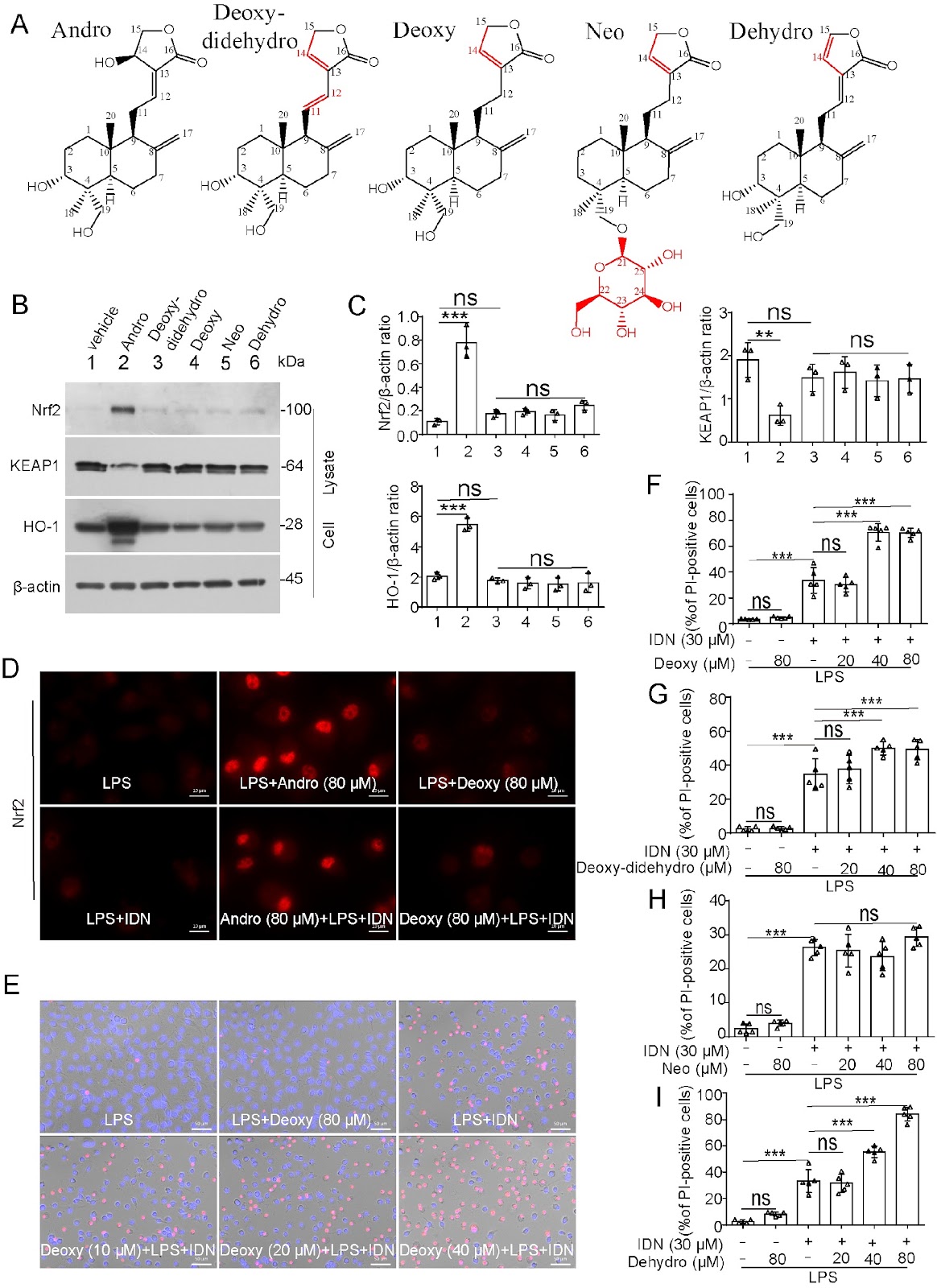
Andrographolide prevents necroptosis by suppressing the generation of reactive oxygen species

Authors: Na Lu, Qing Li, Linghan Duan, Rong Xu, Yaping Li, Fuli Shi, Zhiya Zhou, Yingqing Gan, Bo Hu, Jinhua Li, Xianhui He, Dongyun Ouyang, Qingbing Zha

Andrographolide (Andro), a natural product extracted from the Chinese traditional medicine herb Andrographis paniculata, has been applied for the treatment of diverse inflammatory diseases. However, its effects on necroptosis, a lytic form of cell death implicated in various inflammatory diseases, remain uncharacterized. In the present study, we investigate whether Andro and its derivatives can suppress necroptosis. Our results demonstrate that Andro notably inhibits necroptosis in the in vitro cellular models induced by either lipopolysaccharide (LPS) plus IDN-6556 or a combination of TNF-α, LCL-161 (Smac mimetic) and IDN-6556. In these cellular models, Andro inhibits the phosphorylation of receptor-interacting protein kinase 1 (RIPK1), RIPK3, and mixed lineage kinase domain-like pseudokinase (MLKL), as well as the formation of necrosomes. Specifically, Andro reduces the levels of intracellular reactive oxygen species (ROS) and mitochondrial superoxide (mtROS), preserves the mitochondrial membrane potential during necroptotic induction, and activates the antioxidant transcription factor nuclear factor E2-related factor 2 (Nrf2). Upon necroptotic stimulation, some mitochondrial proteins, such as Bcl-2 and Bak, oligomerize and co-localize with RIPK1, RIPK3, and phosphorylated MLKL (p-MLKL) in necrosomes. However, this process of necrosome formation can be prevented by Andro. In contrast, derivatives, including dehydroandrographolide, neoandrographolide, 14-deoxy-11,12-didehydroandrographolide, and 14-deoxyandrographolide, have no anti-necroptotic effects and fail to upregulate Nrf2. Collectively, our findings demonstrate that Andro specifically inhibits the RIPK1/RIPK3/MLKL signaling axis to suppress necroptosis, highlighting its therapeutic potential against necroptosis-related disorders.

Andrographolide prevents necroptosis by suppressing the generation of reactive oxygen species
Graphical Abstract
Original ResearchVol. 57, Issue 12 • pp. 2034-2047DOI: 10.3724/abbs.2025081

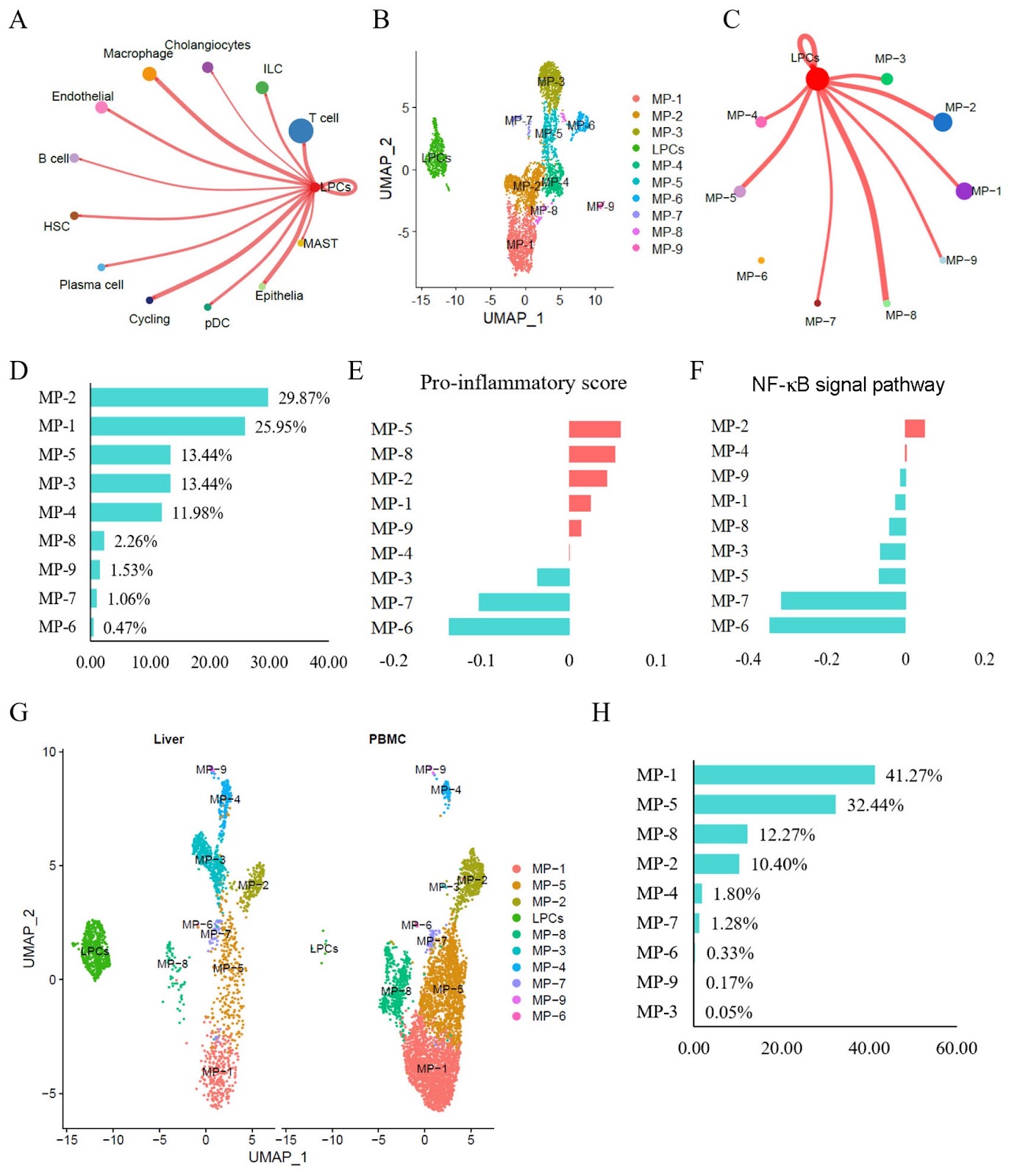
Resident CD24+LCN2+ LPCs aggravate fibrosis and inflammatory progression via the recruitment of TPPP3+COL10A1+ macrophages in NASH

Authors: Min Ding, Xiaoshu Qi, Weijian Huang, Yan Lin, Hexin Yan

Resident CD24+LCN2+ liver progenitor cells (LPCs) reportedly contribute to the expanding ductular reaction and macrophage-mediated inflammation associated with chronic liver damage. Both ductular reactions and macrophage-driven inflammation are associated with liver fibrosis and injury in various mouse liver disorders. This study aims to investigate the molecular phenotypes of LPCs and their regulatory mechanisms in humans with non-alcoholic steatohepatitis (NASH). Single-cell RNA sequencing (scRNA-seq) datasets are used to characterize the status and molecular phenotypes of LPCs in clinical NASH samples. To elucidate the regulatory mechanisms of LPCs, CellChat and NicheNet are employed to assess cell-cell communication between LPCs and other cell types. The findings are validated using RNA sequencing datasets associated with NASH progression, NASH mouse models (CDAHFD and HFD), and human NASH liver samples. Results show that resident CD24+LCN2+ LPCs are identified and found to be significantly enriched in NASH patients. Cell communication analyses predict strong interactions between LPCs and proinflammatory macrophage subtypes. Additionally, in NASH, the liver recruits peripheral blood mononuclear cell (PBMC)-derived macrophages and polarizes them into proinflammatory subtypes. The macrophage subtype MP-2 is identified as the primary recipient of LPC-derived signals, exhibiting marked hyperactivation of the NF-κB pathway and a strong association with liver fibrosis. Finally, the MP-2 markers COL10A1 and TPPP3 are characterized and validated. In summary, this study reveals that resident CD24+LCN2+ LPCs are activated in NASH and contribute to fibrosis progression by promoting the activation of the proinflammatory COL10A1+TPPP3+ macrophage subtype.

Resident CD24+LCN2+ LPCs aggravate fibrosis and inflammatory progression via the recruitment of TPPP3+COL10A1+ macrophages in NASH
Graphical Abstract
Original ResearchVol. 57, Issue 12 • pp. 2074-2093DOI: 10.3724/abbs.2025100

Characterization of the mechanisms underlying sulfasalazine-induced ferroptotic cell death: role of protein disulfide isomerase-mediated NOS activation and NO accumulation

Authors: Yi-Chen Jia, Jia-Ling Zhong, Xiangyu Hao, Bao Ting Zhu

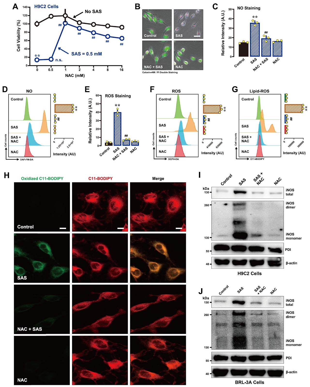
Sulfasalazine (SAS), a clinically utilized anti-inflammatory drug, has been shown to induce ferroptosis by inhibiting system Xc− activity, thereby causing cellular glutathione depletion. Recently, protein disulfide isomerase (PDI) was shown to be an upstream mediator of the oxidative cell death (oxytosis/ferroptosis) induced by glutamate, erastin, RSL3 and SAS. The present study aims to further characterize the detailed biochemical and cellular mechanisms of SAS-induced ferroptosis in two cell lines, i.e., H9C2 rat cardiomyocytes and BRL-3A rat hepatocytes, with a focus on elucidating the critical role of PDI in mediating SAS-induced toxicity. We find that SAS can induce ferroptosis in H9C2 and BRL-3A cells, which is accompanied by a sequential increase in the buildup of cellular nitric oxide (NO), reactive oxygen species (ROS) and lipid-ROS. SAS activates PDI-mediated dimerization of inducible NO synthase (iNOS) and cellular accumulation of NO, and these effects are followed by ROS and lipid-ROS accumulation. Furthermore, SAS markedly upregulates the iNOS protein levels in these cells. Knockdown of PDI or pharmacological inhibition of PDI catalytic activity effectively suppresses SAS-induced iNOS dimerization, abrogates SAS-induced accumulation of NO, ROS and lipid-ROS, and prevents ferroptosis. On the other hand, PDI activation through the use of TrxR1 inhibitors sensitizes these cells to SAS-induced ferroptosis. These findings provide further experimental support for a pivotal role of PDI in SAS-induced cytotoxicity through the activation of the PDI-NOS-NO axis, which then leads to the accumulation of cellular ROS and lipid-ROS and ultimately the induction of oxidative cell death.

Characterization of the mechanisms underlying sulfasalazine-induced ferroptotic cell death: role of protein disulfide isomerase-mediated NOS activation and NO accumulation
Graphical Abstract
Original ResearchVol. 57, Issue 12 • pp. 1983-1998DOI: 10.3724/abbs.2025109

Rhamnose alleviates the proinflammatory response during endotoxemia via the CEACAM1/LGALS9-p38 axis

Authors: Rongjuan Wei, Tao Zhong, Ke Deng, Xianglong Zhang, Dongping Li, Meiling Chen, Ping Chang, Peng Wu, Zhanguo Liu

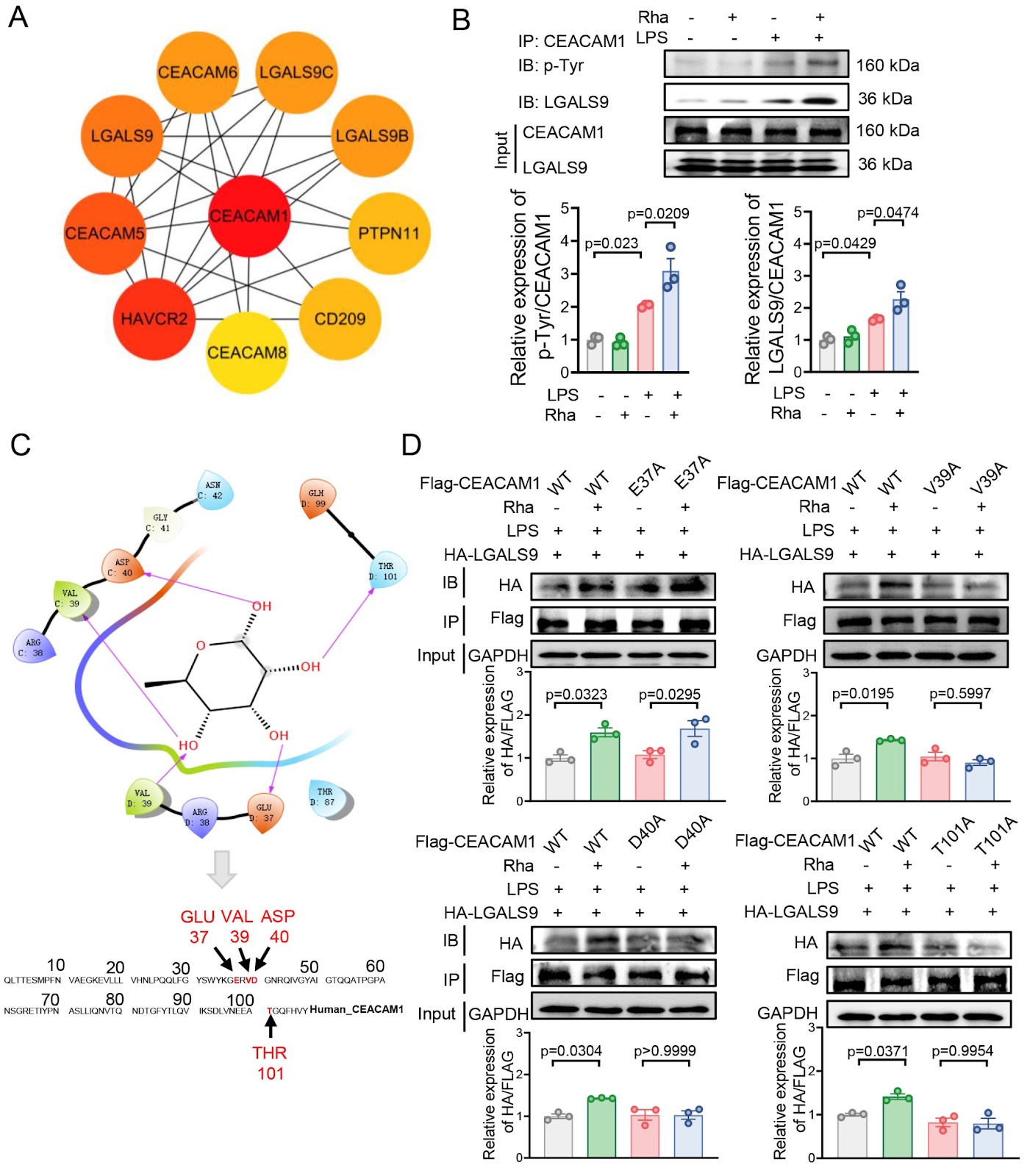
Gut microbiota plays an important role in orchestrating the host immune response. We previously reported that gut microbiota-derived rhamnose enhances the phagocytosis of macrophages, upon which we further asked whether rhamnose has modulatory effects on inflammation. Here, we show that, in an LPS-induced endotoxic mouse model, plasma rhamnose levels are increased. This bacteria-derived sugar alone does not impact inflammatory cytokine homeostasis or cause organ damage. In contrast, it is able to alleviate endotoxin-induced systemic inflammation and organ damage. Mechanistically, in macrophages in vitro, rhamnose binds to the V39, D40, and T101 sites of carcinoembryonic antigen-associated cell adhesion molecule 1 (CEACAM1), subsequently promoting the interaction between CEACAM1 and galectin 9 (LGALS9), which increases the protein level of dual-specificity protein phosphatase 1 (DUSP1). This inhibits p38 phosphorylation and thus attenuates the LPS-triggered expressions of proinflammatory factors. Collectively, our results suggest that rhamnose signals via the CEACAM1/LGALS9-p38 axis, which suppresses endotoxemia-associated inflammation, and that rhamnose is a candidate anti-inflammatory agent for the control of infection-induced organ damage.

Rhamnose alleviates the proinflammatory response during endotoxemia via the CEACAM1/LGALS9-p38 axis
Graphical Abstract
Original ResearchVol. 57, Issue 12 • pp. 1969-1982DOI: 10.3724/abbs.2025145

Angptl4 is upregulated by microenvironmental factors during the wound healing process and promotes epidermal stem cell proliferation via PRL8a6

Authors: Siyuan Yu, Pengxiang Ji, Ting Du, Zuohua Liu, Yuan Yang, Zhenkun Lv, Lei Xu, Qianheng Jin, Weijuan Gong, Yingying Le, Yi Fu, Ruixing Hou

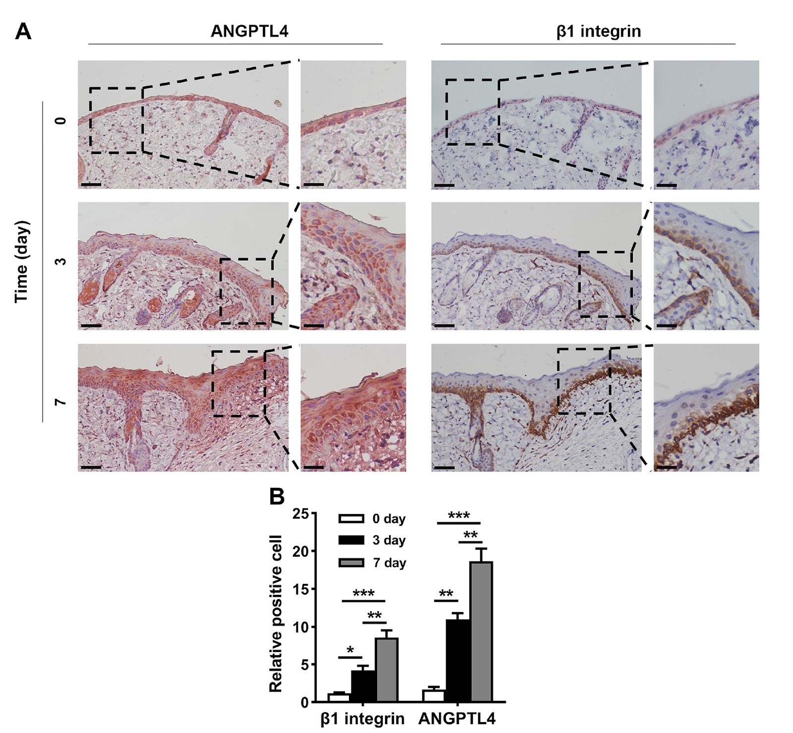
Angiopoietin-like 4 (ANGPTL4) expression is increased in wound tissue and contributes to wound healing. However, the underlying mechanisms are not fully understood. Here, we demonstrate that ANGPTL4 expression is significantly increased in epidermal stem cells (EpSCs) in the periwound epidermis during wound healing in mice. Increased Angptl4 expression is positively correlated with increased expressions of tumor growth factor-α, interleukin-1β, epidermal growth factor, nerve growth factor, fibroblast growth factor 7, and transforming growth factor-β1. Each of these molecules induces Angptl4 expression in mouse EpSCs. RNA sequencing of EpSCs derived from wild-type and Angptl4 knockout (Angptl4–/–) mice reveals altered expressions of genes involved in the cell cycle and cell proliferation in Angptl4–/– EpSCs, including a decrease in cyclin E2/A2/B1 and cyclin-dependent kinase 1 (Cdk1) expression; an increase in Cdk inhibitor 2a (Cdkn2a) and Cdkn2b expression; and a decrease in the prolactin (PRL) family members Prl2a1, Prl8a1, Prl8a9, and Prl8a6. Mechanistic studies reveal that ANGPTL4 stimulates EpSC proliferation via PRL8a6-mediated upregulation of cyclins A2/E2/B1 and Cdk1, downregulation of Cdkn2a, and acceleration of cell cycle progression from the G1 to the S and G2 phases. In vivo studies demonstrate that Prl8a6 mRNA is upregulated by ANGPTL4 in mouse periwound tissue during skin wound healing. Knockdown of Angptl4 or Prl8a6 in periwound skin tissue impairs EpSC proliferation and delays wound re-epithelialization. In conclusion, our study demonstrates that, after skin injury, elevated levels of proinflammatory cytokines and growth factors in periwound tissue stimulate Angptl4 expression in EpSCs and that ANGPTL4 promotes EpSC proliferation by increasing Prl8a6 expression, thereby accelerating wound re-epithelialization.

Angptl4 is upregulated by microenvironmental factors during the wound healing process and promotes epidermal stem cell proliferation via PRL8a6
Graphical Abstract
Original ResearchVol. 57, Issue 12 • pp. 2062-2073DOI: 10.3724/abbs.2025097

Integration of the hammerhead ribozyme into structured RNAs to measure ligand-binding events for riboswitch candidates and aptamers

Authors: Shenglan Zhang, Yinghong Lin, Ting Gao, Binfen Chen, Weibin Wu, Shanshan Fang, Kexin Fan, Yuqing Lai, Yezi Lin, Rongqin Ke, Sanshu Li

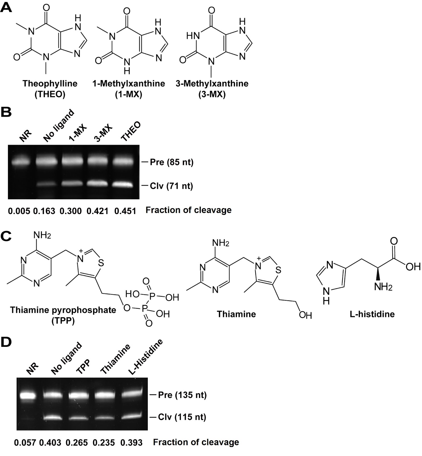
Some structured RNAs, such as riboswitches and aptamers, can bind to their cognate ligands and have been used in biosensors and gene expression control elements. However, current methods for detecting ligand binding to structured RNAs are either severely limited or inconvenient. In this study, we design a multibase pair bridge to integrate a hammerhead ribozyme into structured RNAs to detect ligand binding events. The experimental results demonstrate that the length of the bridge has a significant effect on the cleavage of the ribozyme; optimal cleavage can be achieved with three to six base pairs in the bridge. The dissociation constant (KD) values obtained through this method are in agreement with those determined by in-line probing techniques, and 1 pmol of allosteric ribozyme RNA is sufficient for measurement. We apply this method to evaluate the binding affinity of the riboswitch candidate Motif_9307. Our findings indicate that this motif has no binding affinity for S-adenosylmethionine or several other tested ligands, which is consistent with the results of the in-line probing experiments. Notably, our method reveals an increase in cleavage activity when yeast extract is added as a mixture of ligands, suggesting that the ligand of Motif_9307 is present in the extract. In conclusion, we develop an alternative approach for measuring ligand binding events associated with riboswitch candidates and aptamers.

Integration of the hammerhead ribozyme into structured RNAs to measure ligand-binding events for riboswitch candidates and aptamers
Graphical Abstract
Original ResearchVol. 57, Issue 12 • pp. 1953-1968DOI: 10.3724/abbs.2025089

LPS mediates cuproptosis and inflammation in THP-1 macrophages through HKDC1

Authors: Langlin Ou, Zitong Meng, Jian Mei, Hao Yuan, Xiangrui Zhu, Xiaoying Wang, Ao Shen, Zhaosi Wang, Lixin Zhang, Song Wang, Yingli Chen, Xiangming Pang, Yuxiang Liu, Yadong Xu, Cui Ma

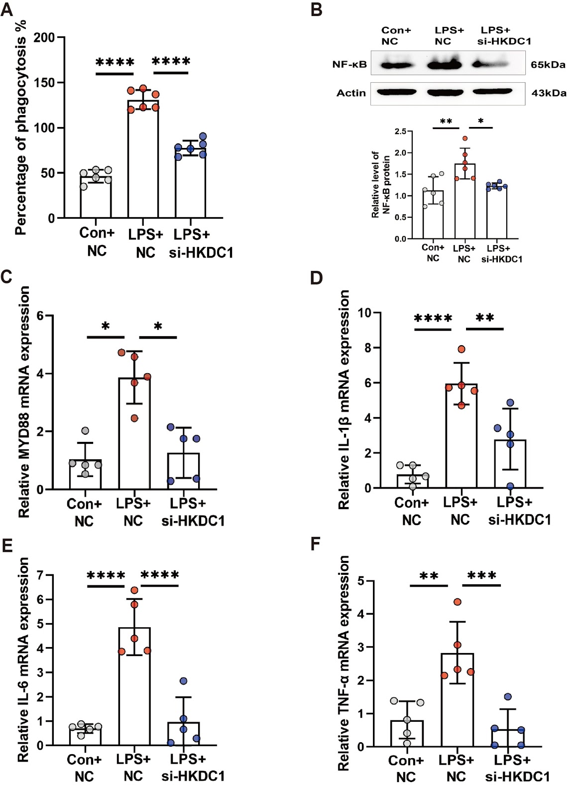
Cuproptosis is a recently identified form of copper-driven cell death characterized by the aggregation of acylated proteins and proteotoxic stress in the mitochondrial tricarboxylic acid cycle, which plays a role in inflammation. Recent studies suggest that hexokinase structural domain protein 1 (HKDC1), a fifth hexokinase, is involved in regulating mitochondrial function. However, the role of HKDC1 in cuproptosis and LPS-induced macrophage inflammation remains unclear. Here, we assess macrophage plasticity using CCK8 viability assays and phagocytosis activity experiments in an in vitro inflammatory model of THP-1 cells. We measure the levels of inflammatory factors and cuproptosis-related proteins using western blot analysis and RT-qPCR. Additionally, we examine the expression and localization of the HKDC1 protein using ChIP-qPCR and immunofluorescence staining. We find that LPS promotes the expressions of inflammatory factors and decreases cuproptosis levels in THP-1-derived macrophages while also activating glycolysis and inducing the expression of HKDC1 via the Toll-like receptor 4 (TLR4) receptor. We further demonstrate that HKDC1 knockdown inhibits glycolysis and induces cuproptosis. Mechanistically, we provide the first evidence that LPS promotes the binding of Yin Yang 1 (YY1) to the HKDC1 promoter, thereby regulating HKDC1 transcription. HKDC1 interacts with heat shock cognate B (HSCB) and ferredoxin 1 (FDX1), leading to increased intracellular copper levels and subsequent cuproptosis. HKDC1 knockdown in vivo alleviates acute sepsis by activating copper-dependent cell death pathways. Collectively, our findings suggest that LPS mitigates cuproptosis and promotes inflammation via HKDC1, suggesting a new cuproptosis-dependent anti-inflammatory strategy.

LPS mediates cuproptosis and inflammation in THP-1 macrophages through HKDC1
Graphical Abstract
Original ResearchVol. 57, Issue 12 • pp. 1939-1952DOI: 10.3724/abbs.2025171

Single-cell transcriptomics reveals apolipoprotein A4-mediated metabolic-immune reprogramming in lymphocytes during early obesity-related chronic kidney disease

Authors: Yang Wei, Ting Zhang, Yingying Jin, Xiaohuan Liu, Jinting Zhou, Na Huang, Yiying Wang

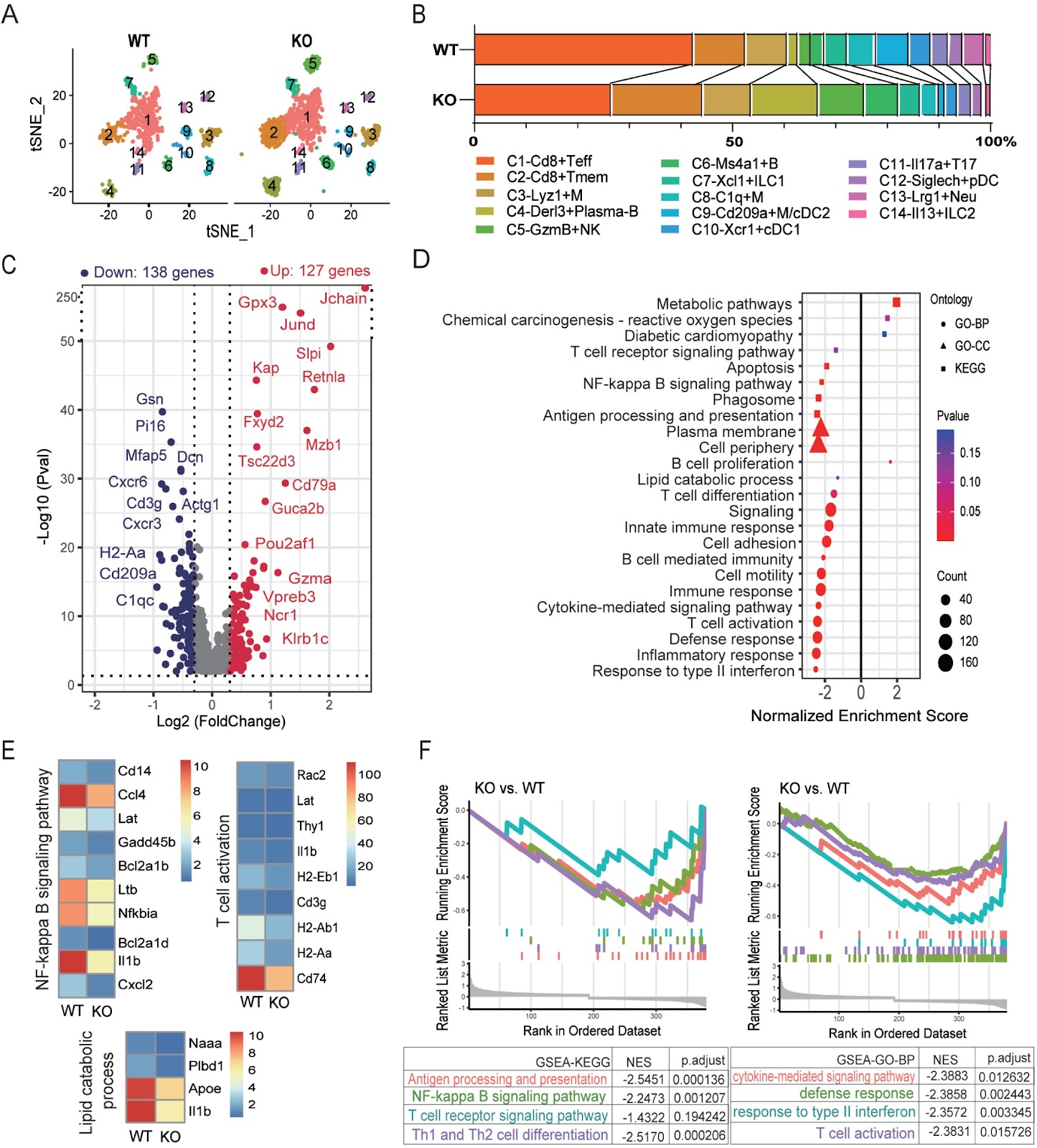
Obesity-induced metabolic inflammation is a key driver of chronic kidney disease (CKD), with immune dysregulation, particularly among lymphocytes, contributing to early disease pathology. To explore the role of apolipoprotein A4 (Apoa4) in regulating immune cell metabolism and function, we establish high-fat diet-induced obese (DIO) models using wild-type and Apoa4-knockout (KO) mice. KO mice exhibit exacerbated insulin resistance and renal lipid accumulation. Single-cell RNA sequencing reveals that Apoa4 deletion remodeled the renal immune-metabolic landscape. This remodeling broadly compromises the immune functions of T, NK, and B cells, even as it expands the proportions of cytotoxic Gzma+ NK cells and Derl3+ plasma cells. Mechanistically, Apoa4 deletion aggravates metabolic dysregulation and oxidative stress and downregulates the expression levels of key effector genes, including Ifng and Il1b. Furthermore, the regulatory network activities of key transcription factors, such as Lef1 and Runx3 in Cd8+ T cells; Irf8, T-bet, and Eomes in NK cells; and Tcf4, Lmo2, and Xbp1 in B cells, are perturbed. CellChat analysis predicts disruptions in pro-inflammatory (IFN-II and IL-1), immunoregulatory (FASLG), and metabolic regulatory (ENHO and ANGPTL) signaling, alongside enhanced IL-2-mediated suppression. These findings are corroborated by flow cytometry, immunofluorescence staining, and qPCR. Our results establish Apoa4 as a crucial regulator of lymphocyte metabolic and immune homeostasis in the early stages of obesity-associated CKD.

Single-cell transcriptomics reveals apolipoprotein A4-mediated metabolic-immune reprogramming in lymphocytes during early obesity-related chronic kidney disease
Graphical Abstract
Original ResearchVol. 57, Issue 12 • pp. 2124-2128DOI: 10.3724/abbs.2025055

Quantitative liquid chromatography-tandem mass spectrometric analysis of 11dH-TXB2 and creatinine in urine

Authors: Chunyan Li, Wuzheng Liu, Yana Xiao, Tenglong Dai, Yu Su, Yubin Wang, Ao Zhang, Ruichen Liu, Xianglong Zhao, Zhao Zhang, Shangqi Yin, Jun Wu

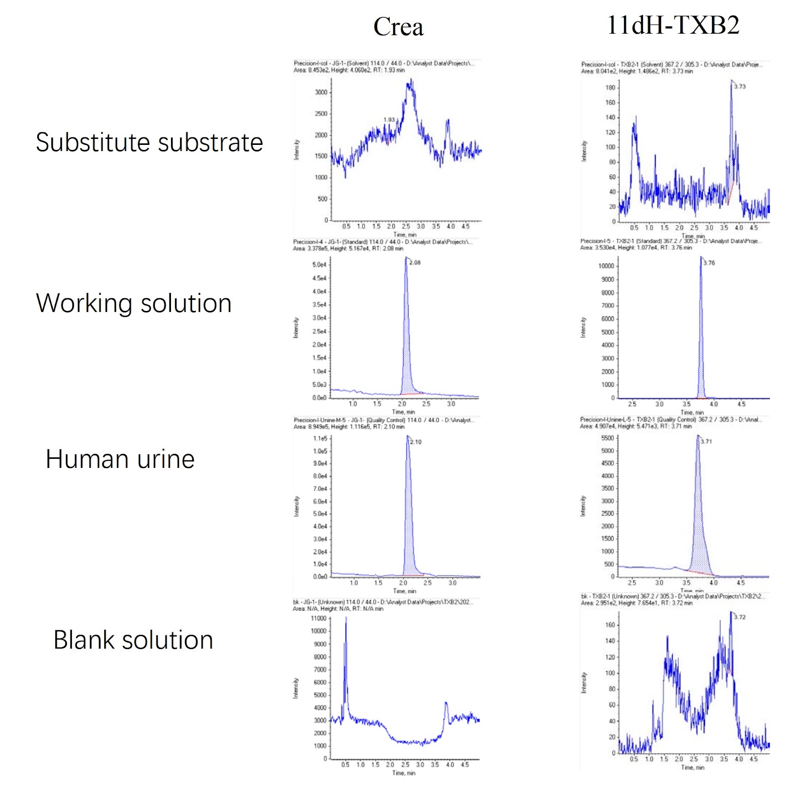
Platelets circulate in an inactive form in the body until they contact with defective areas of endothelial cells or encounter a clotting cascade [1]. Activated platelets release and express bioactive substances and acquire the ability to bind plasma fibrinogen. Abnormal activation of platelets is involved in atherosclerosis and thrombosis [2,3]. When platelets are stimulated and activated, phospholipase A2 is activated at the same time, which then cleaves membrane phospholipids and frees arachidonic acid (AA) [4]. The latter catalyzes thromboxane A2 (TXA2) via thromboxane synthetase [5], which is induced by the cyclooxygenase COX-1 to produce prostaglandins G2 and H2 [6]. TXA2 is highly unstable, with a half-life of only 30 s, and it is rapidly hydrolyzed to relatively stable thromboxane B2 (TXB2), which is then converted in the liver to 11-dehydrothromboxane B2 (11dH-TXB2), which has a longer half-life and is excreted in the urine [7]. Dehydrothromboxane B2 is the final stable metabolite of thromboxane A2, which is derived only from arachidonic acid metabolism and can represent thromboxane A2 level in the body [8]. Specifically, by inhibiting the action of COX-1, the most important enzyme in the process of arachidonic acid metabolism, aspirin inhibits the production of thromboxane A2, that is, the concentration of TXA2 affects the effect of aspirin on platelet aggregation. However, the half-life of TXA2 (including the metabolic intermediate TXB2) is too short to be accurately measured, so the detection of its metabolic end product 11dH-TXB2 can very accurately reflect the sensitivity of the body to aspirin [7]. The concentration of 11dH-TXB2 in the serum correlates well with the concentration of 11dH-TXB2 in the urine, so the determination of 11dH-TXB2 in the urine can more effectively reflect the production of TXA2 in vivo [8]. The 11dH-TXB2 concentration needs to be corrected with the urinary creatinine concentration to rule out the effects of the urine concentration and renal function, so random urine samples can be used for testing [6]. Aspirin can acetylate serine at the key site of cyclooxygenase and thus irreversibly inhibits the activity of COX-1, reduces the synthesis of TXA2, and blocks the production of TXA2 and its induced platelet aggregation. Low-dose aspirin (30–75 mg/day) can effectively inhibit 95% of COX-1 activity [7]. Since the production of TXA2 in serum is largely dependent on platelet COX-1 (a therapeutic target of aspirin), 11dH-TXB2 can be used as a monitor for aspirin-induced platelet inhibition [3]. Creatinine is a metabolic byproduct of muscle metabolism that is primarily excreted via glomerular filtration, and its level is indicative of renal function [9]. The 24-h creatinine clearance can also be used to determine the integrity of the sample or to correct the urine sample concentration with the creatinine ratio [10]. Currently, creatinine detection methods include the Jaffe method, enzymolysis spectrophotometry, HPLC, capillary electrophoresis, capillary zone electrophoresis, gas chromatography tandem mass spectrometry (GC-MS) and liquid chromatography tandem mass spectrometry (LC-MS/MS) [11]. At present, there is no method for the simultaneous detection of 11dH-TXB2 and creatinine. When both analytes are needed, separate tests must be performed, increasing the workload and sample volume requirements. Thus, developing a method that enables the concurrent quantification of 11dH-TXB2 and creatinine in a single assay remains a critical challenge. The aim of this study was to provide a method for the simultaneous detection of 11dH-TXB2 and creatinine and to alleviate the problem that 11dH-TXB2 and creatinine cannot be simultaneously detected. By developing a standardized quantitative approach for measuring 11dHTXB2 and creatinine in human urine, this study aims to provide reliable concentration data, thereby facilitating further clinical research and methodology optimization.
Quantitative liquid chromatography-tandem mass spectrometric analysis of 11dH-TXB2 and creatinine in urine
Graphical Abstract
Original ResearchVol. 57, Issue 12 • pp. 2133-2136DOI: 10.3724/abbs.2025048

Withaferin A combined with ricolinostat: a potent synergistic therapy for cervical cancer through regulating p53 ubiquitination and acetylation

Authors: Tian Chen, Yiting Xu, Kunming Yang, Yutong Du, Zhuan Zhu, Lingling Xu, Xinrong Wang, Yi Yin, Yu Hu, Chengcheng Wang, Ronggui Hu, Chuanyin Li

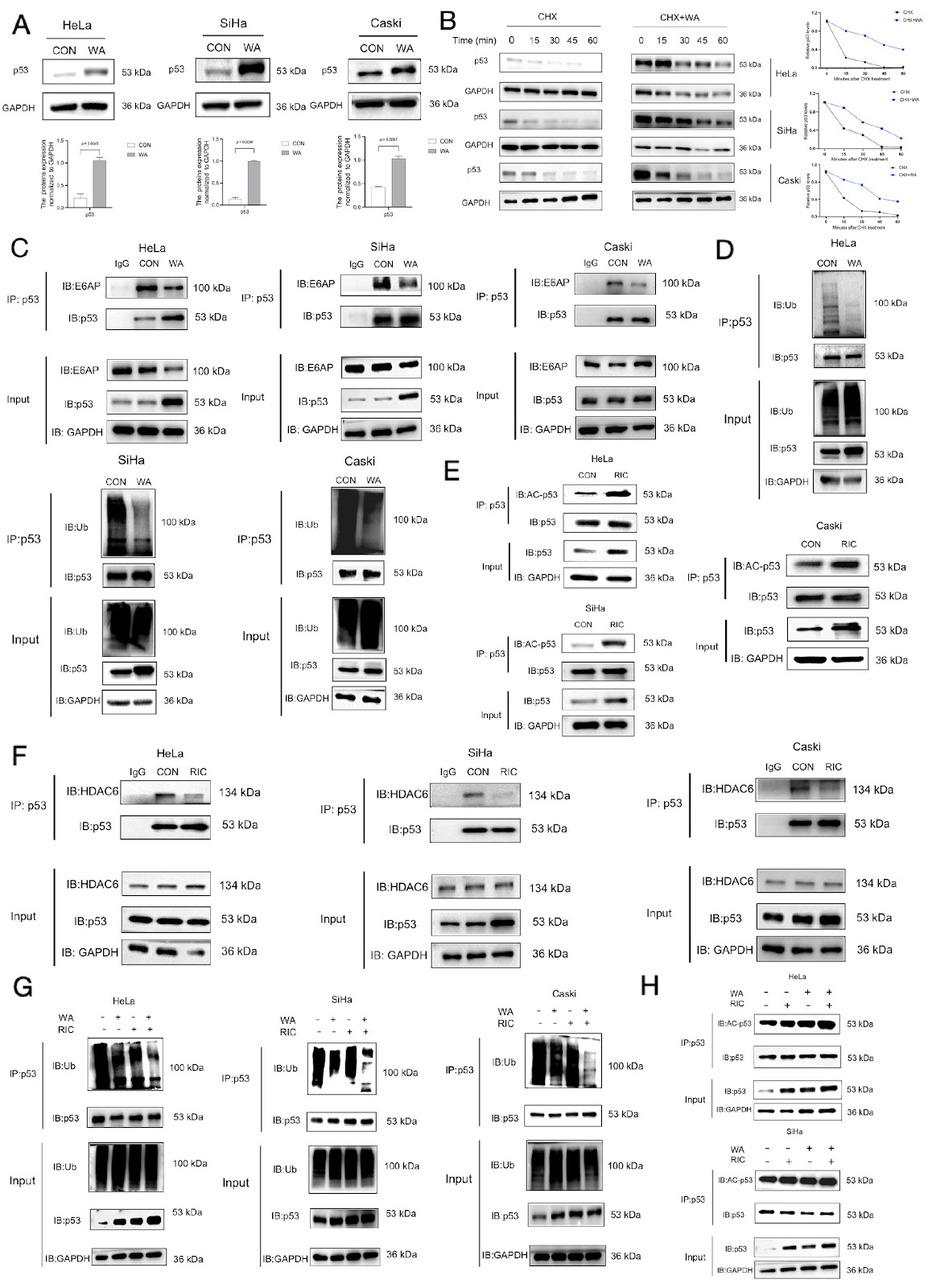
As a classic tumor suppressor gene, p53 has been extensively studied since its discovery in the mid-1980s. Research findings have revealed that p53 protein expression is suppressed in various cancers [1]. For example, in cervical cancer, p53 predominantly exists in a wild-type form to maintain its biological function [2]. Nevertheless, its tumor-suppressive activity is significantly impaired because of rapid protein degradation, short half-life, and low levels. Post-translational modifications (PTMs) of p53, such as ubiquitination, acetylation, phosphorylation and methylation, are critical regulators of its stability, activity, conformation, localization, and interactions with cofactors [3]. Among these, ubiquitination and acetylation play central roles in controlling p53 protein stability and activity [4]. Therefore, targeting p53 PTMs to modulate its ubiquitination and acetylation levels represents an effective strategy to increase its stability and tumor-suppressive function, offering a promising avenue for cervical cancer drug development. In 99% of cervical cancers (high-risk human papillomavirus-positive), E3 ubiquitin ligase E6-associated protein (E6AP) mediates the ubiquitination degradation of p53 [5], whereas histone deacetylase 6 (HDAC6) deacetylates p53. In this study, we explored the possibility of combining the natural product withferin A (WA) with the HDAC6 inhibitor ricolinostat (RIC) to treat cervical cancer cells, with a focus on the ubiquitination and acetylation of p53 and the consequences for its stability. These results suggested that the combination of WA and RIC is more effective than either treatment alone in inhibiting the degradation and increasing the stability of p53, thereby synergistically slowing the onset and progression of cervical cancer.

Withaferin A combined with ricolinostat: a potent synergistic therapy for cervical cancer through regulating p53 ubiquitination and acetylation
Graphical Abstract
Original ResearchVol. 57, Issue 12 • pp. 2137-2138DOI: 10.3724/abbs.2025233

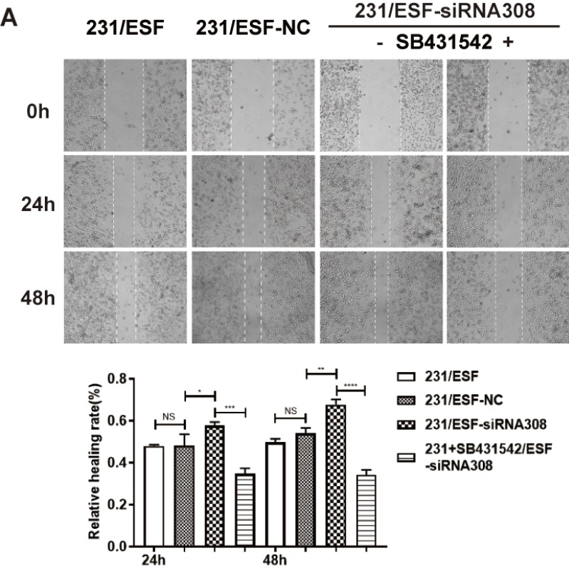
Caveolin-1-deficient fibroblasts promote migration, invasion, and stemness by activating the TGF-β/Smad signaling pathway in breast cancer cells

Authors: Qingyun Huang, Longyuan Wu, Yi Wang, Xinyu Kong, Xinhua Xiao, Qiyuan Huang, Miao Li, Yujia Zhai, Fuxiu Shi, Ruichen Zhao, Junpei Zhong, and Lixia Xiong

This is a corrigendum to the article 'Caveolin-1-deficient fibroblasts promote migration, invasion, and stemness by activating the TGF-β/Smad signaling pathway in breast cancer cells' published in Acta Biochim Biophys Sin 54: 1587–1598. The authors identified inaccuracies in the preparation of several figures (Figure 2D, 4A, and 5A) and have replaced them with corrected versions. The errors are strictly confined to figure presentation and do not impact the underlying data, statistical analysis, or main conclusions. The authors apologize for the oversight.

Caveolin-1-deficient fibroblasts promote migration, invasion, and stemness by activating the TGF-β/Smad signaling pathway in breast cancer cells
Graphical Abstract
Original ResearchVol. 57, Issue 12 • pp. 1923-1938DOI: 10.3724/abbs.2025099

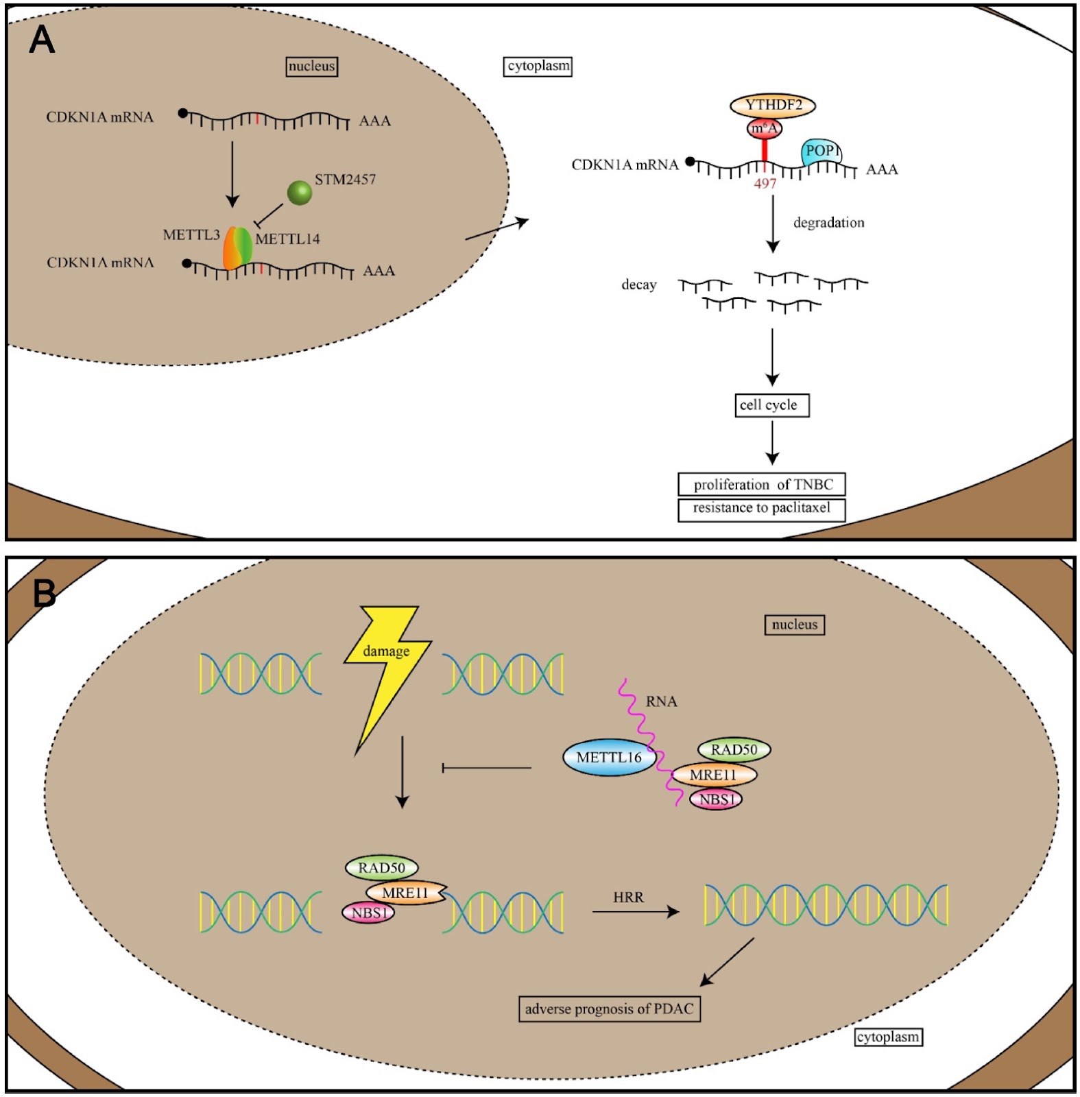
The dual role of RNA-binding proteins: promotion of tumorigenesis, drug resistance, and emerging therapeutic targets

Authors: Zemei Xu, Yukun Cui

Cancer is a complex and multifaceted disease characterized by a multitude of molecular factors. RNA-binding proteins (RBPs) have emerged as pivotal regulators of tumor development, progression, and chemoresistance through their interactions with target transcripts. These interactions regulate a multitude of processes, including alternative splicing, cleavage and polyadenylation, RNA localization, translation, N6-methyladenosine (m6A) RNA modification, and DNA double-strand break repair. The RBP family comprises over 2000 proteins and plays a critical role in oncogene expression, invasion, metastasis, and inhibition of apoptosis. However, the mechanisms by which RBPs selectively recognize RNAs remain an active area of research. In this review, we examine recent advancements in understanding RNA-binding domains and the RNA processes regulated by RBPs in tumorigenesis, summarize and highlight the roles of RNA-binding domains in cancers and the molecular mechanisms of RBPs in chemotherapy resistance, discuss the potential of targeting RBPs for cancer therapy and review RBPs that are dysregulated in cancers. Additionally, we highlight recently developed tools for predicting RBP-RNA binding activities to provide valuable support for ongoing research efforts.

The dual role of RNA-binding proteins: promotion of tumorigenesis, drug resistance, and emerging therapeutic targets
Graphical Abstract
Original ResearchVol. 57, Issue 12 • pp. 1999-2010DOI: 10.3724/abbs.2025066

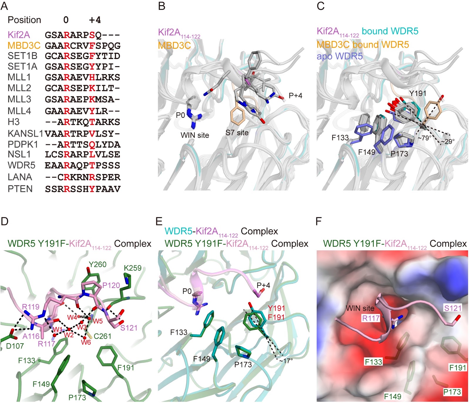
Crystal structures of Kif2A complexed with WDR5 reveal the structural plasticity of WIN-S7 sites

Authors: Yang Yang, Shuting Zhang, Zhangyu Wu, Wenwen Li, Xuefang Sun, Yumi Xuan, Tianrong Hang, Li Xu, Xuemin Chen

Chromosome congression and spindle assembly are essential for genomic stability and proper cell division, with deficiencies in these processes linked to tumorigenesis. WD repeat-containing protein 5 (WDR5), a core component of the mixed lineage leukemia (MLL) methyltransferase complex, directly binds to kinesin family member 2A (Kif2A) to regulate these mitotic events. Despite the importance of this interaction, its structural basis for Kif2A recognition by WDR5 remains unclear. Here, we determine the crystal structure of WDR5 in complex with a Kif2A-derived peptide (residues 114–122) at a resolution of 1.85 Å. Structural analysis reveals that Kif2A engages both the WIN and S7 sites of WDR5 via Arg117 and Ser121, with Ser121 forming hydrogen bonds with WDR5 Tyr191 and Lys259, driving Tyr191 rotation and opening the S7 pocket. Additional structures of WDR5 complexed with truncated or mutated Kif2A peptides and a WDR5 Y191F variant highlight the dynamic nature of Tyr191. Notably, anti-WDR5 compounds exhibit a similar binding mode at the WDR5 WIN-S7 site. The results of mutagenesis combined with isothermal titration calorimetry (ITC) assays underscore the critical roles of Arg117 and Ser121 in mediating the binding of Kif2A to WDR5. In summary, our findings provide atomic-level insights into the molecular mechanisms underlying the non-canonical mitotic function of the MLL/WDR5 complex and highlight WIN-S7 sites as promising therapeutic targets for diseases associated with chromosomal instability, such as cancers.

Crystal structures of Kif2A complexed with WDR5 reveal the structural plasticity of WIN-S7 sites
Graphical Abstract