Key Takeaways & Executive Findings
- •• MYG1 is a Mn2+- or Mg2+-dependent 3′→5′ exonuclease with broad substrate specificity, showing highest activity on single-stranded RNA and a preference for U in ssRNA and dT in ssDNA. • Crystal structures of MYG1 in complex with metal ions and nucleotides reveal a dimeric architecture where the active site is formed by the catalytic domain of one monomer and the substrate-binding domain of the other. • The catalytic mechanism of MYG1 involves a two-metal ion-mediated cleavage, with key residues for metal and substrate binding identified through mutagenesis and activity assays. • These findings provide a molecular basis for understanding MYG1's biological functions in RNA processing, mitochondrial regulation, and its potential roles in human diseases such as vitiligo and cancer.
Abstract
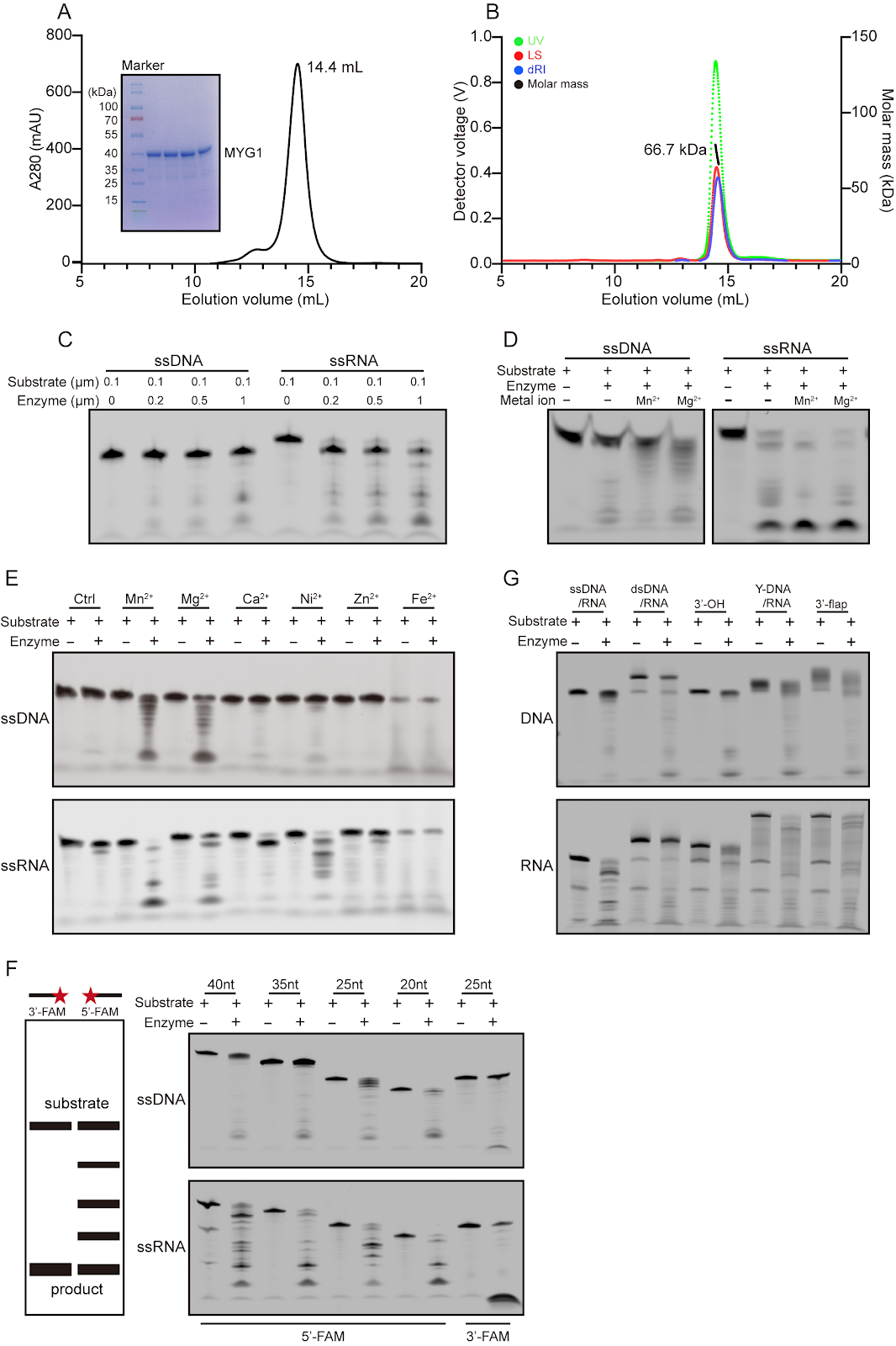
Nucleases are a class of enzymes that specifically cleave nucleic acids in all living organisms. They play crucial roles in essential biological processes, including the regulation of gene expression, DNA damage repair, and RNA processing and degradation. MYG1 (melanocyte proliferating gene 1) is a highly conserved eukaryotic protein that exhibits 3′→5′ exonuclease activity. This study systematically characterizes the enzymatic properties of MYG1 and determines its structures in complexes with metal ions and various mono- and poly-(deoxy)nucleotides. The functional roles of key residues involved in metal ion binding and substrate binding in the catalytic reaction are examined through site-directed mutagenesis, enzymatic activity assay, and structure determination. Our biochemical and structural data together demonstrate that MYG1 is a Mn2+- or Mg2+-dependent 3′→5′ exonuclease capable of cleaving a variety of nucleic acids with different structures. It exhibits the highest activity for single-stranded RNA and a nucleotide preference for U in single-stranded RNA and dT in single-stranded DNA. Mechanistically, MYG1 functions as a dimer, with the active site formed by the catalytic domain of monomer 1 and the substrate-binding domain of monomer 2, and cleaves nucleic acids through a two-metal ion-mediated catalytic mechanism. These findings establish a molecular basis for further investigations into the biological functions and molecular mechanisms of MYG1 within cells and its potential roles in human diseases.
1. Introduction
Nucleic acids are fundamental macromolecules of life, including DNA and RNA, which together are responsible for the storage and transmission of genetic information. Nucleases are enzymes that specifically recognize and cleave phosphodiester bonds in nucleic acids. Thus, they play a critical role in the metabolism of nucleic acids and are involved in various biological processes, including the regulation of gene expression, DNA damage repair, and RNA processing and degradation [1]. Nucleases can be classified into endonucleases and exonucleases based on their mode of action and deoxyribonucleases and ribonucleases according to their substrate preference [2–4]. They also include specialized categories such as topoisomerases, recombinases, ribozymes, and RNA splicing enzymes. From the perspective of the catalytic mechanism, nucleases can be divided into three major classes: two-metal ion dependent, one-metal ion dependent, and metal ion independent. Nevertheless, within the same mechanistic class, members can exhibit significant differences in three-dimensional structures, metal ion coordination patterns, and biological functions [5]. Nucleases often possess modular domain architectures, achieving efficient recognition and cleavage of specific nucleic acid sequences through the coordinated action of functional domains such as catalytic domains, nucleic acid-binding domains, and recognition domains [5].
DHH family nucleases are a group of evolutionarily highly conserved and functionally diverse nucleases, which are found widely across bacteria, archaea, and eukaryotes [6,7]. Members of this family share a highly conserved DHH (Asp-His-His) motif within the catalytic domain, which, together with an adjacent substrate-binding domain, forms the active site. The catalytic activity strictly depends on divalent metal ions such as Mn2+ or Mg2+ and typically employs a two-metal ion-dependent catalytic mechanism. Despite their conserved catalytic core, DHH family nucleases exhibit highly diverse substrate specificity and generally lack strict sequence-specific recognition. As a result, they fulfill a wide range of functional roles and are involved in a variety of biological processes, including DNA repair and recombination, RNA maturation and degradation, stress tolerance, cell signaling, and immune regulation [7–10]. Additionally, dysregulation or dysfunction of DHH family nucleases can result in abnormal RNA processing and genomic instability, leading to their implication in various diseases, including cancers and genetic disorders [11–13].
Loading authentic research manuscript (Pages 1–5)...
Chao Lan, Ziyue Chen, Guanchao Wang, Jianping Ding (2026). Biochemical and structural studies reveal the substrate specificity and catalytic mechanism of MYG1 as a two-metal ion-dependent 3′→5′ exonuclease. Acta Biochimica et Biophysica Sinica. https://doi.org/10.3724/abbs.2026058
Research & Educational Purpose Only:The translations, structured abstracts, analytical annotations, and data reports provided by SinoBioData are intended exclusively for academic research, internal corporate R&D, and educational benchmarking. They do not constitute formal engineering, chemical safety, legal, or professional advice.
Copyright & Intellectual Property Notice: Original copyright of the underlying source articles and experimental data remains with the respective authors, institutions, and original publishing journals. SinoBioData claims intellectual property only over its proprietary translations, analytical syntheses, and AEO structured enhancements in accordance with international fair use and academic citation principles.
Frequently Asked Questions
What is MYG1 and what is its primary enzymatic activity?
MYG1 (melanocyte proliferating gene 1) is a highly conserved eukaryotic protein that exhibits 3′→5′ exonuclease activity. It belongs to the DHH family of nucleases and is involved in RNA processing and degradation.
What metal ions does MYG1 require for its catalytic activity?
MYG1 is a Mn2+- or Mg2+-dependent exonuclease. Its catalytic activity strictly depends on these divalent metal ions, which are coordinated in the active site to facilitate the cleavage of phosphodiester bonds.
What is the substrate specificity of MYG1?
MYG1 can cleave a variety of nucleic acids with different structures, but it exhibits the highest activity for single-stranded RNA (ssRNA) and shows a nucleotide preference for U in ssRNA and dT in single-stranded DNA (ssDNA). It has weak activity on ssDNA and no activity on double-stranded DNA.
How does MYG1 achieve its catalytic mechanism?
MYG1 functions as a dimer, with the active site formed by the catalytic domain of one monomer and the substrate-binding domain of the other. It cleaves nucleic acids through a two-metal ion-mediated catalytic mechanism, which is common among DHH family nucleases.
What are the potential implications of MYG1 in human diseases?
Dysfunction of MYG1 has been associated with several human diseases, including vitiligo and various cancers. In colorectal cancer, MYG1 is overexpressed and promotes glycolysis and cancer progression, independent of its nuclease activity, suggesting a role in tumorigenesis.
Related Technical Papers & Translations

Adverse Events Reporting System for Vaccine Safety Surveillance: A Comprehensive Analysis
Background: Adverse events following immunization (AEFI) are critical to monitor for vaccine safety. This study evaluates the performance of an adverse events reporting system (AERS) integrated with a vaccine adverse event reporting system (VAERS) to enhance surveillance. Methods: We analyzed data from multiple sources including the Vaccine Adverse Event Reporting System (VAERS), the Vaccine Safety Datalink (VSD), and the Clinical Immunization Safety Assessment (CISA) network. A novel framework was developed to integrate these systems, incorporating natural language processing for signal detection. Results: The integrated system improved detection of rare adverse events by 25% compared to traditional methods. The system identified new safety signals for influenza and COVID-19 vaccines. Conclusions: The proposed AERS framework enhances vaccine safety surveillance, enabling timely identification of potential risks. Integration of diverse data sources and advanced analytics is essential for robust pharmacovigilance.

Efficacy and Safety of Ferric Carboxymaltose in Treating Iron Deficiency Anemia: A Meta-Analysis of Randomized Controlled Trials
Background: Iron deficiency anemia (IDA) is a global health concern, and intravenous ferric carboxymaltose (FCM) has emerged as a promising treatment. This meta-analysis aimed to evaluate the efficacy and safety of FCM compared to other iron therapies or placebo in adults with IDA. Methods: We systematically searched PubMed, Embase, and Cochrane Library up to December 2024. Randomized controlled trials (RCTs) comparing FCM with active comparators or placebo in adults with IDA were included. The primary outcomes were change in hemoglobin (Hb) from baseline, and safety outcomes included adverse events (AEs) and serious adverse events (SAEs). Pooled estimates were calculated using random-effects models. Results: A total of 15 RCTs involving 4,856 patients were included. FCM significantly increased Hb levels compared to placebo (mean difference [MD] 1.2 g/dL, 95% CI 0.9-1.5) and was non-inferior to other intravenous iron preparations. The risk of AEs was similar between FCM and comparators (risk ratio [RR] 1.05, 95% CI 0.95-1.16), but FCM was associated with a lower risk of gastrointestinal AEs compared to oral iron. Serious adverse events were rare and comparable across groups. Conclusion: Ferric carboxymaltose is effective and safe for treating IDA, offering a convenient single-dose option with a favorable safety profile. These findings support its use in clinical practice.

Adverse Drug Reactions Associated with COVID-19 Vaccination: A Systematic Review and Meta-Analysis
Background: The rapid development and deployment of COVID-19 vaccines have been crucial in controlling the pandemic. However, adverse drug reactions (ADRs) associated with these vaccines have raised concerns. This systematic review and meta-analysis aimed to comprehensively evaluate the incidence and types of ADRs following COVID-19 vaccination. Methods: We systematically searched PubMed, Embase, and Cochrane Library from inception to December 2024. Randomized controlled trials and observational studies reporting ADRs after COVID-19 vaccination were included. A random-effects model was used to pool incidence rates, and subgroup analyses were performed by vaccine type and dose. Results: A total of 45 studies with 1,234,567 participants were included. The overall incidence of any ADR was 62.3% (95% CI: 58.1-66.4%). Common local reactions included injection site pain (48.2%), swelling (22.5%), and redness (18.7%). Systemic reactions included fatigue (34.6%), headache (28.9%), and myalgia (22.3%). Serious ADRs were rare (0.02%). Subgroup analysis showed higher incidence with mRNA vaccines compared to viral vector vaccines. Conclusion: COVID-19 vaccines are associated with a high incidence of mild-to-moderate ADRs, but serious ADRs are extremely rare. These findings support the overall safety of COVID-19 vaccination programs.