🧬 SinoBioData Academic Portal
🏛️ Indexed Academic JournalOriginal: 干细胞研究与转化

Stem Cell Research & Therapy

Premier Chinese Biomedical Journal indexed in SinoBioData: Stem Cell Research & Therapy (干细胞研究与转化).

Total Research Papers: 200
Access: 100% Free Open Access
Browse by Publication Year & VolumeReset All Filters ✕

Published Research PapersFiltered: Year 2026 • 17 • 1

Showing 9 of 200 peer-reviewed papers with full Graphical Abstracts.

Original ResearchVol. 17, Issue 1 • pp. 1-18DOI: 10.1186/s13287-025-04774-9

Bone marrow–derived mesenchymal stem cells alleviate hepatic lipid metabolism disorders after scald injury: integrating liver transcriptome and metabolome

Authors: Zhian Chen, Ya'e Yang, Xiangwen Shi, Rensheng Yang, Wei Fang, Guangjin Liang, Yun Li, Jing Gao, Lihua Ma, Junchun Yang, Rongqing Pang

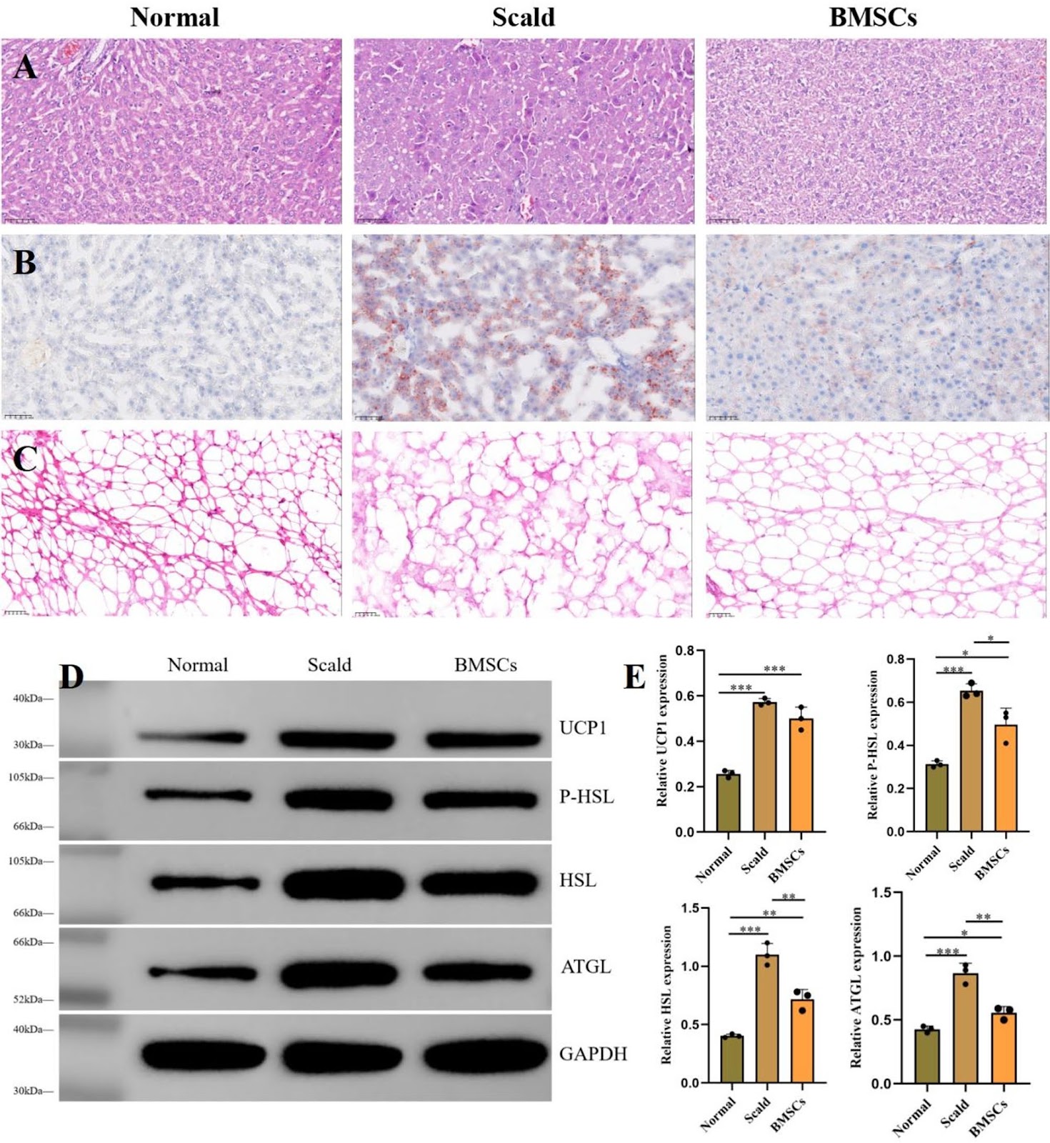
Previous studies have confirmed that scald injuries can lead to disturbances in hepatic lipid metabolism, and bone marrow-derived mesenchymal stem cells (BMSCs) have emerged as a promising therapeutic strategy for alleviating such disorders. However, research focusing on the regulation and restoration of liver lipid metabolic processes remains limited. In this study, we investigated the effects of BMSCs on hepatic lipid metabolism disorders induced by scald injury in rats through integrated transcriptomic and metabolomic analyses. The results demonstrated that portal vein infusion of BMSCs markedly improved body weight recovery, reduced hepatic lipid accumulation, normalized serum lipid profiles, and attenuated liver injury following scalding. Combined transcriptomic and metabolomic data further suggested that the therapeutic mechanism may involve inhibition of NF-κB/Gadd45a signaling in hepatocytes, restoration of sphingolipid metabolism, enhancement of hepatic lipid conversion, and suppression of adipocyte lipolysis. Overall, this study provides a theoretical basis for the potential clinical application of BMSCs in treating hepatic lipid metabolism disorders secondary to severe burn injury.
Bone marrow–derived mesenchymal stem cells alleviate hepatic lipid metabolism disorders after scald injury: integrating liver transcriptome and metabolome
Graphical Abstract
Original ResearchVol. 17, Issue 1 • pp. 182DOI: 10.1186/s13287-026-05006-4

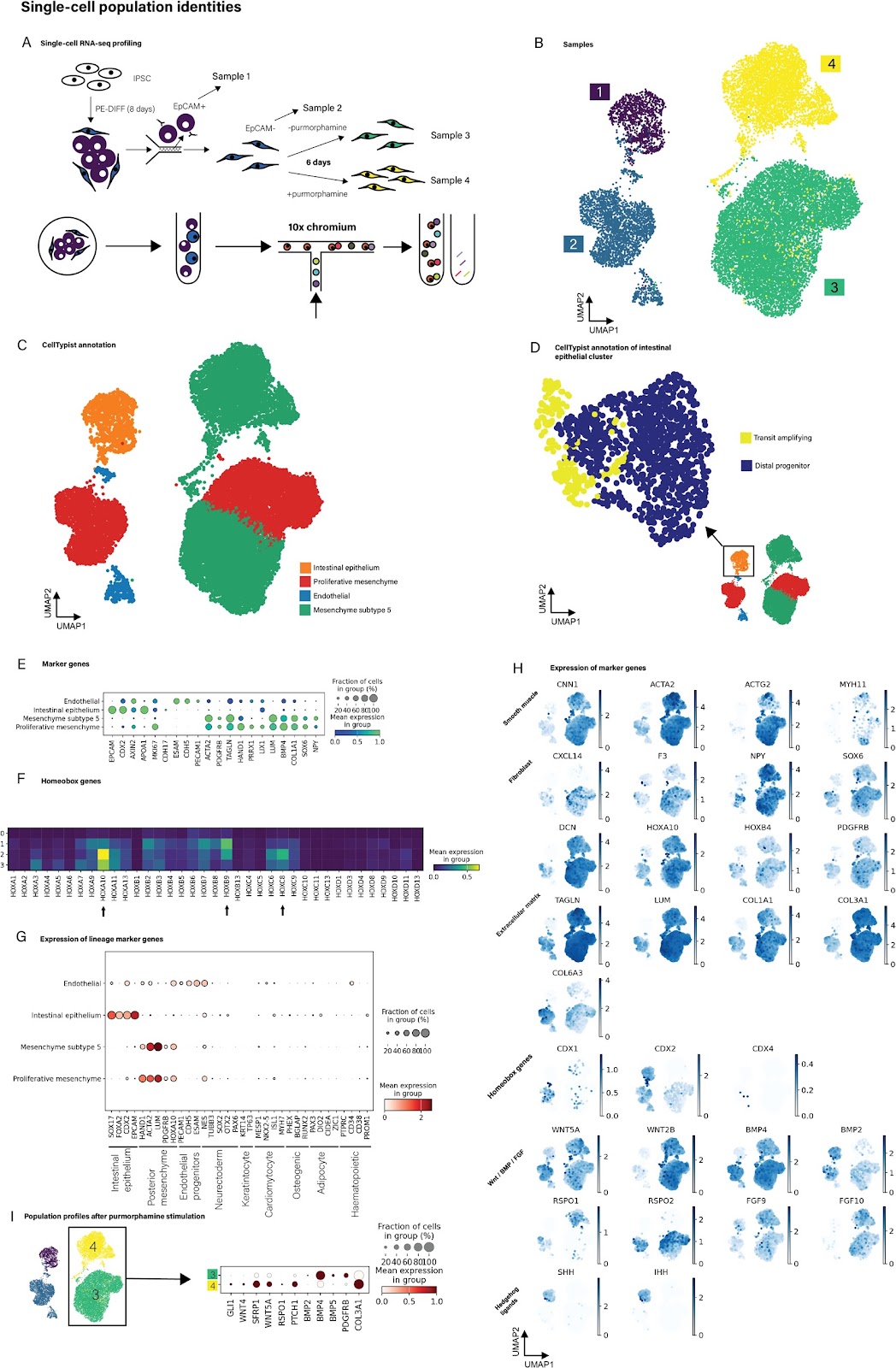
Generation of biologically responsive colon-like intestinal tissue patches from human induced pluripotent stem cells using a rapid co-differentiation platform

Authors: William Dalleywater, Alexander V. Predeus, Batuhan Cakir, Pavel Mazin, Jayakumar Vadakekolathu, Sergio Rutella, Marian L. Meakin, Alison A. Ritchie, Shamir Montazid, Sara Cuevas Ocaña, Nadine Holmes, Victoria Wright, Fei Sang, Silvia Santoni, Adam Bills, Declan Sculthorpe, Rasa Elmentaite, Sarah A. Teichmann, Shazia Irshad, Ian Tomlinson, Andrew Silver, Ricky D. Wildman, Nicholas R. F. Hannan, Felicity R. A. J. Rose, Mohammad Ilyas

The intestinal mucosa is a complex functional layer which is formed from a diverse range of cell types that include epithelial cells (within crypts and villi) and an array of mesenchymal cells. Many intestinal diseases involve loss of the surface mucosa which can be difficult to restore, and which delays healing and return to normal function. We reason that development of a transplantable intestinal mucosal tissue graft may be a potential therapeutic strategy to aid healing. To be clinically useful, such a tissue graft would need to be capable of rapid production, avoid the risk of host rejection and be demonstrably safe. To create a potential intestinal graft, we developed a novel early-stage human induced pluripotent stem cell (hiPSC) co-differentiation platform capable of generating multiple intestinal cell lineages (epithelial, mesenchymal and endothelial) in 8 days. This protocol is simple to implement, serum-free and greatly reduces the use of animal products. We confirmed the identity of cells by demonstrating that these cells had RNA and protein expression profiles typical of intestinal cell lineages. In particular, we used bulk and single-cell RNA sequencing to characterise global cellular transcriptional profiles robustly and showed that the cells have intestinal identity with early polarisation towards colonic differentiation. The results were replicated across multiple hiPSC lines and in an independent centre. We further cultured the derived cells on collagen hydrogels to form colon-like intestinal patches (CL-IPs). When transplanted into mouse subcutis, CL-IPs formed into colon-like tissue structures, including crypts, stromal and muscle layers. They also developed human-origin vasculature which underwent anastomosis with the murine vasculature to transport murine blood into the graft. Teratoma assays and molecular analyses showed no evidence of residual pluripotency. While at an early stage, this platform shows great potential for further development as a potential source for novel intestinal mucosal regeneration therapy. In addition, the platform is physiologically relevant and thus shows promise as the basis for a new generation of in vitro models of intestinal pathobiology.

Generation of biologically responsive colon-like intestinal tissue patches from human induced pluripotent stem cells using a rapid co-differentiation platform
Graphical Abstract
Original ResearchVol. 17, Issue 1 • pp. 79DOI: 10.1186/s13287-026-04900-1

Hybrid endometrial-derived hydrogel and human endometrial organoids synergize for uterine regeneration in an immunocompetent murine model

Authors: María Gómez-Álvarez, Clara Bueno-Fernandez, Emilio Francés-Herrero, Marcos Agustina-Hernández, Paula Alonso-Frías, Nadaya Corpas, Amparo Faus, Ana Díaz, Antonio Pellicer, Hortensia Ferrero, Irene Cervelló

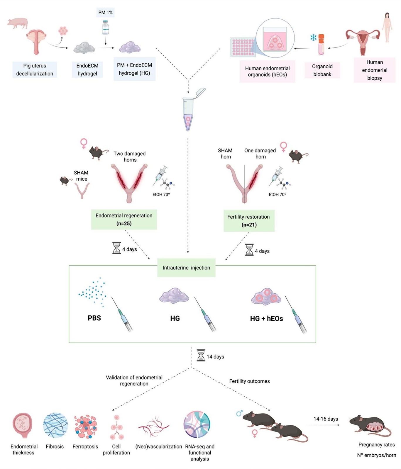
Background The human endometrium is a regenerative tissue essential for fertility, but pathological conditions like Asherman syndrome, endometrial atrophy, and thin endometrium can impair its function. Current therapies lack efficacy, driving demand for innovative regenerative therapies. In this context, endometrial-derived hydrogels and organoids have shown promise individually for tissue regeneration, but their combined therapeutic potential has not been previously evaluated in vivo. This study explores a dual regenerative strategy combining a hybrid hydrogel — composed of synthetic PuraMatrix® and endometrial extracellular matrix hydrogel — with human endometrial organoids in an immunocompetent murine model with uterine damage. Methods Endometrial damage model was established in female C57BL/6 mice (n = 46) via uterine injury using 70° ethanol. After 4 days of endometrial damage, human endometrial organoids were co-injected with the hybrid hydrogel into the uterine horns. Two weeks post-injection, a subset of mice (n = 25) was sacrificed for biocompatibility, histological, and transcriptomic analyses. Functional recovery of the endometrium was assessed in the remaining animals (n = 21) through fertility outcome evaluation. For endometrial regeneration analyses, normally distributed data were analyzed by one-way ANOVA and Tukey’s multiple comparisons, while non-normally distributed data were analyzed by the Kruskal–Wallis test with Dunn’s multiple comparisons. For fertility outcomes, t-test or Mann–Whitney U tests for 2-by-2 comparisons were performed.

Hybrid endometrial-derived hydrogel and human endometrial organoids synergize for uterine regeneration in an immunocompetent murine model
Graphical Abstract
Original ResearchVol. 17, Issue 1 • pp. 86DOI: 10.1186/s13287-026-04942-5

Correction: Development of a robust induced pluripotent stem cell atrial cardiomyocyte differentiation protocol to model atrial arrhythmia

Authors: Jordan Thorpe, Matthew D. Perry, Osvaldo Contreras, Emily Hurley, George Parker, Richard P. Harvey, Adam P. Hill, Jamie I. Vandenberg

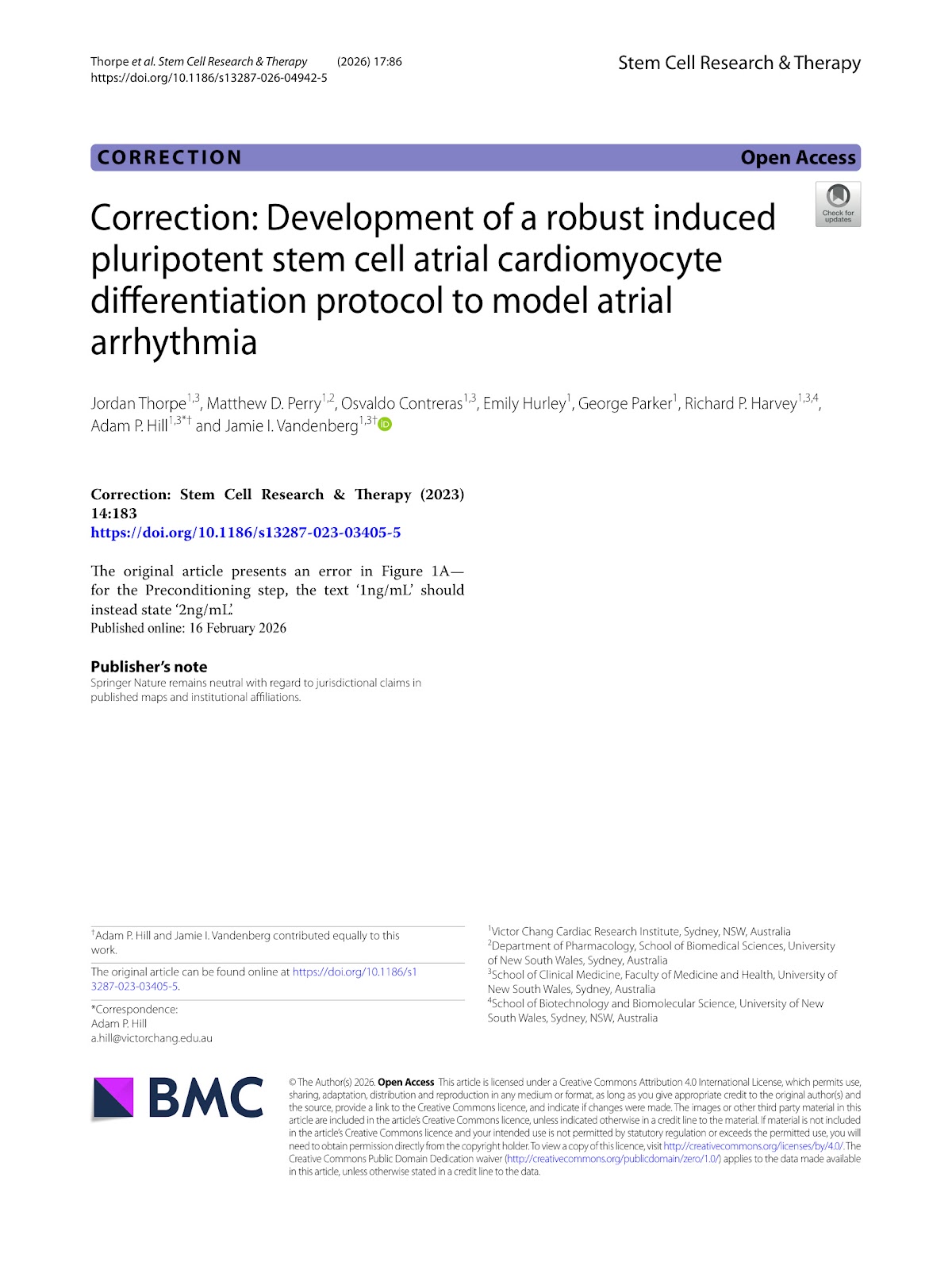
The original article presents an error in Figure 1A—for the Preconditioning step, the text ‘1ng/mL’ should instead state ‘2ng/mL’.

Correction: Development of a robust induced pluripotent stem cell atrial cardiomyocyte differentiation protocol to model atrial arrhythmia
Graphical Abstract
Original ResearchVol. 17, Issue 1 • pp. 67DOI: 10.1186/s13287-025-04883-5

Mesenchymal stromal cells alleviate pulmonary arterial hypertension by suppressing pulmonary arterial adventitial fibroblast activation and extracellular matrix remodeling via the SOCS3/STAT3 pathway

Authors: Jiaojiao Wang, Jing Jin, Mengni Zhang, Xinyuan Chen, Sheng Du, Xiaoxiao Mao, Changlei Bao, Jinsheng Zhu, Xinyu Song, Shiyue Li

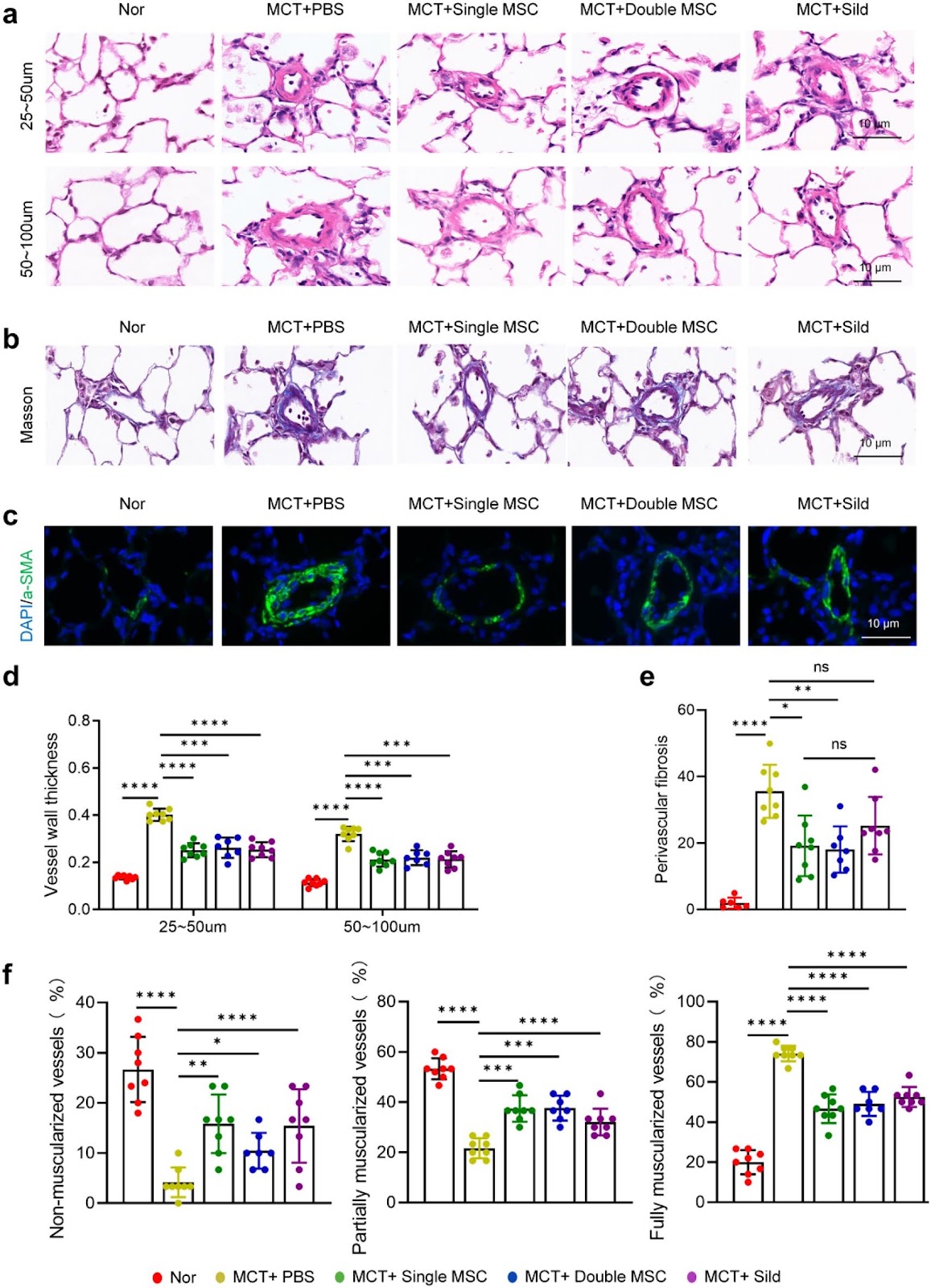
Background Pulmonary arterial hypertension (PAH) is a fatal condition characterized by progressive vascular remodeling in the pulmonary arteries, eventually leading to right heart failure and death. Dysregulated extracellular matrix (ECM) remodeling is central to PAH pathogenesis and represents a potential therapeutic target. Mesenchymal stromal cells (MSCs) have shown promise in preclinical studies; however, the optimal therapeutic window, dosing frequency, and mechanistic basis for their regulation of vascular ECM remain unclear. Methods We employed a monocrotaline (MCT)-induced rat model of PAH to evaluate different MSC treatment regimens, including early administration (day 1 post-MCT), delayed administration (days 7 and 14), and repeated dosing (days 1 and 11). Additionally, we combined in vivo and in vitro approaches to investigate how MSCs modulate the activation of pulmonary arterial adventitial fibroblasts (PAAFs) and influence ECM remodeling. Results Biodistribution studies indicated that MSC retention in lung tissue peaked within 24 h and gradually declined by day 21. A single early dose of MSCs (on day 1) significantly ameliorated PAH progression, increasing the 28-day survival rate, reducing right ventricular systolic pressure (RVSP), improving right ventricular function, and attenuating small pulmonary vascular remodeling, including reductions in medial thickening, excessive muscularization, and collagen deposition. Repeated MSC administration did not provide additional therapeutic benefit. Both in animal models and cell cultures, MSCs effectively suppressed PAAF activation and reduced ECM protein production. This anti-fibrotic effect was mediated via the SOCS3/STAT3 signaling pathway. Conclusions Early single-dose MSC therapy effectively alleviates PAH by suppressing PAAF activation and ECM remodeling through the SOCS3/STAT3 pathway, offering a potential therapeutic strategy for PAH.

Mesenchymal stromal cells alleviate pulmonary arterial hypertension by suppressing pulmonary arterial adventitial fibroblast activation and extracellular matrix remodeling via the SOCS3/STAT3 pathway
Graphical Abstract
Original ResearchVol. 17, Issue 1 • pp. 37DOI: 10.1186/s13287-025-04851-z

Intratracheal administration of mesenchymal stem cells ameliorates hyperoxia-induced bronchopulmonary dysplasia by inhibiting NLRP3 inflammasome activation: the critical role of Aldh1a2

Authors: Xuejing Xu, Linghong Liu, Na Dong, Tianqing Xin, Qing Shi, Dong Li, Xiuli Ju

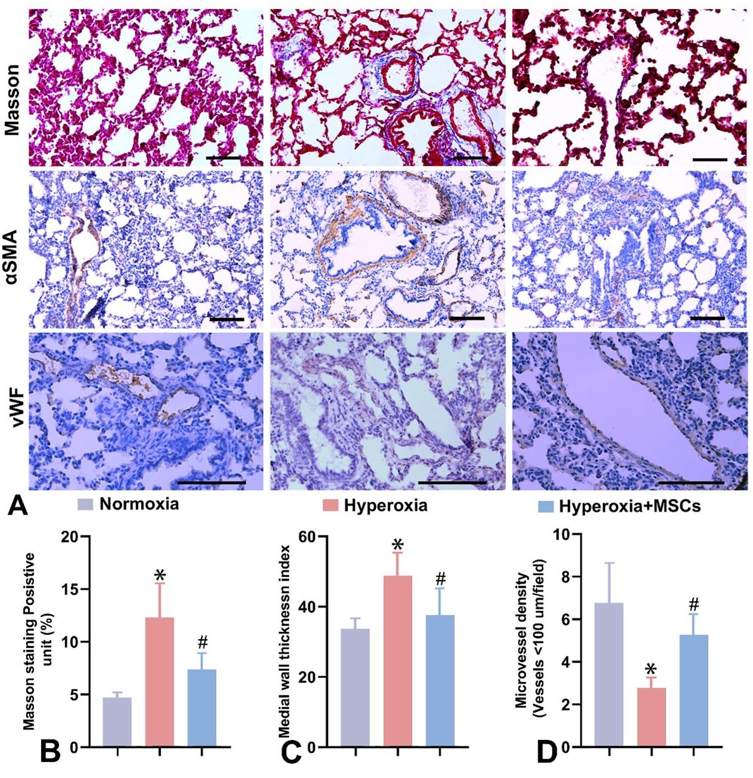
Background Bronchopulmonary dysplasia (BPD) is a chronic lung disease driven by inflammation and oxidative stress. Mesenchymal stem cells (MSCs) have shown protective effects against hyperoxic lung injury. However, few studies have thoroughly examined the significantly differentially expressed genes (DEGs) in the lungs before and after MSC treatment. In this study, we analyzed the significant DEGs in lung tissues during both in vivo and vitro umbilical cord-derived mesenchymal stem cells (UCMSCs)-mediated repair of hyperoxic lung injury and investigated their potential mechanisms of action. Methods Neonatal rats were exposed to hyperoxia and subsequently treated with UCMSCs. Inflammatory responses were quantified via ELISA and RT‒qPCR, while Western blotting (WB) and immunohistochemistry (IHC) were used to examine NLRP3 inflammasome and IL-1β expression. Transcriptomic analysis of UCMSC-mediated lung repair revealed 46 DEGs, which were validated by RT‒qPCR, and WB verified the significant differential expression of ALDH1A2. In RLE-6TN cells, Aldh1a2 expression was reduced during MSC-mediated repair of H2O2-induced oxidative stress injury. Functional evaluations were performed. WB further analyzed NLRP3 inflammasome and IL-1β expression in these processes. A recombinant adenoviral overexpression vector was intratracheally administered to hyperoxia-exposed neonatal rats. Arterial blood gas and RT‒qPCR were performed, and ELISA, WB, and IHC were used to evaluate the impact of Aldh1a2 overexpression on lung inflammation and oxidative stress, focusing on the NLRP3 inflammasome. Results UCMSCs ameliorated hyperoxia-induced alveolar simplification and microvessel loss, reduced inflammation and oxidative stress injury, and inhibited the expression of the NLRP3 inflammasome. RT‒qPCR and WB analyses revealed significant differential expression of Aldh1a2 in UCMSC-treated hyperoxia-induced lung injury. UCMSCs also mitigated H2O2-induced oxidative stress injury in RLE-6TN cells. Inhibition of Aldh1a2 expression exacerbated oxidative stress, upregulated NLRP3 inflammasome and IL-1β expression, and impaired the reparative effects of UCMSCs. Conversely, Aldh1a2 overexpression or UCMSC intervention ameliorated hyperoxia-induced alveolar simplification and microvascular abnormalities, suppressed inflammation, and enhanced lung ventilation and angiogenesis. These findings indicated that Aldh1a2 overexpression inhibits NLRP3 inflammasome activation and IL-1β release. Conclusions Aldh1a2 was significantly differentially expressed in UCMSC-mediated repair of hyperoxic lung injury, and its overexpression ameliorates BPD by inhibiting NLRP3 inflammasome activation, suggesting a novel therapeutic target for BPD.

Intratracheal administration of mesenchymal stem cells ameliorates hyperoxia-induced bronchopulmonary dysplasia by inhibiting NLRP3 inflammasome activation: the critical role of Aldh1a2
Graphical Abstract
Original ResearchVol. 17, Issue 1 • pp. 39DOI: 10.1186/s13287-025-04862-w

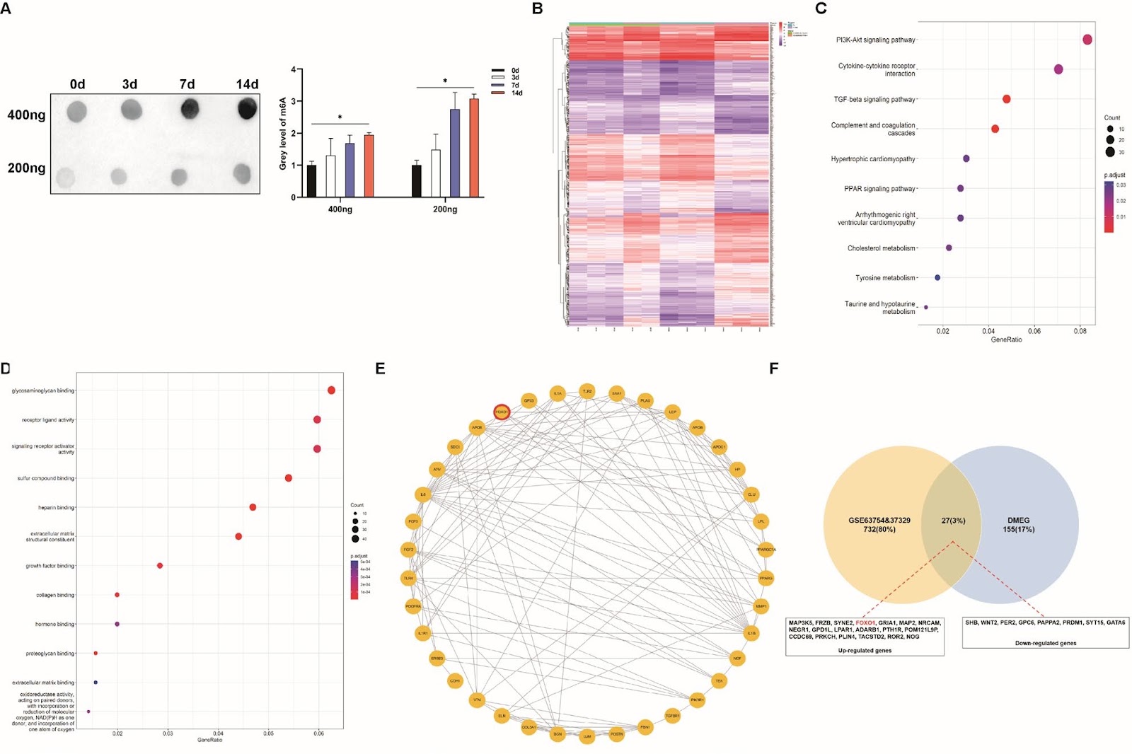
FTO-mediated m6A modification regulates the osteogenic differentiation of ADSCs by targeting FOXO1

Authors: Zhaohua Wang, Si Wen, Huizheng Li, Xiaosu Wang, Shu Guo, Shude Yang

Using adipose-derived stem cells (ADSCs) has recently become a crucial approach for treating bone defects owing to their ease of accessibility and substantial differentiation potential. N6-methyladenosine (m6A) modification greatly influences biological processes and determines the differentiation fate of stem cells. However, the specific mechanisms by which m6A modification influences the osteogenic differentiation of ADSCs remain unclear. We identified FOXO1 as the key m6A-modified gene during the osteogenesis of ADSCs. Furthermore, demethylase FTO enhanced RUNX2 expression while inhibiting PPARG expression by modifying FOXO1, thereby facilitating ADSC osteogenesis. FTO knockdown inhibited ADSC migration and proliferation and impaired osteogenesis by suppressing FOXO1. At the mechanistic level, we first revealed that FTO was exported to the cytoplasm and then directly bound with FOXO1 mRNA at its 1760th bp site. Consistent use of non-steroidal anti-inflammatory drugs (NSAIDs) containing FTO inhibitors impeded ADSC-mediated bone formation both in vivo and in vitro. In summary, our study reveals the role of m6A modification based on the FTO–FOXO1–RUNX2/PPARG axis in regulating the osteogenic differentiation of ADSCs, thereby improving the clinical use of ADSCs and providing strategies for related drug applications in bone regeneration.

FTO-mediated m6A modification regulates the osteogenic differentiation of ADSCs by targeting FOXO1
Graphical Abstract
Original ResearchVol. 17, Issue 1 • pp. 31DOI: 10.1186/s13287-025-04852-y

Mesenchymal stem cell-derived extracellular vesicles for disease therapy by regulating ferroptosis: focus on diabetes mellitus and diabetic complications

Authors: Jiayuan Wu, Zixuan Zhou, Hui Qian

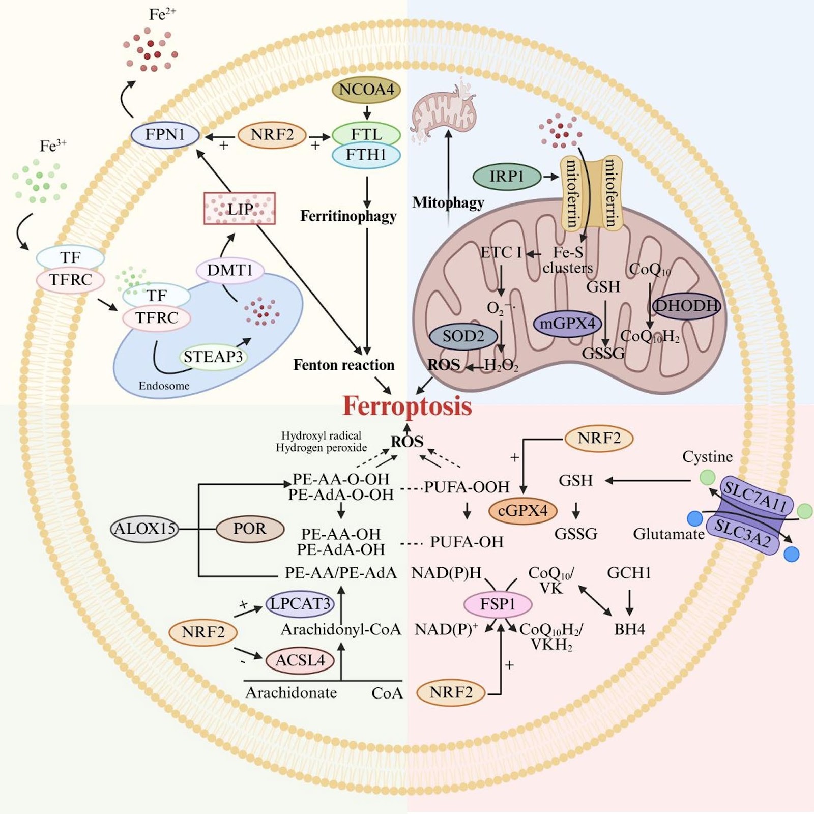
Ferroptosis is a novel form of programmed cell death, which has been demonstrated to play a pivotal role in various pathological processes due to its association with iron overload, lipid peroxidation, and dysregulation of the antioxidant system. In recent years, mesenchymal stem cell-derived extracellular vesicles (MSC-EVs) have garnered significant attention as a promising cell-free therapeutic strategy for modulating ferroptosis. This article elucidates the biological characteristics of MSC-EVs and the molecular mechanisms underlying ferroptosis, with a focus on how MSC-EVs regulate ferroptosis through three key pathways: iron metabolism, lipid metabolism, and the antioxidant defense system. Additionally, the therapeutic potential of both natural and engineered MSC-EVs in treating ferroptosis-related diseases is discussed, particularly highlighting their efficacy in diabetes mellitus and diabetic complications. Finally, this article evaluates the challenges and opportunities in translating MSC-EVs-based ferroptosis modulation therapies into clinical applications, providing valuable insights for future research and therapeutic development.

Mesenchymal stem cell-derived extracellular vesicles for disease therapy by regulating ferroptosis: focus on diabetes mellitus and diabetic complications
Graphical Abstract
Original ResearchVol. 17, Issue 1 • pp. 20DOI: 10.1186/s13287-025-04819-z

Mesenchymal stem cell-derived exosomes ameliorate gentamicin-induced vestibular hair cell injury by regulating the SNARE pathway and enhancing autophagy

Authors: Runnan Han, Ning Yu, GuoWei Qi, Jing Wang, Yanan Wu, Chuan Qin, Lin Shi, Liang Wang

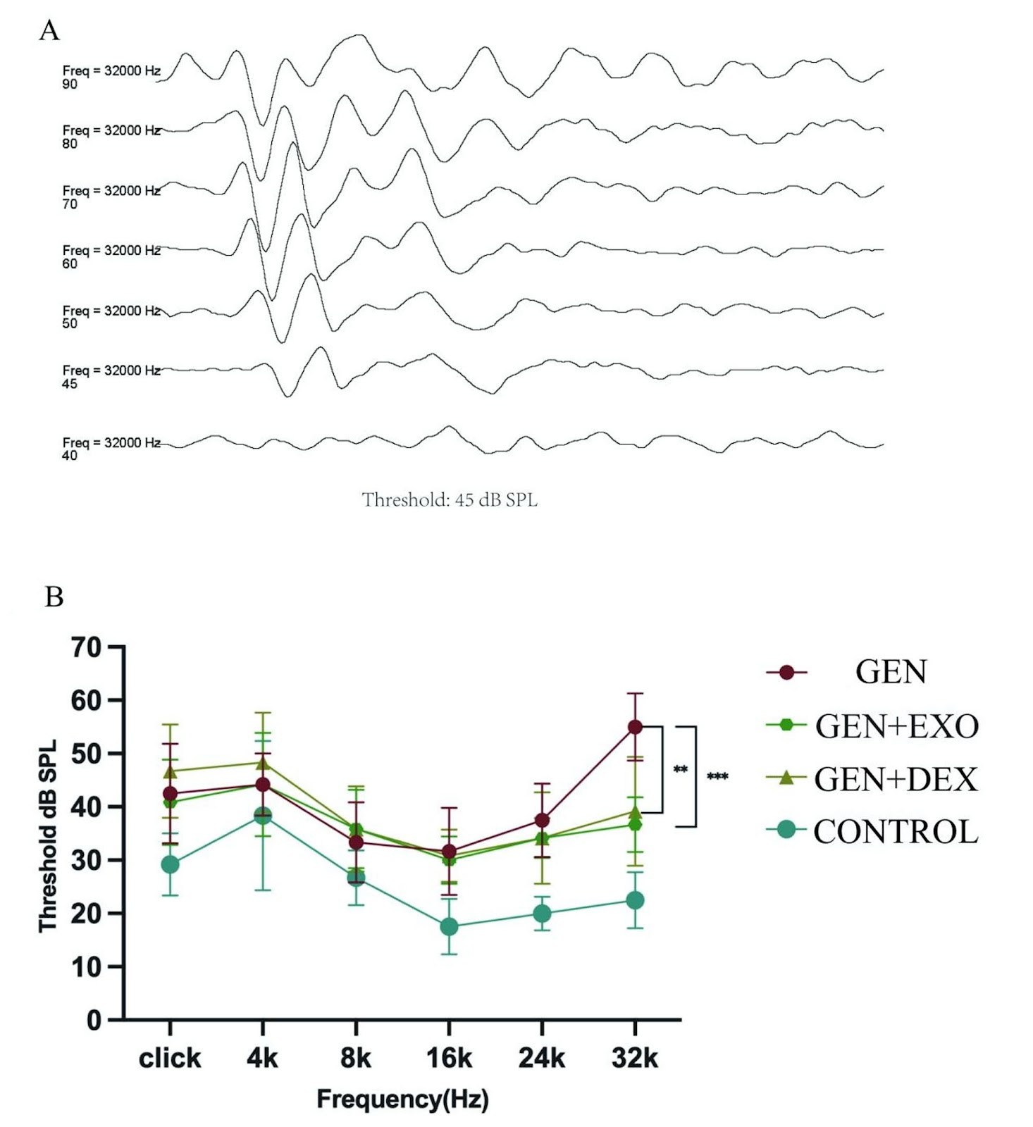
Objective To investigate the delivery efficiency of human umbilical cord mesenchymal stem cell-derived exosomes (hucMSC-EXOs) via intratympanic injection into vestibular end organs, evaluate their protective effects against gentamicin-induced vestibular dysfunction and hearing loss on gentamicin-induced vestibular dysfunction and hearing loss, and explore their regulatory mechanisms on hair cell apoptosis and autophagy. Methods Exosome characteristics were identified by transmission electron microscopy, nanoparticle tracking analysis, and Western blot. PKH26 labeling was used to trace their distribution in the vestibule. SD rats were randomly divided into four groups: control group, gentamicin group (GEN group), gentamicin + exosome group (GEN + EXO group), and gentamicin + dexamethasone group (GEN + DEX group). On day 6 after administration, vestibular function was assessed via open-field test and beam balance test. On day 7, high-frequency hearing (32 kHz) was detected by auditory brainstem response (ABR). The quantity and structural changes of hair cells were analyzed by immunofluorescence staining and scanning electron microscopy. Proteomics was used to analyze differentially expressed proteins in vestibular tissues treated with dexamethasone or hucMSC-EXOs. The regulatory effects on Caspase-3 (apoptosis) and LC3 (autophagy) were validated by immunofluorescence. Results hucMSC-EXOs administered via intratympanic injection were found to target the utricle, saccule, and crista ampullaris. Behavioral studies showed that the GEN + EXO group exhibited significant suppression of gentamicin-induced reduction in total movement distance (p < 0.05) and movement speed (p < 0.05, superior to the GEN + DEX group), with a 60.5% reduction in beam balance test passage time (p < 0.05). ABR results revealed that the auditory threshold at 32 kHz in the GEN + EXO group was 18.3 dB SPL lower than that in the injury group (p < 0.01), with no statistical difference compared to the GEN + DEX group. Hair cell counting showed significant protective effects of exosomes in reducing hair cell loss in the utricular striola (+25%), saccular striola (+44%), and central crista ampullaris

Mesenchymal stem cell-derived exosomes ameliorate gentamicin-induced vestibular hair cell injury by regulating the SNARE pathway and enhancing autophagy
Graphical Abstract