🧬 SinoBioData Academic Portal
🏛️ Indexed Academic JournalOriginal: 中国病理生理杂志

Chinese Journal of Pathophysiology

Premier Chinese Biomedical Journal indexed in SinoBioData: Chinese Journal of Pathophysiology (中国病理生理杂志).

Total Research Papers: 10
Access: 100% Free Open Access
Browse by Publication Year & Volume

Published Research Papers

Showing 10 of 10 peer-reviewed papers with full Graphical Abstracts.

Original ResearchVol. 41, Issue 9 • pp. 1674-1684DOI: 10.3969/j.issn.1000-4718.2025.09.002

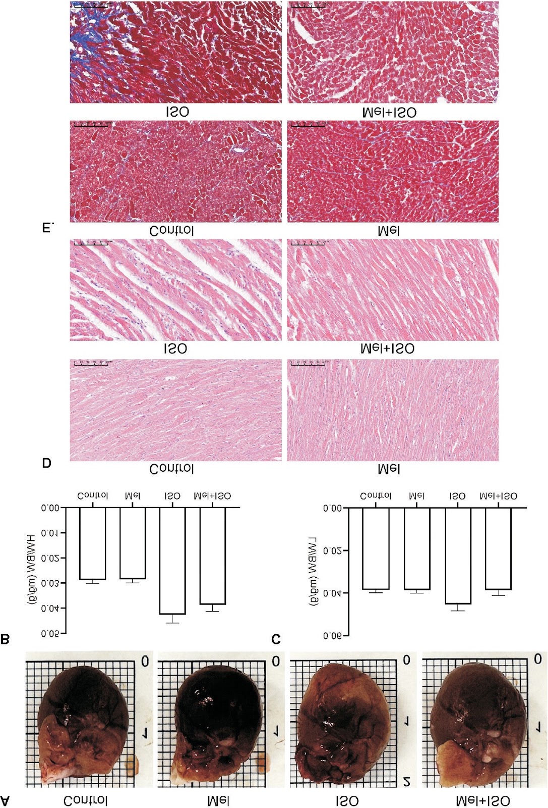
Melatonin alleviated acute myocardial infarction by inhibiting ferroptosis

Authors: HUANG Xiaohui, WEN Weixing, CHEN Peng, LI Weiwen, LI Jiahuan, CAO Yue, HU Yunzhao, HUANG Yuli

AIM: To investigate whether melatonin can ameliorate acute myocardial infarction (AMI) by inhibiting ferroptosis. METHODS: H9C2 cells were cultured in AnaeroPack system with low sugar and serum-free medium for 10 h to construct a cell model of AMI. Then cells were treated with melatonin and ferroptosis inducer erastin. The cell activity, reactive oxygen species (ROS), lipid peroxidation, mitochondrial membrane potential (MMP), and ferroptosis related protein expression were detected. A rat model of AMI induced by isoprenaline (ISO) injection was established to evaluate the effects of melatonin, in which the myocardial infarction size, cardiac injury, pathological changes, oxidative stress, iron ion and ferroptosis related protein expression were examined. RESULTS: Melatonin decreased the oxidative stress, lipid peroxidation and expression of ferroptosis protein in cardiomyocytes induced by hypoxia, but these effects could be impeded by the ferroptosis inducer erastin. Furthermore, in vivo experiments, we also found that melatonin improved the myocardial infarction size, cardiac injury, pathological changes, oxidative stress, and alleviated iron ion accumulation and ferroptosis. CONCLUSION: The cardioprotective effects of melatonin in AMI are associated with the inhibition of ferroptosis.

Melatonin alleviated acute myocardial infarction by inhibiting ferroptosis
Graphical Abstract
Original ResearchVol. 41, Issue 6 • pp. 1041-1054DOI: 10.3969/j.issn.1000-4718.2025.06.001

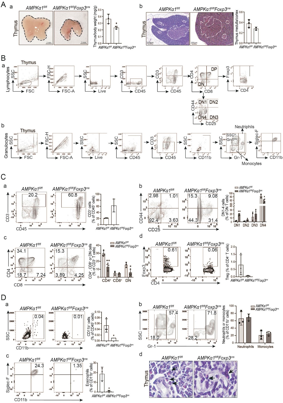
Treg-specific AMPKα1 deficiency alters immune cell compositions in immune organs of mice

Authors: RUAN Zhang, YANG Wenjing, YU Tianli, LI Pinxian, ZHANG Shunhui, LIN Caixia, ZHENG Lingyun, WANG Lijing

AIM: Regulatory T cells (Tregs) are a specialized subset of CD4+ T cells primarily involved in immunosuppressive functions. AMP-activated protein kinase (AMPK) serves as a metabolic sensor that governs the differentiation, maturation, and immune functions of Tregs through metabolic reprogramming. However, the impact of AMPKα1 (the catalytic subunit of AMPK) knockout specifically in Tregs on the host's immune microenvironment remains largely unexplored. METHODS: Histological changes in immune organs were assessed using HE staining. The types of immune cells and their relative population percentages in immune organs and blood were quantified through flow cytometry in both AMPKα1flox/flox (AMPKα1fl/fl) mice and Treg-specific AMPKα1 knockout mice (AMPKα1fl/flFoxp3cre mice). RESULTS: Compared to AMPKα1fl/fl mice, the percentage of eosinophils in the bone marrow of AMPKα1fl/flFoxp3cre mice was significantly reduced. Additionally, while the thymus of AMPKα1fl/flFoxp3cre mice exhibited normal structure, both its size and the ratio of thymus weight to body weight were significantly decreased. The knockout of AMPKα1 in Tregs led to a notable reduction in the total percentage of immature double-negative (DN) cells. Consequently, the percentage of CD4+ T cells derived from these DN cells also decreased, even though the percentages of DN1 and DN4 cells were higher in the thymus of AMPKα1fl/flFoxp3cre mice compared to AMPKα1fl/fl mice. Importantly, the proportion of Siglec-F+ CD11b+ eosinophils in the thymus was significantly lower in AMPKα1fl/flFoxp3cre mice. Knockout of AMPKα1 in Tregs resulted in a marked increase in the percentage of CD4+ T cells in peripheral blood, alongside a decrease in the proportion of mature CD8+ T cells. Similarly, the proportion of CD4+ T cells in the spleen of AMPKα1fl/flFoxp3cre mice was elevated compared to AMPKα1fl/fl mice. In contrast, the proportion of neutrophils significantly decreased, while mononuclear cell proportions increased in the spleen of AMPKα1fl/flFoxp3cre mice. In lymph nodes, the medullary boundaries in AMPKα1fl/flFoxp3cre mice were blurred, and the lymphoid follicles were missing, a feature not observed in AMPKα1fl/fl mice. Furthermore, the knockout of AMPKα1 in Tregs reduced the CD3+ T cell population, particularly the CD8+ T cell population, in lymph nodes. Although the mature Treg cell population was significantly lower in AMPKα1fl/flFoxp3cre mice, the percentage of CD4+ T cells was markedly increased. In contrast, there was no statistically significant difference in granulocyte populations between AMPKα1fl/flFoxp3cre and AMPKα1fl/fl mice. CONCLUSION: The populations of mature Tregs, CD8+ T cells and eosinophils in various immune organs were significantly altered in mice with Treg-specific AMPKα1 knockout, suggesting a potential remodeling of the host immune microenvironment in response to inflammatory stimuli.

Treg-specific AMPKα1 deficiency alters immune cell compositions in immune organs of mice
Graphical Abstract
Original ResearchVol. 41, Issue 12 • pp. 2289-2298DOI: 10.3969/j.issn.1000-4718.2025.12.001

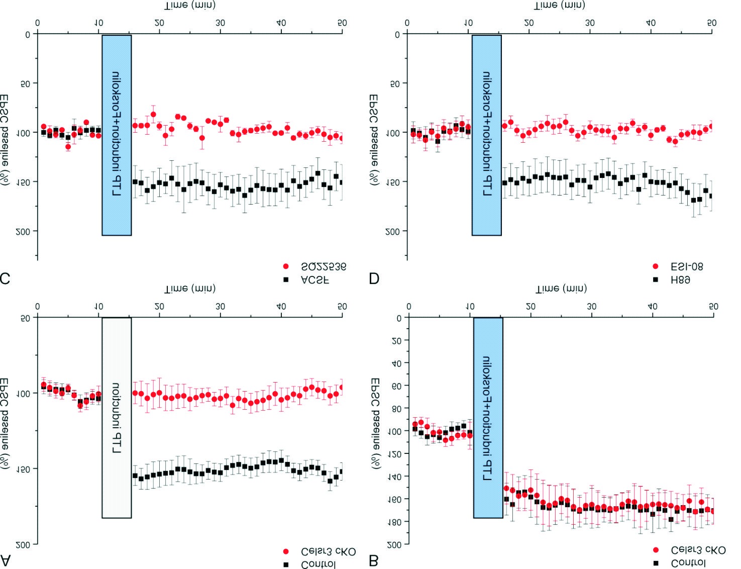
Forskolin ameliorates ataxia-like behavior in Purkinje cell-Celsr3 cKO mice via cAMP/Epac signaling pathway

Authors: WU Chuyue, GUO Jing, HONG Tu, HUANG Yu, CHEN Shengli, ZHOU Qinji

AIM: To evaluate the function and mechanisms of forskolin in treating ataxia-like behavior in Celsr3 conditional knockout (cKO) mice. METHODS: The efficiency of intraperitoneally administered forskolin was evaluated by behavioral tests, and the molecular mechanisms were investigated by patch-clamp experiments. RESULTS: The loss of Celsr3 led to ataxia-like behavior, accompanied by impaired miniature excitatory postsynaptic currents (mEPSCs) and postsynaptic long-term potentiation (LTP) in PCs. The cAMP activator forskolin ameliorated ataxia-like behavior and abrogated the mEPSCs impairment and LTP in model mice. Interestingly, the effects of forskolin could be blocked by SQ22536 (a cAMP antagonist) and ESI-08 (exchange protein activated by cAMP antagonist; Epac) but the H89 (a PKA antagonist) could not block the effects. CONCLUSION: Celsr3 plays an important role in motor coordination by modulating synaptic function, and forskolin may be a valuable therapeutic drug for certain types of inherited cerebellar ataxia.

Forskolin ameliorates ataxia-like behavior in Purkinje cell-Celsr3 cKO mice via cAMP/Epac signaling pathway
Graphical Abstract
Original ResearchVol. 40, Issue 8 • pp. 1488-1496DOI: 10.3969/j.issn.1000-4718.2024.08.016

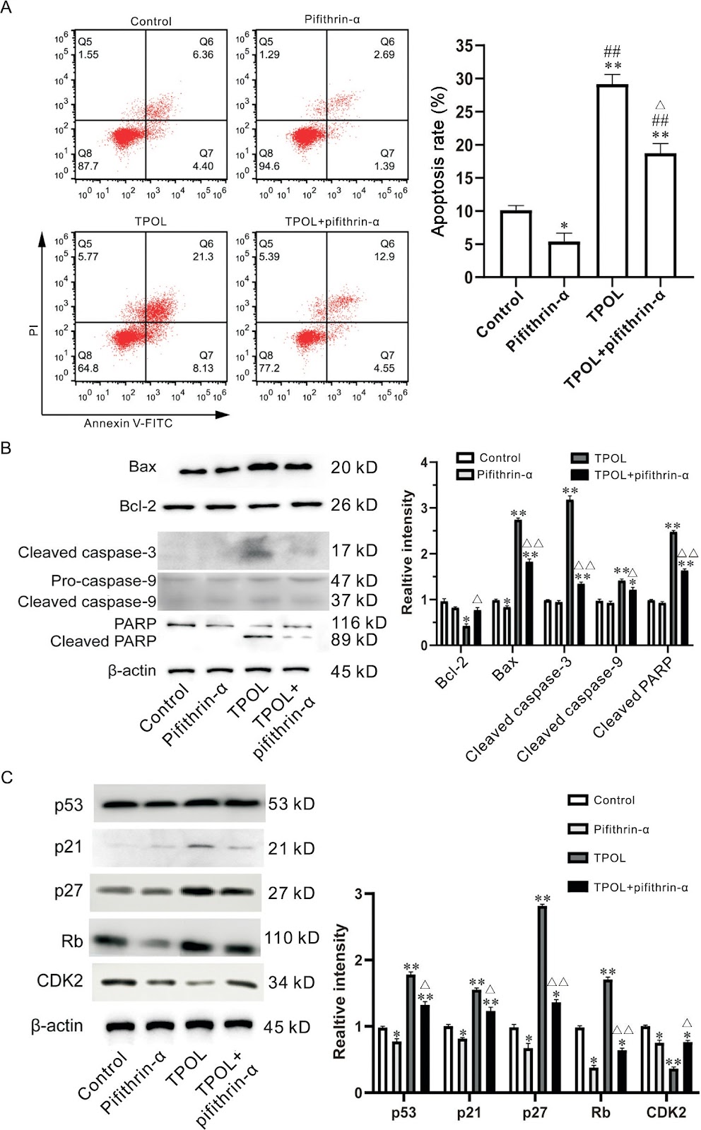
TPOL triggers apoptosis with mitochondrial injury through activating a ROS-dependent p53/p21/p27/Rb/Bax/Cyto C/caspase-mediated signaling

Authors: CHENG Zongwei, ZENG Boning, XING Feiyue

AIM: To explore the influence of ethyl (2,4,6-trimethylbenzoyl)phenylphosphinate (TPOL) on cell apoptosis and its potential mechanism. METHODS: HEK293T cells sensitive to TPOL were treated with different concentrations of TPOL with or without exposure to light radiation, before treatment with various inhibitors, N-acetyl-L-cysteine (NAC), pifithrin-α and Z-DVED-FMK. Cell viability was measured by CCK-8 assay. Annexin V/propidium iodide staining was used to count the number of apoptotic cells. DCFH-DA staining was used to detect reactive oxygen species (ROS) levels, and JC-1 staining was used to assess mitochondrial membrane potential by flow cytometry. The expression of apoptosis-related proteins and cell cycle-regulated molecules was measured by Western blot. RESULTS: TPOL enhanced the apoptosis of HEK293T cells in a dose-dependent manner (P<0.05), with a decrease in Bcl-2 and increases in Bax and cytochrome C (Cyto C), followed by up-regulation of activated caspase-9 and caspase-3, and the cleavage of PARP (P<0.05). The TPOL-enhanced cleavage of caspase-3 and PARP was rescued by Z-DVED-FMK (P<0.01). TPOL also led to a rapid increase in ROS, a reduction in mitochondrial membrane potential, and the release of Cyto C (P<0.01), all of which could be reversed by the ROS scavenger NAC. Moreover, the TPOL-caused alterations in p21, p27, Rb, and CDK2 were also recovered by the p53 inhibitor pifithrin-α (P<0.05). The TPOL-induced changes in Bax, Bcl-2, cleaved caspase-9, activated caspase-3, and cleaved PARP were subsequently rescued by pretreatment with pifithrin-α (P<0.05). CONCLUSION: TPOL can induce cellular apoptosis with ROS-mediated mitochondrial membrane damage through the activation of a ROS-dependent p53/p21/p27/Rb/Bax/Cyto C/caspase-mediated signal axis.

TPOL triggers apoptosis with mitochondrial injury through activating a ROS-dependent p53/p21/p27/Rb/Bax/Cyto C/caspase-mediated signaling
Graphical Abstract
Original ResearchVol. 40, Issue 4 • pp. 594-601DOI: 10.3969/j.issn.1000-4718.2024.04.003

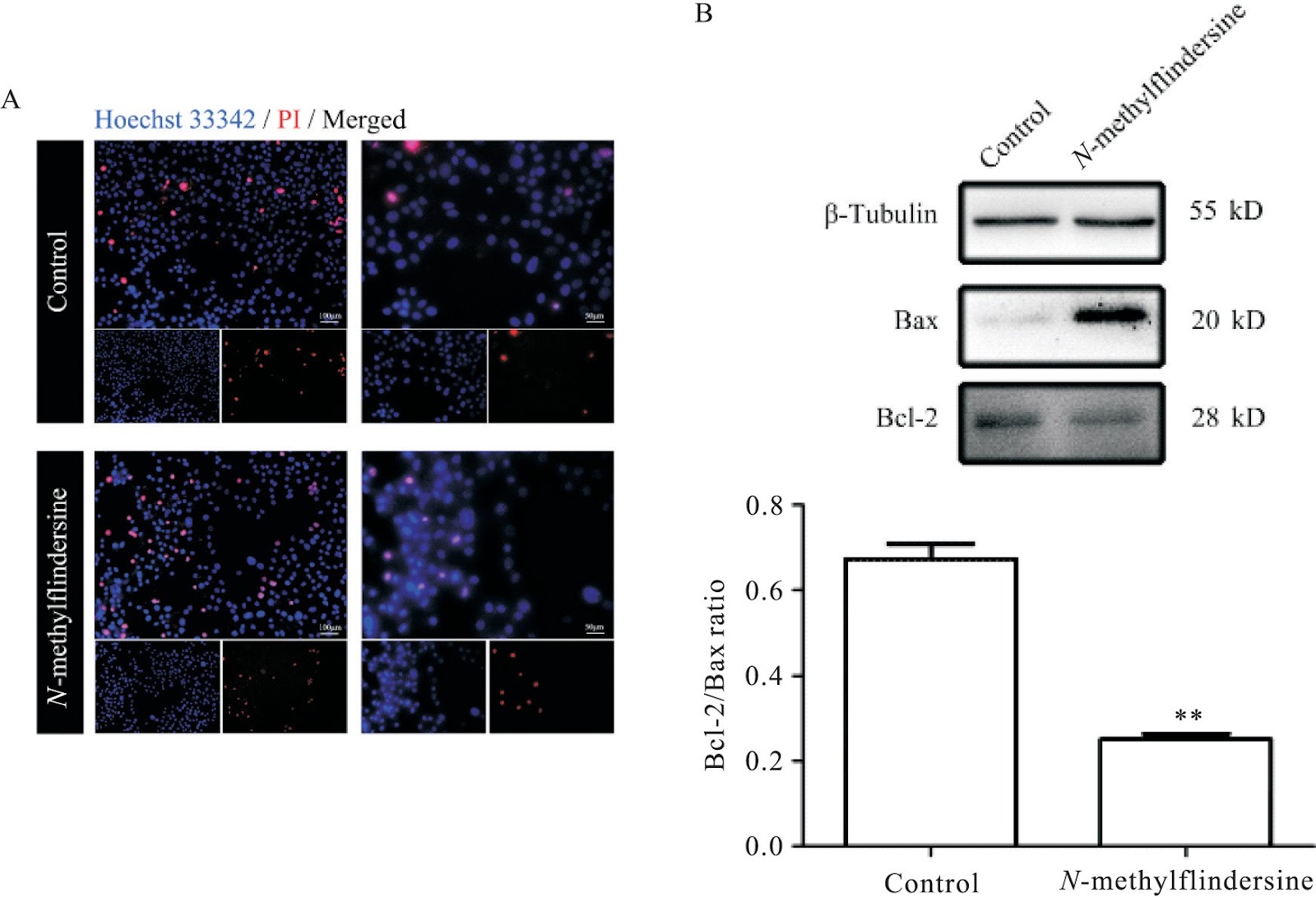
Induction of apoptosis and autophagy in human glioblastoma cells by N-methylflindersine: insights into regulatory role of ERK pathway

Authors: LI Siyuan, LUO Yuyou, LUO Xiongming, WANG Zhongyu, CHEN Huitong, FAN Dong, YUAN Xingyi, CHEN Le, TANG Pei, LIU Jing, WANG Zongming, WANG Xin

AIM: Mangrove-associated plants are known for producing natural compounds with antitumor activity. Despite the potential therapeutic value of these compounds, the molecular mechanisms underlying their antitumor effects remain unclear. This study aimed to investigate the antitumor properties of N-methylflindersine, an alkaloid derived from the mangrove-associated plant, Micromelum falcatum (Lour.) Tan., and its effects on U87 human glioblastoma cells. METHODS: We identified and isolated 15 compounds from the stem bark of Micromelum falcatum. Among these, we screened N-methylflindersine for its potential inhibitory effects on U87 cell growth. Various assays, including wound healing, Hoechst 33342/PI staining, and protein expression analysis, were conducted to investigate the compound's impact on cell migration, apoptosis, and autophagy-related proteins. RESULTS: Within 24 h, N-methylflindersine demonstrated the ability to reduce U87 cell migration and increase the apoptotic U87 cell population. Furthermore, it downregulated the anti-apoptosis protein Bcl-2 expression, upregulated the pro-apoptosis protein Bax expression, and elevated the ratio of autophagy-related protein LC3-II/LC3-I in U87 cells. Additionally, the ERK signaling pathway was found to be down-regulated following N-methylflindersine treatment. CONCLUSION: N-methylflindersine appears to induce both apoptosis and autophagic cell death in U87 cells, resulting in reduced cell growth. This effect seems to be associated with the downregulation of the ERK signaling pathway.

Induction of apoptosis and autophagy in human glioblastoma cells by N-methylflindersine: insights into regulatory role of ERK pathway
Graphical Abstract
Original ResearchVol. 40, Issue 5 • pp. 796-805DOI: 10.3969/j.issn.1000-4718.2024.05.004

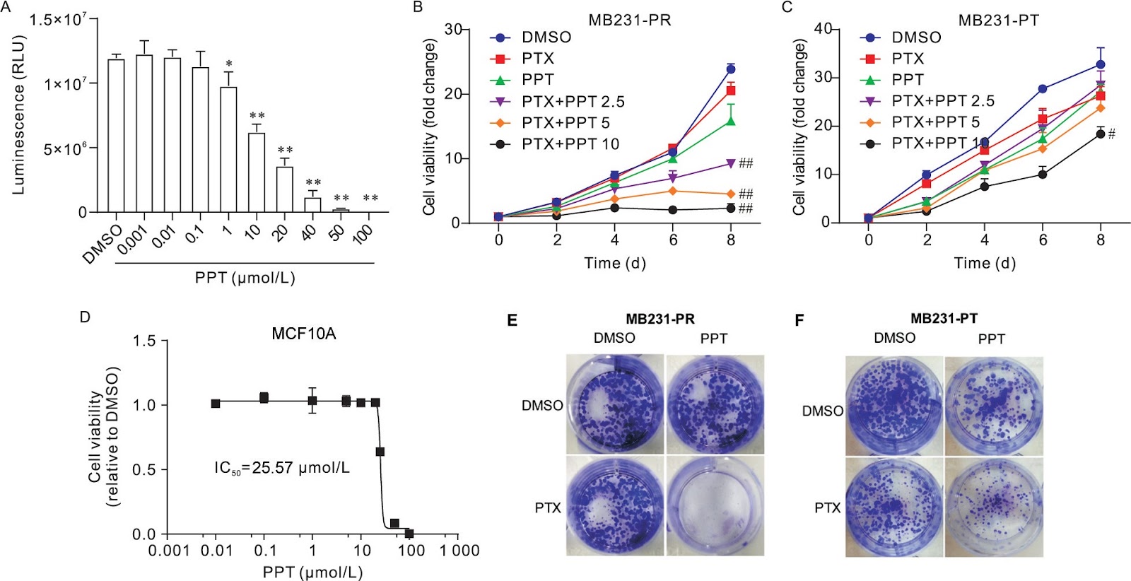
Mechanism of protopanaxatriol attenuating paclitaxel resistance in MDA-MB-231 cells

Authors: LI Lingyu, YE Qianyun, LI Yan, HAN Li, WANG Panpan, ZHANG Ronghua

AIM: To investigate the effect of protopanaxatriol (PPT) on the drug resistance of paclitaxel (PTX)-resistant human breast cancer MDA-MB-231 cells (MB231-PR cells). METHODS: The MB231-PR cells were constructed as cell models. They were treated with PPT, and incubated for a certain period of time according to the experimental settings. CellTiter-Glo was used to determine the viability of MB231-PR cells and MDA-MB-231 parental cells (MB231-PT cells). The change of sub-G1 phase was detected by flow cytometry. Western blot was used to evaluate the apoptosis-related proteins, such as cleaved caspase-3, cleaved poly(ADP-ribose) polymerase (PARP), B-cell lymphoma-2 (Bcl-2), Bcl-2-associated X protein (Bax) and survivin. The activity of nuclear factor-κB (NF-κB) was detected by luciferase reporter assay and immunofluorescence assay. The mRNA expression levels of interleukin-6 (IL-6), IL-8, chemokine CXC motif ligand 1 (CXCL1), chemokine CC motif ligand 2 (CCL2), CD44, NANOG, octamer-binding transcription factor 4 (OCT4), sex-determining region Y-box 2 (SOX2) and aldehyde dehydrogenase 1 (ALDH1) were detected by qPCR. The protein levels of IL-6 and IL-8 were measured by ELISA. Tumor sphere formation assay was used to evaluate the characteristics of stem cells. RESULTS: (1) The viability of MB231-PR cells was suppressed by PPT treatment in a dose-dependent manner compared with MB231-PT cells (P<0.01). Besides, the viability of MB231-PR cells was decreased after combined treatment with PPT and PTX (P<0.01), the accumulation of sub-G1 phase was induced (P<0.01), the ratio of Bax/Bcl-2 was elevated (P<0.01), and the protein levels of survivin, cleaved PARP and cleaved caspase-3 were increased (P<0.05). (2) After PPT treatment combined with PTX, the mRNA expression of inflammatory cytokines (IL-6, IL-8, CXCL1 and CCL2) and cancer stem cell-related markers (OCT4, SOX2, NANOG, ALDH1 and CD44) was reduced (P<0.05), and the protein levels of IL-6 and IL-8 were decreased (P<0.01). The activity of NF-κB in MB231-PR cells was suppressed (P<0.05), and the growth of tumor spheres from MB231-PR cells was damaged (P<0.05). (3) Immunofluorescence assay showed that PTX induced nuclear p-p65 expression, but this effect was attenuated by PPT. CONCLUSION: Combined treatment with PPT and PTX could attenuate PTX resistance of MB231-PR cells by inhibiting inflammatory cytokines and cancer stem cells.

Mechanism of protopanaxatriol attenuating paclitaxel resistance in MDA-MB-231 cells
Graphical Abstract
Original ResearchVol 42, Issue 6 • pp. 100-112DOI: 10.3969/j.issn.1000-4718.2026.06.013Jan 15, 2026

Enterococcus faecalis Promotes Chemoresistance in Colorectal Cancer via Lactate-Mediated MOB3B Down-Regulation

Authors: QI Jingru, ZHANG Weiyang, YANG Longan, LI Yuxuan, SHI Zhuoran, LIN Chuman, HUANG Yiyan, HUA Xing, ZHOU Rui, YU Lina

AIM: To investigate the role of Enterococcus faecalis (E. faecalis) in colorectal cancer (CRC) chemoresistance and elucidate the underlying molecular mechanisms. METHODS: Conditioned media (CM) were collected from cultures of E. faecalis treated with oxaliplatin or 5-fluorouracil (5-FU). The effects of these media on CRC chemoresistance were evaluated using in vitro functional assays and in vivo xenograft models in nude mice. Bioinformatics analysis was conducted to identify candidate genes associated with E. faecalis-induced chemoresistance. Gain- and loss-of-function experiments were performed to assess the role of MOB3B in regulating CRC cell proliferation and drug sensitivity. RT-qPCR, Western blot, and immunohistochemistry were used to validate the molecular mechanisms involved. Metabolomic profiling identified key metabolites in E. faecalis-oxaliplatin CM, and their roles in drug resistance were also confirmed. RESULTS: Compared with oxaliplatin treatment alone, E. faecalis-oxaliplatin CM significantly promoted CRC cell growth and chemoresistance in vitro (P<0.01). Tumors treated with E. faecalis-oxaliplatin CM exhibited significantly larger volumes and faster growth in vivo (P<0.01). Mechanistically, down-regulation of MOB3B mediated the chemoresistance-promoting effects of E. faecalis-oxaliplatin CM (P<0.01). Overexpression of MOB3B inhibited CRC cell proliferation and enhanced chemosensitivity, whereas MOB3B knockdown produced the opposite effect (P<0.01). Metabolomic analysis revealed elevated lactate levels in the E. faecalis-oxaliplatin CM (P<0.01). Lactate inhibition significantly reduced CRC cell proliferation, reversed chemoresistance, and restored MOB3B expression (P<0.01). CONCLUSION: E. faecalis promotes chemoresistance in CRC through lactate-mediated down-regulation of MOB3B, highlighting MOB3B as a potential therapeutic target for overcoming CRC chemoresistance.

Original ResearchVol 42, Issue 5 • pp. 100-112DOI: 10.3969/j.issn.1000-4718.2026.05.014Jan 15, 2026

A Spatial Atlas of Neuroimmune and Epigenetic Microenvironment in Psoriasis

Authors: XIAN Haiyan, DAI Jingfang, WU Minghui, QIU Xinmin, WANG Maojie, ZHANG Ge, LIU Huazhen, CHEN Yonggen, ZHU Ying, FENG Bing, SU Zuqing, LU Chuanjian, ZHENG Guangjuan, TANG Lipeng

AIM: Psoriasis is a chronic inflammatory skin disease characterized by keratinocyte hyperproliferation and immune dysregulation, yet the spatial epigenetic and neuroimmune features within the skin remain poorly understood. This study aims to construct a spatial atlas of the neuroimmune and epigenetic microenvironment in psoriasis. METHODS: Formalin-fixed, paraffin-embedded skin tissue samples from five psoriasis patients and four healthy controls were stained with a 33-metal antibody panel targeting immune and epigenetic markers. Imaging data were processed to analyze immune cell composition, spatial relationships, and epigenetic marker distribution in psoriatic lesions. RESULTS: Analysis of over 163,000 cells from five psoriasis patients and four healthy controls revealed that psoriatic lesions have a more complex cellular composition than normal skin, including diverse immune subsets, endothelial cells, keratinocytes, and nerve fibers. Neighborhood analysis showed enrichment of multiple immune cells, such as CD14+ monocytes, CD4+/CD8+ T-lymphocytes (T cells), CD68+ macrophages, CD69+ tissue-resident memory T cells and CD20+ B-lymphocytes (B cells), and nerve fibers around keratinocytes. Notably, positive interactions were observed between cutaneous nerve fibers and specific immune cells (CD8+ T cells and CD68+ macrophages) as well as blood vessels. Additionally, histone H3 lysine 27 trimethylation (H3K27me3) modification was mapped across cell types and found in immune cells adjacent to keratinocytes. CONCLUSION: Imaging mass cytometry delineated the psoriatic microenvironment's multicellular structure integrating epigenetic and neuroimmune components, offering new insights into psoriasis pathogenesis.

Original ResearchVol 42, Issue 6 • pp. 100-112DOI: 10.3969/j.issn.1000-4718.2026.06.009Jan 15, 2026

Enterococcus faecalis Promotes Chemotherapy Resistance by Down-regulating MOB3B in Colorectal Cancer

Authors: ZHANG Weiyang, QI Jingru, ZHAO Zhuoyang, YANG Longan, YAN Yongrong, LI Yuxuan, SHUAI Xinran, WU Gongfa, SONG Jiawei, HUA Xing, ZHOU Rui, YU Lina

AIM: To investigate the contribution of Enterococcus faecalis (E. faecalis) to chemoresistance in colorectal cancer (CRC) and uncover the underlying mechanisms. METHODS: Bioinformatics analyses were performed to evaluate the expression of MOB3B, an Mps-one binder coactivator (MOB) protein family member, and its clinical implications in CRC patients. E. faecalis was co-cultured with CRC cells to assess its effect on MOB3B expression. MOB3B was overexpressed or silenced in CRC cells to determine its effects on cell viability and chemosensitivity. The LRRC19-dependent mechanism was investigated through additional bioinformatics analyses. Immunohistochemical staining of clinical CRC tissues was performed to correlate MOB3B expression with Tumour Regression Grade. RESULTS: MOB3B down-regulation was associated with adverse clinicopathological characteristics and poor prognosis in CRC patients. Co-culture with E. faecalis down-regulated MOB3B expression in CRC cells. MOB3B overexpression decreased cell viability, while its silencing increased viability. MOB3B overexpression also reversed chemoresistance in CRC cells. Bioinformatics analyses revealed that MOB3B modulated resistance to oxaliplatin and 5-fluorouracil in an LRRC19-dependent manner. Low MOB3B expression correlated with high Tumour Regression Grade in clinical tissues. CONCLUSION: These findings indicate that Enterococcus faecalis promotes chemotherapy resistance by down-regulating MOB3B in colorectal cancer, and that MOB3B may serve as a potential marker for evaluating chemosensitivity and prognosis in CRC patients. The role of E. faecalis abundance as a clinical biomarker requires further validation in prospective cohorts.

Original ResearchVol 42, Issue 4 • pp. 100-112DOI: 10.3969/j.issn.1000-4718.2026.04.010Jan 15, 2026

Calcium Sensitivity, but Not Its Level, Determines Hypoxic Constriction of Porcine Coronary Arteries

Authors: FAN Jinxia, WU Zhuozhi, NAN Yan, YAN Haochen, YAN Jiazhen, XIE Junjun, YING Lei, WANG Yang

AIM: Acute hypoxia can induce transient contraction of coronary arteries, leading to myocardial ischemia and even cardiac dysfunction. However, the precise regulatory mechanisms remain unclear. In this study, we applied various interventions to isolated porcine coronary arteries by modulating cytoplasmic calcium concentrations mediated by calcium channels on the plasma membrane and sarcoplasmic reticulum, aiming to investigate the relationship between hypoxic contraction and intracellular calcium levels as well as calcium sensitization effects. METHODS: Isolated rings of the porcine left anterior descending coronary artery served as the experimental model. Based on distinct intervention targets, four core experimental groups were established. The specific grouping, sample size (n) for each group, and treatments were as follows: (1) nitric oxide (NO)-soluble guanylyl cyclase (sGC) pathway and energy metabolism intervention groups including control (n=5), nitric oxide synthase inhibitor nitro-L-arginine (NLA, 10^-4 mol/L; n=4 to 5), soluble guanylyl cyclase (sGC) antagonist 1H-[1,2,4]oxadiazolo[4,3-a]quinoxalin-1-one (ODQ, 3×10^-5 mol/L; n=5), endothelium-denuded (n=5), normal glucose incubation (n=3), and glucose-free incubation (n=3) groups; (2) calcium source intervention groups including normal calcium control (n=4), calcium-free incubation with 5×10^-3 mol/L ethylene glycol tetraacetic acid (EGTA; n=4), L-type calcium channel antagonist nifedipine (10^-6 mol/L; n=5 to 7), non-selective cation channel inhibitor NiCl2 (5×10^-5 mol/L; n=5 to 7), sarcoplasmic reticulum Ca²⁺-ATPase inhibitor thapsigargin (2×10^-6 mol/L; n=5 to 7), and inositol trisphosphate (IP3) receptor antagonist 2-aminoethoxydiphenyl borate (2-APB, 10^-4 mol/L; n=5 to 7) groups; (3) myosin light chain kinase (MLCK) pathway intervention groups including control (n=4) and MLCK-specific inhibitor 1-(5-iodonaphthalene-1-sulfonyl)-1H-hexahydro-1,4-diazepine hydrochloride (ML-7, 10^-5 mol/L; n=6) groups; (4) myosin light chain phosphatase (MLCP) activity and endothelium-dependence intervention groups including endothelium-intact (n=4) and mechanically endothelium-denuded (n=6) groups. All arterial rings were pre-contracted with either U46619 (3×10^-7 mol/L) or KCl (6×10^-2 mol/L) and then subjected to 10 minutes of hypoxia (95% N2+5% CO2). Changes in vascular tension were continuously monitored and recorded using a multi-channel physiological signal acquisition system. Furthermore, combined with Western blotting, the phosphorylation level of myosin light chain (MLC) and the activity of MLCP were determined; the phosphorylation levels of MLC and MLCP were also compared between endothelium-intact and endothelium-denuded coronary arteries under hypoxic conditions. RESULTS: (1) Hypoxic constriction of porcine coronary arteries is dependent on the activation of endothelium-derived nitric oxide (NO) and sGC in vascular smooth muscle cells. (2) Hypoxic contraction in porcine coronary arteries is independent of extracellular Ca²⁺ influx. (3) Hypoxic contraction in porcine coronary arteries does not rely on intracellular Ca²⁺ release from the sarcoplasmic reticulum. (4) Hypoxic contraction in porcine coronary arteries leads to inhibition of myosin light chain phosphatase activity, suggesting increased calcium sensitization in coronary artery smooth muscle. CONCLUSION: The mechanism underlying acute hypoxia-induced vasoconstriction exhibits distinct characteristics: it does not rely on extracellular calcium influx mediated by plasma membrane calcium channels, nor is it associated with intracellular calcium mobilization from sarcoplasmic reticulum stores. Instead, it is mediated by a significant enhancement in calcium sensitivity regulated by myosin light chain phosphatase, a process referred to as calcium sensitization.