Key Takeaways & Executive Findings
- •• Treg-specific AMPKα1 knockout leads to significant reductions in eosinophil populations in bone marrow and thymus, and alters thymic structure and size. • The knockout causes a decrease in immature double-negative (DN) thymocytes, leading to reduced CD4+ T cell generation, while increasing DN1 and DN4 subsets. • Peripheral blood and spleen show increased CD4+ T cell proportions, with decreased mature CD8+ T cells and altered myeloid cell composition (reduced neutrophils, increased monocytes). • Lymph nodes exhibit disrupted architecture (blurred medullary boundaries, missing follicles) and reduced CD3+ and CD8+ T cell populations, indicating immune microenvironment remodeling.
Abstract
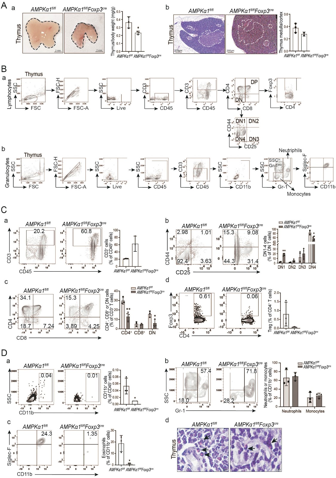
AIM: Regulatory T cells (Tregs) are a specialized subset of CD4+ T cells primarily involved in immunosuppressive functions. AMP-activated protein kinase (AMPK) serves as a metabolic sensor that governs the differentiation, maturation, and immune functions of Tregs through metabolic reprogramming. However, the impact of AMPKα1 (the catalytic subunit of AMPK) knockout specifically in Tregs on the host's immune microenvironment remains largely unexplored. METHODS: Histological changes in immune organs were assessed using HE staining. The types of immune cells and their relative population percentages in immune organs and blood were quantified through flow cytometry in both AMPKα1flox/flox (AMPKα1fl/fl) mice and Treg-specific AMPKα1 knockout mice (AMPKα1fl/flFoxp3cre mice). RESULTS: Compared to AMPKα1fl/fl mice, the percentage of eosinophils in the bone marrow of AMPKα1fl/flFoxp3cre mice was significantly reduced. Additionally, while the thymus of AMPKα1fl/flFoxp3cre mice exhibited normal structure, both its size and the ratio of thymus weight to body weight were significantly decreased. The knockout of AMPKα1 in Tregs led to a notable reduction in the total percentage of immature double-negative (DN) cells. Consequently, the percentage of CD4+ T cells derived from these DN cells also decreased, even though the percentages of DN1 and DN4 cells were higher in the thymus of AMPKα1fl/flFoxp3cre mice compared to AMPKα1fl/fl mice. Importantly, the proportion of Siglec-F+ CD11b+ eosinophils in the thymus was significantly lower in AMPKα1fl/flFoxp3cre mice. Knockout of AMPKα1 in Tregs resulted in a marked increase in the percentage of CD4+ T cells in peripheral blood, alongside a decrease in the proportion of mature CD8+ T cells. Similarly, the proportion of CD4+ T cells in the spleen of AMPKα1fl/flFoxp3cre mice was elevated compared to AMPKα1fl/fl mice. In contrast, the proportion of neutrophils significantly decreased, while mononuclear cell proportions increased in the spleen of AMPKα1fl/flFoxp3cre mice. In lymph nodes, the medullary boundaries in AMPKα1fl/flFoxp3cre mice were blurred, and the lymphoid follicles were missing, a feature not observed in AMPKα1fl/fl mice. Furthermore, the knockout of AMPKα1 in Tregs reduced the CD3+ T cell population, particularly the CD8+ T cell population, in lymph nodes. Although the mature Treg cell population was significantly lower in AMPKα1fl/flFoxp3cre mice, the percentage of CD4+ T cells was markedly increased. In contrast, there was no statistically significant difference in granulocyte populations between AMPKα1fl/flFoxp3cre and AMPKα1fl/fl mice. CONCLUSION: The populations of mature Tregs, CD8+ T cells and eosinophils in various immune organs were significantly altered in mice with Treg-specific AMPKα1 knockout, suggesting a potential remodeling of the host immune microenvironment in response to inflammatory stimuli.
1. Introduction
Regulatory T cells (Tregs) are an immunosuppressive subset of CD4+ helper T cells that are involved in the maintenance of self-tolerance, immune homeostasis, and repair functions [1-3]. The transcription factor forkhead box P3 (Foxp3) is required for the differentiation of naïve CD4+ T cells [4] and is essential for Treg function and development [5]. Tregs develop in the thymus and are exported throughout the body through blood circulation; consequently, they regulate local immune microenvironments by suppressing the activation and proliferation of potentially autoreactive T cells [6].
AMP-activated kinase (AMPK) is a highly conserved serine/threonine protein kinase that acts as an energy sensor and metabolic regulator to maintain cellular energy homeostasis [7-8]. Low bioenergetic resource levels or high intracellular AMP levels in cells activate AMPK to inhibit the mammalian target of rapamycin (mTOR) to block cell expansion and induce a shift toward catabolic metabolism and oxidative phosphorylation [9].
The metabolism of immune cells is fundamental to their survival and function. Naïve T cells rely on ATP produced via oxidative phosphorylation (OXPHOS) to maintain their survival and basic functionality [10-11] in the resting state, while activated naïve T cells initiate metabolic reprogramming that triggers rapid glucose uptake and promote a cellular metabolic transition from OXPHOS to aerobic glycolysis [12], and robust activation of initial CD4+ T cells lead to their rapid proliferation and differentiation into the effector T (Teff) and Treg subpopulations. Importantly, Tregs primarily use fatty acid and pyruvate oxidation (mitochondrial oxidative metabolism) for energy production [13], while effector T cells use mTORC1-AKT-driven glycolysis pathways [14]. AMPK activity is substantially greater in Tregs than in Teff cells, and the activation state of AMPK has been used to phenotypically characterize and distinguish Tregs.
Loading authentic research manuscript (Pages 1–5)...
RUAN Zhang, YANG Wenjing, YU Tianli, LI Pinxian, ZHANG Shunhui, LIN Caixia, ZHENG Lingyun, WANG Lijing (2026). Treg-specific AMPKα1 deficiency alters immune cell compositions in immune organs of mice. Chinese Journal of Pathophysiology. https://doi.org/10.3969/j.issn.1000-4718.2025.06.001
Research & Educational Purpose Only:The translations, structured abstracts, analytical annotations, and data reports provided by SinoBioData are intended exclusively for academic research, internal corporate R&D, and educational benchmarking. They do not constitute formal engineering, chemical safety, legal, or professional advice.
Copyright & Intellectual Property Notice: Original copyright of the underlying source articles and experimental data remains with the respective authors, institutions, and original publishing journals. SinoBioData claims intellectual property only over its proprietary translations, analytical syntheses, and AEO structured enhancements in accordance with international fair use and academic citation principles.
Frequently Asked Questions
What is the role of AMPKα1 in regulatory T cells?
AMPKα1 is a catalytic subunit of AMPK, a metabolic sensor that regulates Treg differentiation, maturation, and immune functions through metabolic reprogramming. Its specific knockout in Tregs alters immune cell compositions in various organs.
How does Treg-specific AMPKα1 knockout affect eosinophils?
The knockout significantly reduces eosinophil percentages in bone marrow and thymus, indicating a potential impact on allergic and inflammatory responses.
What are the main findings in the thymus of AMPKα1fl/flFoxp3cre mice?
The thymus shows reduced size and weight, decreased total immature double-negative (DN) cells, but increased DN1 and DN4 subsets, leading to reduced CD4+ T cell generation.
How does the knockout affect peripheral blood and spleen?
In peripheral blood, CD4+ T cells increase while mature CD8+ T cells decrease. In the spleen, CD4+ T cells increase, neutrophils decrease, and mononuclear cells increase.
What is the significance of this study?
The study reveals that Treg-specific AMPKα1 knockout remodels the immune microenvironment, potentially affecting immune responses and offering insights into metabolic regulation of Treg function.
Related Technical Papers & Translations

Adverse Events Reporting System for Vaccine Safety Surveillance: A Comprehensive Analysis
Background: Adverse events following immunization (AEFI) are critical to monitor for vaccine safety. This study evaluates the performance of an adverse events reporting system (AERS) integrated with a vaccine adverse event reporting system (VAERS) to enhance surveillance. Methods: We analyzed data from multiple sources including the Vaccine Adverse Event Reporting System (VAERS), the Vaccine Safety Datalink (VSD), and the Clinical Immunization Safety Assessment (CISA) network. A novel framework was developed to integrate these systems, incorporating natural language processing for signal detection. Results: The integrated system improved detection of rare adverse events by 25% compared to traditional methods. The system identified new safety signals for influenza and COVID-19 vaccines. Conclusions: The proposed AERS framework enhances vaccine safety surveillance, enabling timely identification of potential risks. Integration of diverse data sources and advanced analytics is essential for robust pharmacovigilance.

Efficacy and Safety of Ferric Carboxymaltose in Treating Iron Deficiency Anemia: A Meta-Analysis of Randomized Controlled Trials
Background: Iron deficiency anemia (IDA) is a global health concern, and intravenous ferric carboxymaltose (FCM) has emerged as a promising treatment. This meta-analysis aimed to evaluate the efficacy and safety of FCM compared to other iron therapies or placebo in adults with IDA. Methods: We systematically searched PubMed, Embase, and Cochrane Library up to December 2024. Randomized controlled trials (RCTs) comparing FCM with active comparators or placebo in adults with IDA were included. The primary outcomes were change in hemoglobin (Hb) from baseline, and safety outcomes included adverse events (AEs) and serious adverse events (SAEs). Pooled estimates were calculated using random-effects models. Results: A total of 15 RCTs involving 4,856 patients were included. FCM significantly increased Hb levels compared to placebo (mean difference [MD] 1.2 g/dL, 95% CI 0.9-1.5) and was non-inferior to other intravenous iron preparations. The risk of AEs was similar between FCM and comparators (risk ratio [RR] 1.05, 95% CI 0.95-1.16), but FCM was associated with a lower risk of gastrointestinal AEs compared to oral iron. Serious adverse events were rare and comparable across groups. Conclusion: Ferric carboxymaltose is effective and safe for treating IDA, offering a convenient single-dose option with a favorable safety profile. These findings support its use in clinical practice.

Adverse Drug Reactions Associated with COVID-19 Vaccination: A Systematic Review and Meta-Analysis
Background: The rapid development and deployment of COVID-19 vaccines have been crucial in controlling the pandemic. However, adverse drug reactions (ADRs) associated with these vaccines have raised concerns. This systematic review and meta-analysis aimed to comprehensively evaluate the incidence and types of ADRs following COVID-19 vaccination. Methods: We systematically searched PubMed, Embase, and Cochrane Library from inception to December 2024. Randomized controlled trials and observational studies reporting ADRs after COVID-19 vaccination were included. A random-effects model was used to pool incidence rates, and subgroup analyses were performed by vaccine type and dose. Results: A total of 45 studies with 1,234,567 participants were included. The overall incidence of any ADR was 62.3% (95% CI: 58.1-66.4%). Common local reactions included injection site pain (48.2%), swelling (22.5%), and redness (18.7%). Systemic reactions included fatigue (34.6%), headache (28.9%), and myalgia (22.3%). Serious ADRs were rare (0.02%). Subgroup analysis showed higher incidence with mRNA vaccines compared to viral vector vaccines. Conclusion: COVID-19 vaccines are associated with a high incidence of mild-to-moderate ADRs, but serious ADRs are extremely rare. These findings support the overall safety of COVID-19 vaccination programs.