🧬 SinoBioData Academic Portal
🏛️ Indexed Academic JournalOriginal: 生物化学与生物物理学报

Acta Biochimica et Biophysica Sinica

Premier Chinese Biomedical Journal indexed in SinoBioData: Acta Biochimica et Biophysica Sinica (生物化学与生物物理学报).

Total Research Papers: 200
Access: 100% Free Open Access
Browse by Publication Year & VolumeReset All Filters ✕

Published Research PapersFiltered: Year 2026 • xx

Showing 15 of 200 peer-reviewed papers with full Graphical Abstracts.

Original ResearchVol. xx, Issue xx • pp. xx–xxDOI: 10.3724/abbs.2026084

HDAC6 promotes osimertinib resistance evolution in non-small cell lung cancer by activating EGFR degradation through the ubiquitin-proteasome pathway

Authors: Cheng Yang, Danlei Xu, Zifang Zhu, Zhen Kang, Kangkang Ren, Yihan Yang, Ye Huang, Wei Zhang, Jun Che, Xinping Xu, Xiaolei Li

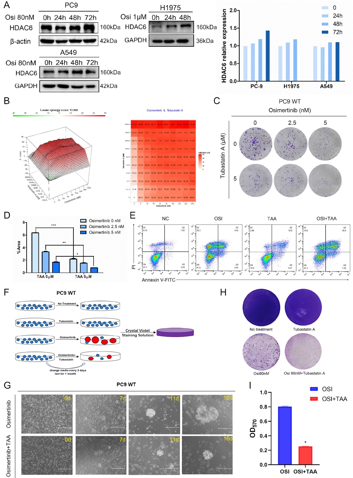
Osimertinib is the standard first-line treatment for patients with non-small cell lung cancer (NSCLC) harboring EGFR-sensitive mutations. However, drug resistance inevitably develops, highlighting the critical need for strategies to overcome this resistance and prolong therapeutic efficacy. Understanding the mechanisms underlying drug resistance is essential, and drug-resistant cell models serve as valuable tools for investigating acquired resistance. In this study, we establish an osimertinib resistance evolution model in vitro by continuous high-dose drug induction and identify cell lines exhibiting “permanent” resistance to osimertinib (osimertinib resistant, OR). Transcriptome sequencing (RNA-seq), gain- and loss-of-function assay, including lentiviral-mediated overexpression and RNAi knockdown, pharmacological inhibition, and protein degradation analysis reveal significant alterations in genes associated with epigenetic regulation, notably a marked upregulation of histone deacetylase 6 (HDAC6) in OR cells. Knockdown of HDAC6 or pharmacological inhibition of HDAC6 restores the sensitivity of OR cells to osimertinib, whereas overexpression of HDAC6 in sensitive cells reduces drug efficacy and accelerates the onset of resistance. Furthermore, we find that HDAC6 upregulation promotes EGFR degradation, thereby contributing to resistance. Collectively, our findings demonstrate the utility of drug resistance evolution models in identifying key resistance factors. HDAC6 plays a pivotal role in osimertinib resistance, and targeting HDAC6 may represent a novel therapeutic strategy to overcome resistance and enhance treatment efficacy.
HDAC6 promotes osimertinib resistance evolution in non-small cell lung cancer by activating EGFR degradation through the ubiquitin-proteasome pathway
Graphical Abstract
Original ResearchVol. xx, Issue xx • pp. xx-xxDOI: 10.3724/abbs.2026049

circ_0006156 promotes esophageal squamous cell carcinoma progression via activation of the TGFβ/Smad pathway

Authors: Zhanpeng Tang, Jian Li, Rongyang Li, Luyuan Ma, Renchang Zhao, Zhenguo Sun, Hui Tian

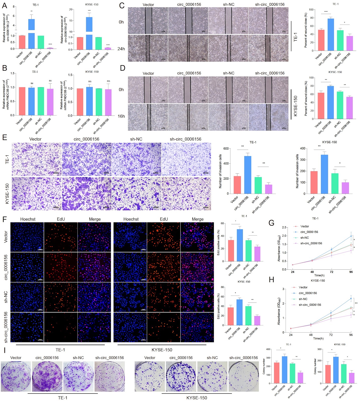
Esophageal squamous cell carcinoma (ESCC) represents a common malignancy of the digestive system. Circular RNAs (circRNAs) are a distinct class of single-stranded non-coding RNAs that are essential in the progression of various tumors given that they can act as microRNA (miRNA) sponges in a manner similar to that of mRNAs. In this study, circ_0006156 is screened to be highly expressed in ESCC tissues through high-throughput sequencing and quantitative real-time polymerase chain reaction. Subsequent in vitro and in vivo experiments are conducted to validate its biological functions. Furthermore, the regulatory relationships among circ_0006156, miR-202-5p, and TGFBR1 are investigated using RNA antisense purification, miRNA sequencing, RNA immunoprecipitation, fluorescence in situ hybridization, dual-luciferase reporter assay, and bioinformatics analyses. The results show significant overexpression of circ_0006156 in ESCC tissues, with relatively shorter overall survival observed in patients with high circ_0006156 expression. circ_0006156 is further identified to directly bind to miR-202-5p. miR-202-5p inhibits the proliferation, migration, and invasion of ESCC cells in vitro and partially rescues the effects induced by circ_0006156. Consistent results are reported by subcutaneous xenograft tumor experiments in nude mice. In addition, circ_0006156 is confirmed to act as an endogenous sponge for miR-202-5p, which results in a relieved suppression of its target gene TGFβR1. In summary, circ_0006156 can regulate TGFβR1 expression by sponging miR-202-5p, which may further activate the TGFβ/Smad pathway and promote ESCC progression. Collectively, circ_0006156 functions as a novel oncogenic RNA in ESCC and may serve as a potential tumor marker.

circ_0006156 promotes esophageal squamous cell carcinoma progression via activation of the TGFβ/Smad pathway
Graphical Abstract
Original ResearchVol. xx, Issue xx • pp. xx-xxDOI: 10.3724/abbs.2025251

Efficient production of recombinant mAbs mediated by a MAR-enhanced transposon vector combined with blasticidin selection in CHO cells

Authors: Xiaoyin Wang, Xuelian Han, Ting Liu, Shiqi Zhang, Zimeng Han, Ruoyuan Feng, Tianyun Wang

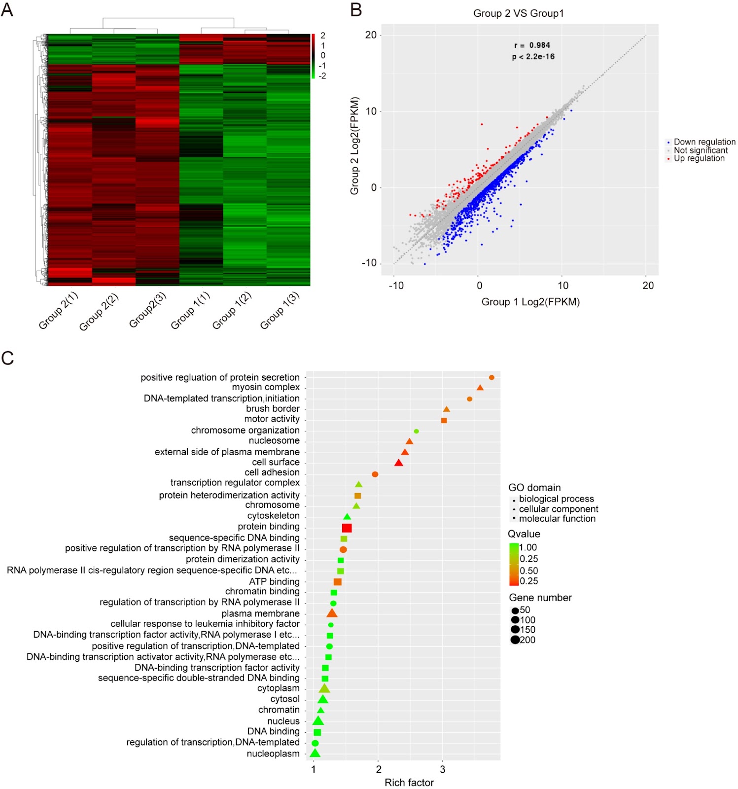
Recombinant antibodies, primarily produced in Chinese hamster ovary (CHO) cells, are widely used to treat various diseases. For industrial production, a rapid and efficient method to screen stable, high-expressing clones is essential. However, conventional screening based on random integration is often cumbersome and labor intensive. This study establishes a novel strategy for generating stable, high-yielding clones by combining a MAR-based piggyBac (PB) transposon semitargeted integration system with blasticidin (BSD) selection. Compared to the random integrated vector pMAR-mAb, the MAR-PB system increases the titers (3.95- to 5.61-fold) and specific protein productivity (Qp; 4.28- to 6.07-fold) of four monoclonal antibodies in stable cell pools. When compared to PB-only vectors, the MAR-PB transposon system enhances the titers (by up to 2.50-fold) and Qp (1.96- to 2.77-fold), respectively. The increased antibody production correlates with elevated mRNA expression. Furthermore, this approach increases the proportion of high-expressing clones by more than 10-fold and significantly improves volumetric yield. Importantly, this approach promotes the long-term stability of recombinant mAb expression for over 60 generations. Transcriptome analysis reveals that the system modulates genes involved in DNA binding, transcriptional regulation, and protein binding. In conclusion, the MAR-based PB transposon system combined with BSD selection presents a significant improvement for efficiently generating high-yielding and stable CHO cell clones, offering a valuable tool for recombinant antibody production.

Efficient production of recombinant mAbs mediated by a MAR-enhanced transposon vector combined with blasticidin selection in CHO cells
Graphical Abstract
Original ResearchVol. xx, Issue xx • pp. xx-xxDOI: 10.3724/abbs.2026087

A bifunctional aptamer-siRNA chimera targeting ACE2 for the inhibition of SARS-CoV-2 S pseudovirus entry and replication

Authors: Tao Jiang, Zhiqiang Chen, Wei Li, Xiaohua Ni, Wen Pan, Qihan Wu

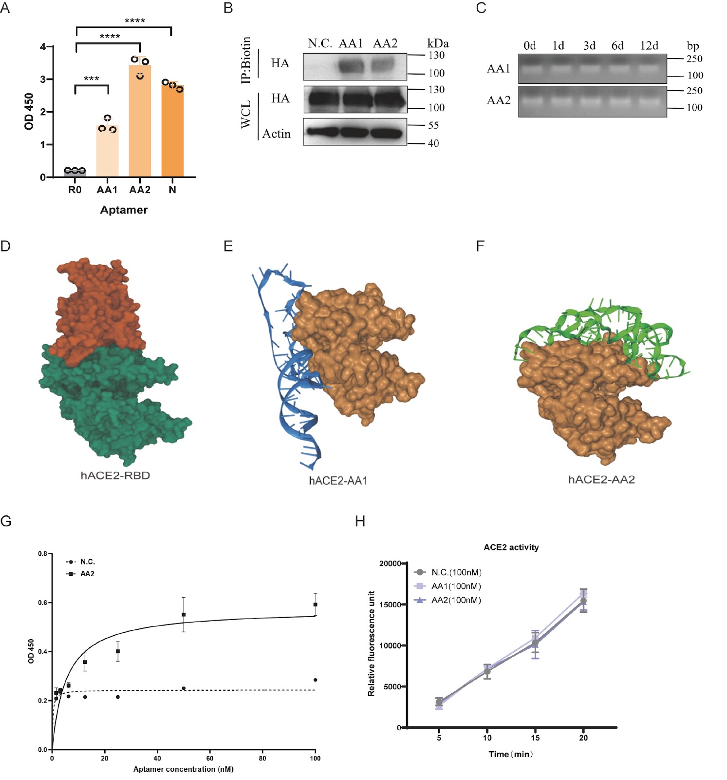
The relentless evolution of severe acute respiratory syndrome coronavirus 2 (SARS-CoV-2) and the emergence of immune-evasive variants underscore an urgent need for novel therapeutic strategies that are resilient to viral mutations. Targeting conserved host factors essential for viral entry represents a promising approach to overcome this challenge. Here, we report the development of a bifunctional therapeutic platform targeting the primary human receptor for SARS-CoV-2, angiotensin-converting enzyme 2 (ACE2). Using systematic evolution of ligands by exponential enrichment (SELEX), we isolate a high-affinity DNA aptamer, designated AA2, that binds to human ACE2 with a dissociation constant (Kd) of 5.41 ± 1.23 nM. Molecular docking and competitive binding assays confirm that AA2 sterically hinders the interaction between the viral spike receptor-binding domain (RBD) and ACE2. Consequently, AA2 demonstrates potent neutralization of SARS-CoV-2 S pseudovirus entry into host cells. To achieve a synergistic antiviral effect, we engineer an aptamer-siRNA chimera (AsiC) by conjugating AA2 to a short interfering RNA (siRNA) targeting the GFP coding region of the pseudovirus genome. This AsiC construct significantly represses viral replication compared to aptamer or siRNA treatment alone, validating a dual mechanism of action that combines receptor blockade with targeted gene silencing. This study establishes a robust proof-of-concept for an ACE2-targeted AsiC, representing a new class of dual-function antiviral therapeutics with the potential to effectively combat current and future ACE2-dependent coronaviruses.

A bifunctional aptamer-siRNA chimera targeting ACE2 for the inhibition of SARS-CoV-2 S pseudovirus entry and replication
Graphical Abstract
Original ResearchVol. xx, Issue xx • pp. xx-xxDOI: 10.3724/abbs.2025237

Altered gut microbial dynamics and the antivascular remodeling effect of carnosine in hypobaric hypoxic pulmonary hypertension rats

Authors: Huaying Wei, Shikun Guo, Wenjing Ding, Yifan Yang, Xinyu Hu, Ailifeila Aili, Xiaolan Chen, Xinying Xue, Lei Pan

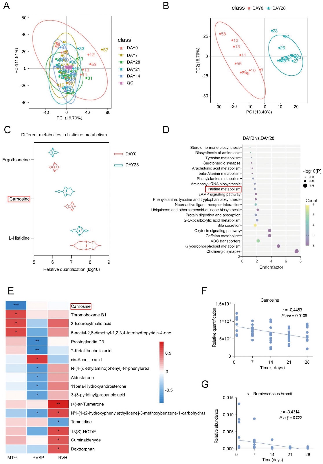
Exposure to chronic hypobaric hypoxia provokes marked alterations in the gut microbiota and its metabolome, yet the functional significance of histidine-derived metabolites in hypobaric hypoxic pulmonary hypertension (PH) remains underexplored. Here, we employ 16S rDNA, metagenomic, and untargeted metabolomic sequencing to characterize longitudinal shifts in the fecal microbiota and metabolites during hypobaric hypoxic PH development in Sprague-Dawley rats. Fecal carnosine levels and the abundance of its producer, Ruminococcus bromii, both decrease significantly over 28 days of hypobaric hypoxia (P < 0.05). Spearman correlation shows that carnosine is inversely correlated with the percentage of pulmonary arteriole media thickness (MT%; r = –0.8741, P < 0.001). Therapeutic supplementation with carnosine restores systemic and pulmonary antioxidant defenses and attenuates vascular remodeling without altering right ventricular pressures. In vitro, carnosine inhibits hypoxia-induced pulmonary artery smooth muscle cell (PASMC) proliferation and migration and suppresses nuclear factor erythroid 2-related factor 2 (Nrf2) accumulation. These findings reveal dynamic gut-lung crosstalk in hypobaric hypoxic PH and nominate carnosine as a metabolite-based intervention to mitigate hypoxia-driven pulmonary vascular remodeling.

Altered gut microbial dynamics and the antivascular remodeling effect of carnosine in hypobaric hypoxic pulmonary hypertension rats
Graphical Abstract
Original ResearchVol. xx, Issue xx • pp. xx–xxDOI: 10.3724/abbs.2025245

SPP1 facilitates sorafenib resistance in hepatocellular carcinoma by upregulating aerobic glycolysis in endothelial cells

Authors: Wufei Ye, Tao Chen, Yueran Li, Jiajie Luan, Kui Yang, Sheng Wang

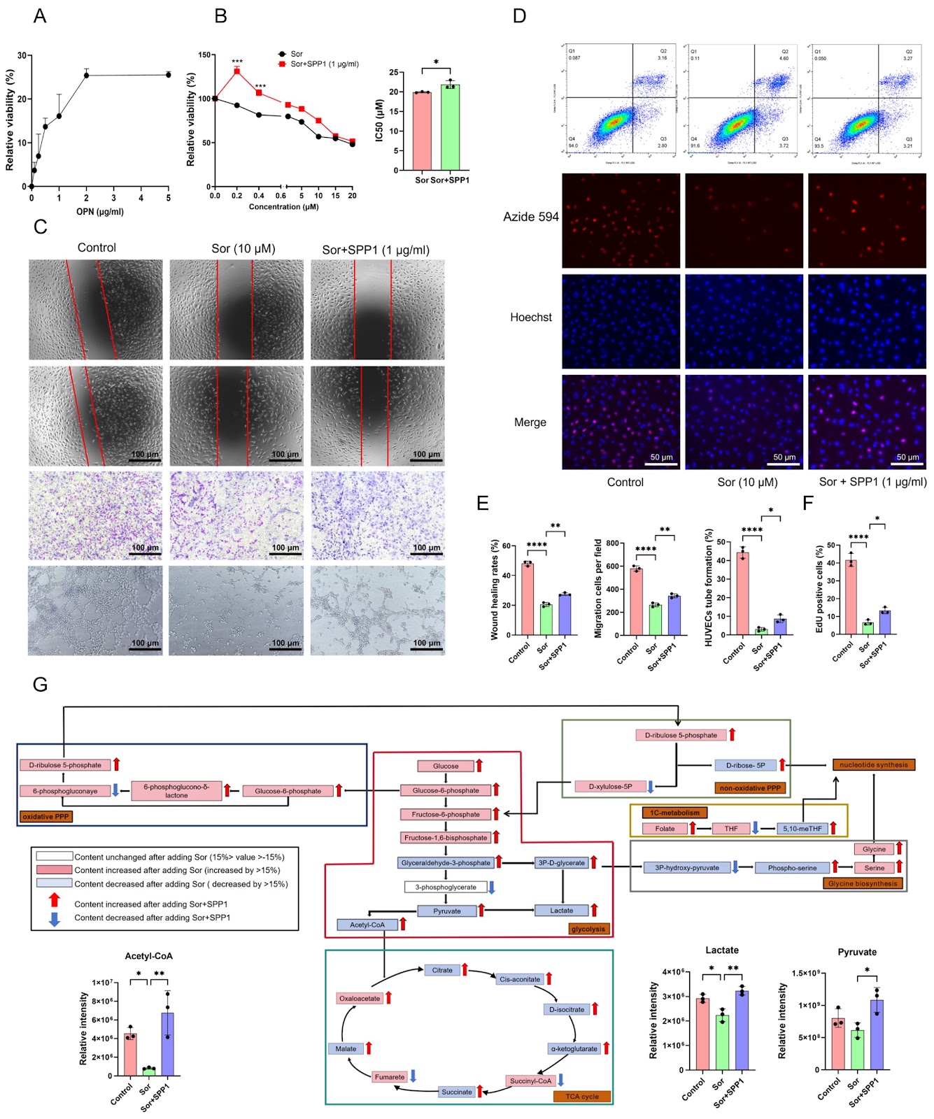
The occurrence of resistance to sorafenib, a first-line treatment for hepatocellular carcinoma (HCC), significantly limits its clinical efficacy. Therefore, investigating the potential mechanism of sorafenib resistance in HCC is highly important for developing HCC treatment strategies. In the present study, we identify that SPP1 (encoding osteopontin; OPN) is significantly elevated in sorafenib-resistant HCC. Furthermore, the upregulation of SPP1 is related to vascular invasion, advanced disease stage and poor prognosis in HCC patients. As the IC50 value of sorafenib increases in HepG2 cells, the SPP1 protein secreted by the cells is significantly upregulated, which subsequently facilitates the proliferation of human umbilical vein endothelial cells (HUVECs) and resistance to sorafenib. Further studies reveal that SPP1 induces resistance to sorafenib in HepG2 cells by upregulating glycolysis in HUVECs and further producing lactate. Mechanistically, SPP1 increases the expressions of the glucose transporter GLUT1 and the key glycolytic enzymes PFK1 and PKM2 in HUVECs, resulting in lactate accumulation, which in turn promotes the phosphorylation levels of BRAF and ERK as well as HIF-1α expression in HepG2 cells, leading to sorafenib resistance in HCC. Notably, SPP1 silencing can inhibit the proliferation and invasion of sorafenib-resistant HepG2 cells both in vitro and in vivo. Importantly, lactate derived from HUVECs plays a more dominant role in sorafenib resistance than does SPP1 in HepG2 cells. In summary, SPP1 enhances sorafenib resistance in HepG2 cells through promoting aerobic glycolysis in HUVECs, suggesting that the SPP1-aerobic glycolysis axis might be a prognostic biomarker as well as a potential therapeutic target for sorafenib-resistant HCC.

SPP1 facilitates sorafenib resistance in hepatocellular carcinoma by upregulating aerobic glycolysis in endothelial cells
Graphical Abstract
Original ResearchVol. xx, Issue xx • pp. xx-xxDOI: 10.3724/abbs.2026043

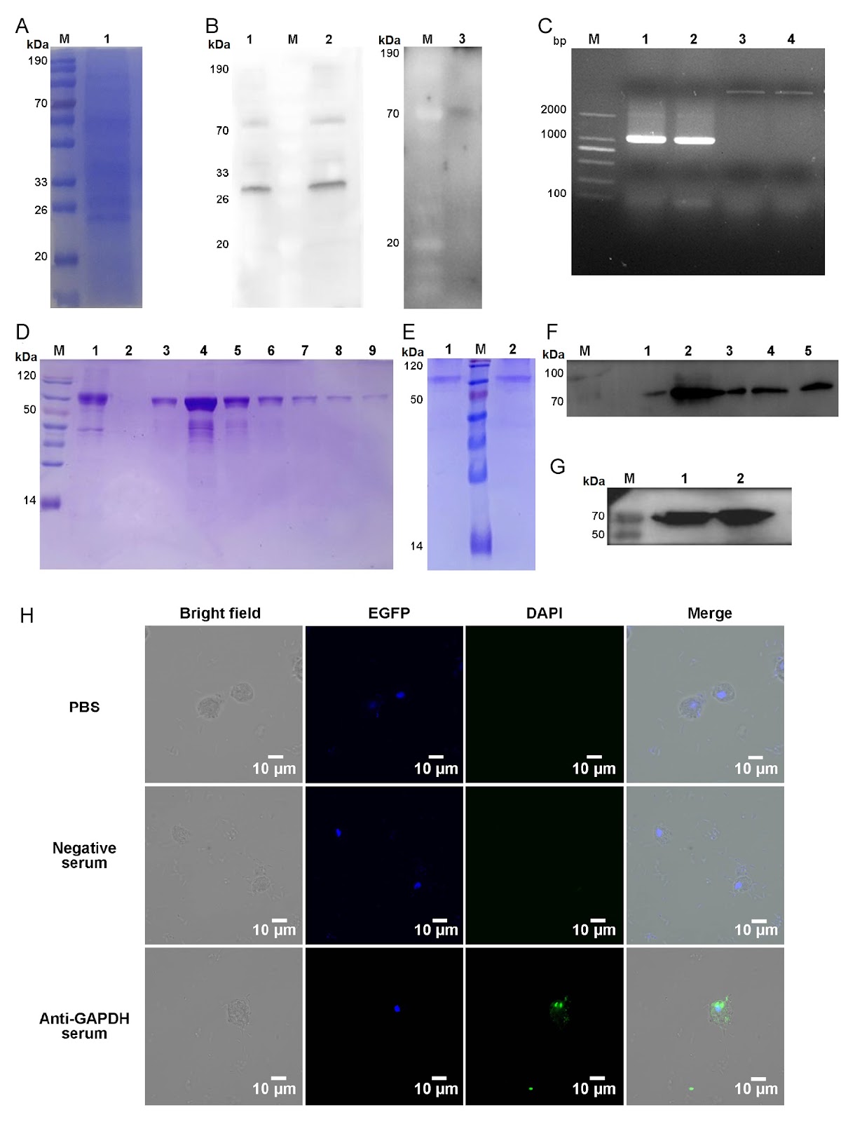
Development of a colloidal gold immunochromatographic strip based on GAPDH for Pentatrichomonas hominis in dogs

Authors: Qian Zhai, Xuejiao Chen, Xichen Zhang, Jianhua Li, Pengtao Gong, Xiaocen Wang, Xin Li, Xu Zhang, Nan Zhang

Pentatrichomonas hominis is a zoonotic protozoan belonging to the family Trichomonadidae that primarily inhabits the cecum and colon. Although traditionally regarded as an opportunistic pathogen, P. hominis is increasingly recognized for its pathogenic potential, including roles in animal diarrheal disease and the induction of intestinal epithelial damage and chronic inflammation in mice. A previous study further identified a significant correlation between P. hominis infection and colorectal cancer, underscoring its growing clinical and public health significance. This parasite infects a wide range of hosts, among which dogs, displaying infection rates as high as 47.4%, are regarded as a potential zoonotic reservoir because of their close contact with humans. Consequently, establishing reliable detection methods for P. hominis in dogs is essential for veterinary practice and public health surveillance. Current methods for detecting P. hominis infections in dogs mainly include direct smear microscopy and polymerase chain reaction (PCR)-based techniques. Although direct smear microscopy is straightforward, it frequently exhibits low sensitivity. In comparison, PCR demonstrates high sensitivity and specificity, yet it relies on specialized equipment, trained operators, and extended processing time. Recently, reported nucleic acid detection approaches, such as recombinase polymerase amplification coupled with lateral flow dipstick (RPA-LFD) and RPA-CRISPR/Cas12a assays, have enhanced the efficiency and accessibility of molecular detection for P. hominis. However, these methods still necessitate nucleic acid extraction, controlled temperature conditions, and operational complexity. In addition, some emerging detection technologies, such as microfluidic chips and nanozyme-based detection systems, offer advantages of high throughput and sensitivity but have not yet been widely applied in the field of detection of parasitic infection. Although immunoassays, including enzyme-linked immunosorbent assay (ELISA) and colloidal gold immunochromatographic strips, have been successfully used for the detection of infections of some intestinal protozoans, such as Giardia, there have been no reports on their application for detecting P. hominis infections, primarily due to the lack of specific detection antigens. To address the detection need, liquid chromatography-tandem mass spectrometry (LC-MS/MS) was used as the primary discovery tool to identify specific detected antigens. Using this targeted proteomics approach, we precisely identified immunoreactive proteins and selected glyceraldehyde 3 phosphate dehydrogenase (GAPDH), an immunogenic and species-specific antigen in related parasites, as the candidate antigen. Based on this identified antigen, we subsequently developed and evaluated the detection performance in both indirect ELISA and colloidal gold immunochromatographic strips using recombinant GAPDH. This study aimed to identify novel antigens for immunodetection of P. hominis and to establish a practical, on-site method for detecting dog infections, thereby facilitating further epidemiological and clinical research.

Development of a colloidal gold immunochromatographic strip based on GAPDH for Pentatrichomonas hominis in dogs
Graphical Abstract
Original ResearchVol. xx, Issue xx • pp. xx-xxDOI: 10.3724/abbs.2026104

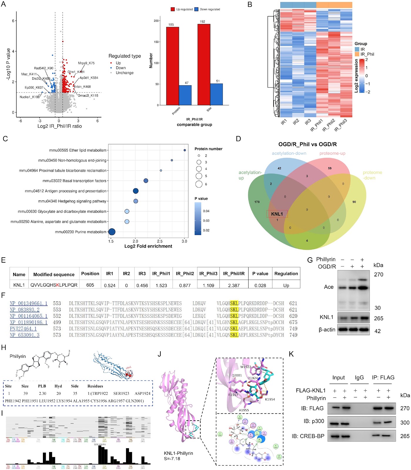
Phillyrin protects against myocardial ischemia/reperfusion injury by promoting KNL1 K605 acetylation to inhibit the p53/p21 pathway

Authors: Dongsheng He, Zhipeng Ren, Shangxuan Li, Ziqiang Dai, Gen Zhang, Huan Wang, Guanzheng Cui, Dianyuan Li

Reperfusion therapy is critical for acute myocardial infarction but is often accompanied by myocardial ischemia/reperfusion injury (MIRI). Phillyrin, a natural lignan from Forsythia suspensa, exerts anti-inflammatory and antioxidant effects; however, its role and mechanism in MIRI remain unclear. In this study, HL-1 cardiomyocytes are subjected to oxygen-glucose deprivation/reperfusion (OGD/R). Cell viability, apoptosis, oxidative stress, and inflammation are measured after phillyrin treatment. Multiomics (mRNA-seq, proteomics, and acetylproteomics) is used to identify key targets and pathways. Molecular docking, co-immunoprecipitation, site-directed mutagenesis, and western blot analysis are used to validate posttranslational regulation. A mouse MIRI model is established to confirm the in vivo cardioprotective effects of phillyrin. Phillyrin preserves cell viability and reduces apoptosis, oxidative stress, and inflammation in OGD/R-injured HL-1 cells. Multiomics integration reveals that phillyrin acts primarily through posttranslational regulation and highlights kinetochore scaffold 1 (KNL1) as the only protein that is both upregulated and hyperacetylated at lysine 605 (K605). Mechanistically, phillyrin may bind to the KNL1 C-terminus and enhance the interaction between KNL1 and acetyltransferase p300/CBP. KNL1 K605R mutation and Knl1 knockdown reduce KNL1 protein expression and reverse the inhibitory effects of phillyrin on p53 pathway-mediated apoptosis, oxidative stress, and inflammation. In mouse MIRI models, phillyrin reduces infarct size, myocardial damage, and cardiomyocyte apoptosis; these effects are abolished by knockdown of Knl1. Therefore, phillyrin promotes KNL1 acetylation at K605 to increase KNL1 protein expression, thereby inhibiting p53 signaling and alleviating apoptosis, oxidative stress, and inflammation in MIRI. This study identifies KNL1 acetylation at K605 as a novel posttranslational modification target for cardioprotection.

Phillyrin protects against myocardial ischemia/reperfusion injury by promoting KNL1 K605 acetylation to inhibit the p53/p21 pathway
Graphical Abstract
Original ResearchVol. xx, Issue xx • pp. xx-xxDOI: 10.3724/abbs.2026102

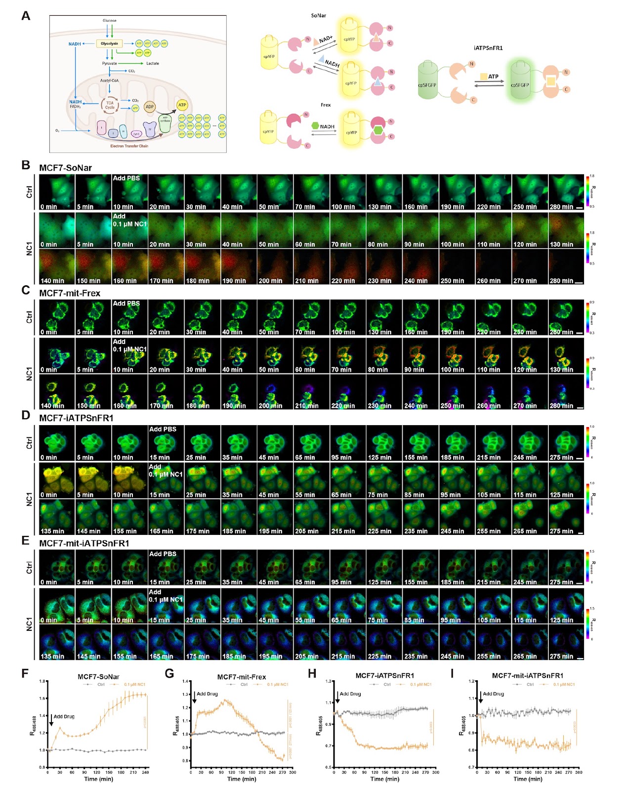
Integrating genetically encoded fluorescent sensors to elucidate the spatiotemporal choreography of necrosis by sodium overload

Authors: Yike Song, Yale Xia, Yin Fu, Jing Yao, Lian Zeng, Yuxi Duan, Ni Su, Xie Li, Xiawei Cheng, Yuzheng Zhao, Yi Yang, Yejun Zou

Necrosis by sodium overload (NECSO) is a distinct cell death modality induced by the chemical regulator necrocide 1 (NC1), which targets the transient receptor potential melastatin 4 (TRPM4) channel to drive excessive sodium influx and potassium efflux. This unique necrosis is characterized by the exchange of monovalent ions, a restrained ATP supply, redox disorder and a final membrane rupture as main features. Real-time monitoring of metabolic and redox causality during the whole process in living cells has been technically challenging. To bridge this gap, we integrate an advanced toolset of genetically encoded fluorescent sensors to monitor monovalent ions, energy metabolites, and redox equivalents with high spatiotemporal resolution. By directing these sensors to specific subcellular compartments, we successfully capture the real-time choreography of potassium loss specifically induced by NC1 via TRPM4. Furthermore, we establish a precise detection paradigm for evaluating energy currency by integrating sensors for NADH and ATP that are capable of subcellular imaging. We observe general and rapid NADH accumulation along with an ATP shortage in the mitochondria and cytosol. A concomitant reduction in mitochondrial oxidative stress is observed. This study not only elucidates the metabolic progression of a peculiar type of necrosis but also establishes a robust methodological framework for applying genetically encoded sensors to broader physiological and toxicological research.

Integrating genetically encoded fluorescent sensors to elucidate the spatiotemporal choreography of necrosis by sodium overload
Graphical Abstract
Original ResearchVol. xx, Issue xx • pp. xx-xxDOI: 10.3724/abbs.2026081

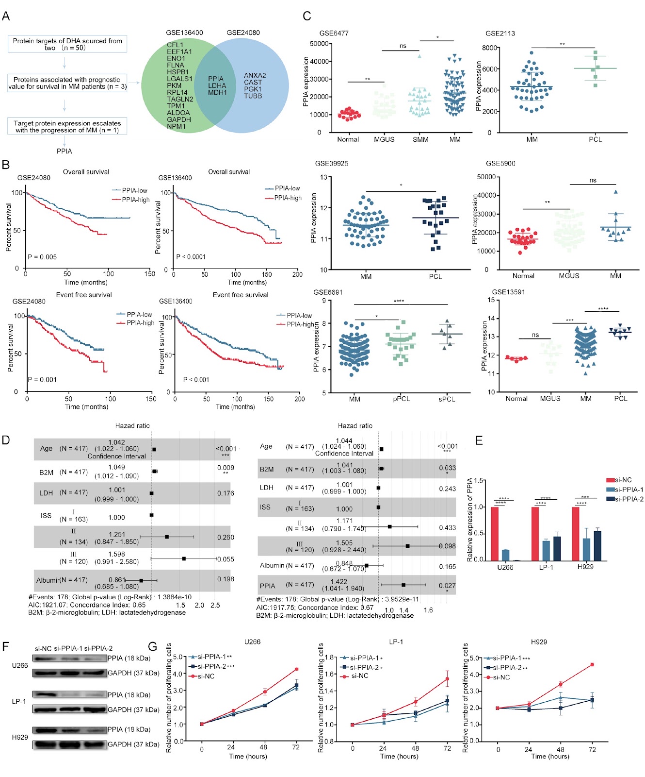
PPIA as a central regulator in a novel cell death pathway activated by iron homeostasis and redox disruption in multiple myeloma

Authors: Zhiming Wang, Chuan He, Qi Li, Xinyun Zhang, Si Chen, Lexin He, Yali Chai, Meifang Zhao, Linlin Qin, Menglu Chen, Hongxia Cui, Hao Xu, Yuchen Zhang, Bingzong Li, Wenzhuo Zhuang

Multiple myeloma (MM) patients frequently experience relapse, disease progression, and drug resistance, necessitating novel therapeutic strategies. This study investigates the role of peptidylprolyl isomerase A (PPIA) in mediating dihydroartemisinin (DHA)-induced ferroptosis in MM. Building on our previous work establishing the prognostic relevance of ferroptosis in MM, we elucidate the mechanism by which DHA triggers this form of cell death through the disruption of iron metabolism and redox homeostasis. DHA significantly reduces the viability of MM cell lines and primary CD138+ cells derived from patient bone marrow samples and attenuates tumor burden in orthotopic MM models. Mechanistically, DHA upregulates the iron-regulatory genes transferrin receptor 1 (TFRC) and heme oxygenase-1 (HMOX1), thereby perturbing iron homeostasis and promoting ferroptosis. Crucially, DHA targets the oxidized form of PPIA, a redox-sensitive chaperone, binding to it and preventing its reduction, which elevates intracellular reactive oxygen species (ROS). Combined treatment with DHA and erastin, which concurrently disrupt iron and amino acid metabolism, exerts synergistic cytotoxicity and enhances MM inhibition. Furthermore, in a syngeneic mouse model, DHA promotes T-cell activation and augments tumor suppression. Collectively, these findings underscore PPIA’s pivotal role in a novel ferroptotic cell death pathway and reveal new therapeutic opportunities for MM.

PPIA as a central regulator in a novel cell death pathway activated by iron homeostasis and redox disruption in multiple myeloma
Graphical Abstract
Original ResearchVol. xx, Issue xx • pp. xx–xxDOI: 10.3724/abbs.2026025

Sanguinarine exerts anti-hepatocellular carcinoma activity by targeting FDX1 to induce FDX1/LIAS/DLAT/HSP70 axis-dependent cuproptosis

Authors: Xiaoyan Hao, Yiqiao Qiu, Ling Li, Siqi Chen, Yuxuan Gao, Ketao Ma, Lili Wei, Qiang Zhang, Liang Zhang

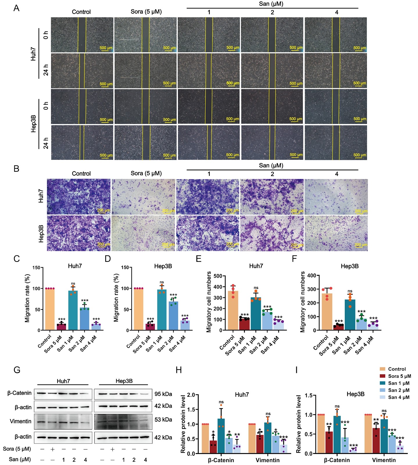
Hepatocellular carcinoma (HCC), the predominant type of primary liver cancer, represents an extremely aggressive malignancy. The induction of cuproptosis has developed into a favorable therapeutic direction for HCC, considering its strong association with HCC. Sanguinarine (San), a benzophenanthridine alkaloid derived from traditional herbs such as Chelidonium majus L., demonstrates broad-spectrum anticancer activities against various cancer cell types. However, the precise molecular mechanisms underlying its effects in the treatment of HCC remain largely undefined. This investigation seeks to examine the anti-HCC effects of San and to explore the mechanisms underlying these effects through the induction of cuproptosis. In vitro experiments demonstrate that San markedly inhibits the proliferation, movement, and epithelial-mesenchymal transition of HCC cells while enhancing their apoptosis. In vivo, San notably impedes tumor growth and upregulates the cuproptosis signature markers ferredoxin 1 (FDX1), oligomeric dihydrolipoamide S-acetyltransferase (DLAT), and heat shock protein 70 (HSP70) in HCC xenograft tumor models. Mechanistically, San induces proteotoxic stress and cuproptosis in HCC cells by increasing copper concentration, upregulating the expression of FDX1, lipoic acid synthetase (LIAS), HSP70, and lipoylated DLAT aggregation, and simultaneously reducing mitochondrial membrane potential and intracellular glutathione and pyruvate levels. Moreover, the combination of San with copper ionophores (Elesclomol-CuCl2) exhibits synergistic effects in promoting cuproptosis. FDX1 silencing markedly diminishes San-induced suppression of cell proliferation and FDX1 and HSP70 levels in HCC cells. Additionally, molecular docking analysis predicts that San exhibits the highest potential for binding with FDX1. Surface plasmon resonance experiments and cellular thermal shift assay confirm that San strongly interacts with FDX1 and markedly enhances the thermostability of FDX1. In conclusion, our findings indicate that San substantially inhibits the progression of HCC by targeting FDX1/LIAS/DLAT/HSP70 axis-dependent cuproptosis.

Sanguinarine exerts anti-hepatocellular carcinoma activity by targeting FDX1 to induce FDX1/LIAS/DLAT/HSP70 axis-dependent cuproptosis
Graphical Abstract
Original ResearchVol. xx, Issue xx • pp. xx–xxDOI: 10.3724/abbs.2025196

OAZ1/CASP8AP2 double knockout enhances recombinant protein production in HEK293 cells through metabolic reprogramming and antiapoptotic effects

Authors: Junhe Zhang, Liao Zhang, Lu Hou, Weidong Li, Shaolei Geng, Xiaoyin Wang, Tianyun Wang

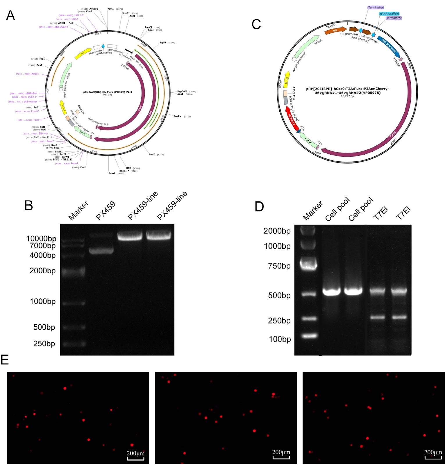
Human embryonic kidney (HEK) 293 cells are widely used for recombinant protein production because of their efficient posttranslational modification capabilities. However, their large-scale culture is often limited by metabolic stress and early apoptosis, leading to insufficient protein yields. In this study, we aim to increase protein expression through the coordinated modulation of metabolic and apoptotic pathways. Using CRISPR/Cas9 technology, we target and knockout the genes of ornithine decarboxylase antizyme 1 (OAZ1), which regulates polyamine metabolism, and caspase 8-associated protein 2 (CASP8AP2), an apoptosis-related protein. We successfully construct an OAZ1/CASP8AP2 double-knockout HEK293 cell line. Following transfection with the knockout vector and screening of single-cell clones, multiple levels of validation confirm the successful gene knockout. The results show that the double-knockout cells exhibit significantly reduced apoptosis rates. Furthermore, the production of recombinant secreted alkaline phosphatase (SEAP) and vitronectin (VN) increases by 2.1 folds and 2.9 folds, respectively, compared with those in wild-type cells. Metabolic profiling reveals that the cell cycle is arrested in the G1/G0 phase, accompanied by increased specific consumption and production rates of key metabolites. This study demonstrates that concurrent inhibition of apoptosis and optimization of metabolism effectively enhances recombinant protein production in HEK293 cells, suggesting a novel strategy for improving HEK293 cell-based expression.

OAZ1/CASP8AP2 double knockout enhances recombinant protein production in HEK293 cells through metabolic reprogramming and antiapoptotic effects
Graphical Abstract
Original ResearchVol. xx, Issue xx • pp. xx-xxDOI: 10.3724/abbs.2026027

PCIF1 modulates glioblastoma cell migration and invasion by altering PI(3,4)P2 levels through the PI5-phosphatase INPP5B

Authors: Preethi Priyanka Musunuru, Yihan Li, Yu Zhou, Gai Liu, Zhuang Sha, Zhiyuan Hu, Junbo Zhou, Naveena Konduru, Qingming Meng, Shangfeng Gao, Rutong Yu

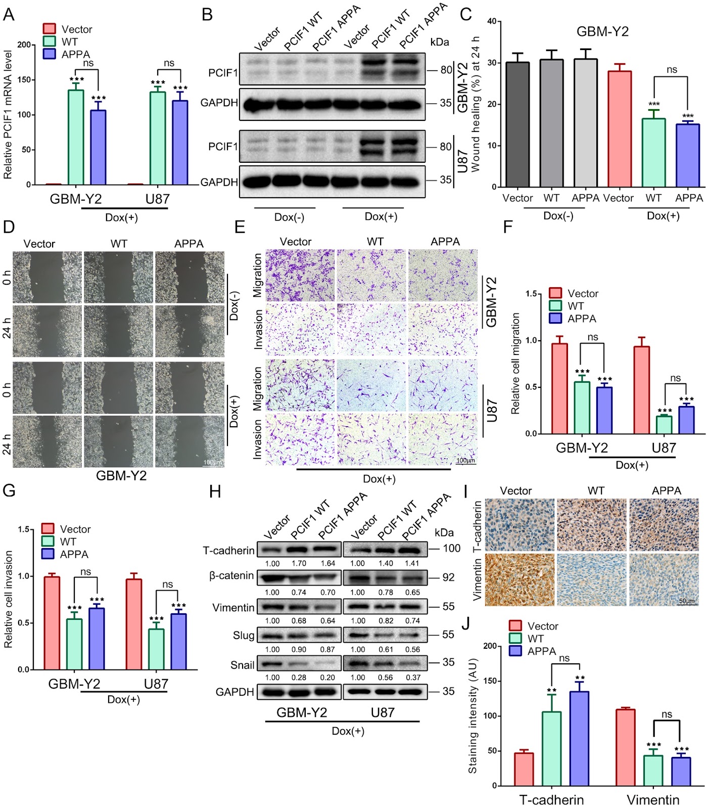
Phosphorylated CTD Interacting Factor 1 (PCIF1) is the exclusive methyltransferase responsible for the N6,2-O-dimethyladenosine (m6Am) modification in mammalian mRNA. Our previous research identified PCIF1 as a potent tumor suppressor in glioma, demonstrating its ability to impair cell proliferation, induce G2/M phase arrest, and promote apoptosis. However, its role in glioma cell migration and invasion remains unclear. In this study, we investigate how PCIF1 regulates glioma cell migration and invasion. Overexpression of PCIF1 inhibits migration and invasion, whereas PCIF1 knockdown enhances these behaviors. Corresponding changes are observed in mesenchymal markers (Vimentin, β-catenin, Snail, Slug) and the epithelial marker T-cadherin, indicating that PCIF1 suppresses epithelial-to-mesenchymal transition (EMT)-mediated glioma invasion. Mechanistically, PCIF1 modulates the AKT pathway by promoting proteasomal degradation of AKT while increasing phosphorylated AKT (p-AKT) levels, revealing a complex regulatory mechanism. PCIF1 knockdown upregulates INPP5B, a lipid phosphatase, causing accumulation of PI(3,4)P2 and enhanced AKT activation. Conversely, PCIF1 overexpression increases PI(3,4,5)P3 production, elevating p-AKT levels. This bidirectional regulation suggests that PCIF1 influences phosphoinositide signaling and AKT activation. Our findings highlight PCIF1 as a key modulator of glioblastoma cell migration and invasion through phosphoinositide signaling, positioning it as a potential biomarker and therapeutic target in glioma.

PCIF1 modulates glioblastoma cell migration and invasion by altering PI(3,4)P2 levels through the PI5-phosphatase INPP5B
Graphical Abstract
Original ResearchVol. xx, Issue xx • pp. xx-xxDOI: 10.3724/abbs.2026074

Tryptophan-substituted antimicrobial peptide temporin-1CEb: in vitro and in vivo antibacterial activity against clinically isolated multidrug-resistant Klebsiella pneumonia

Authors: Fengquan Jiang, Yanjun Ma, Yunfei Zhang, Dejing Shang, Weibing Dong

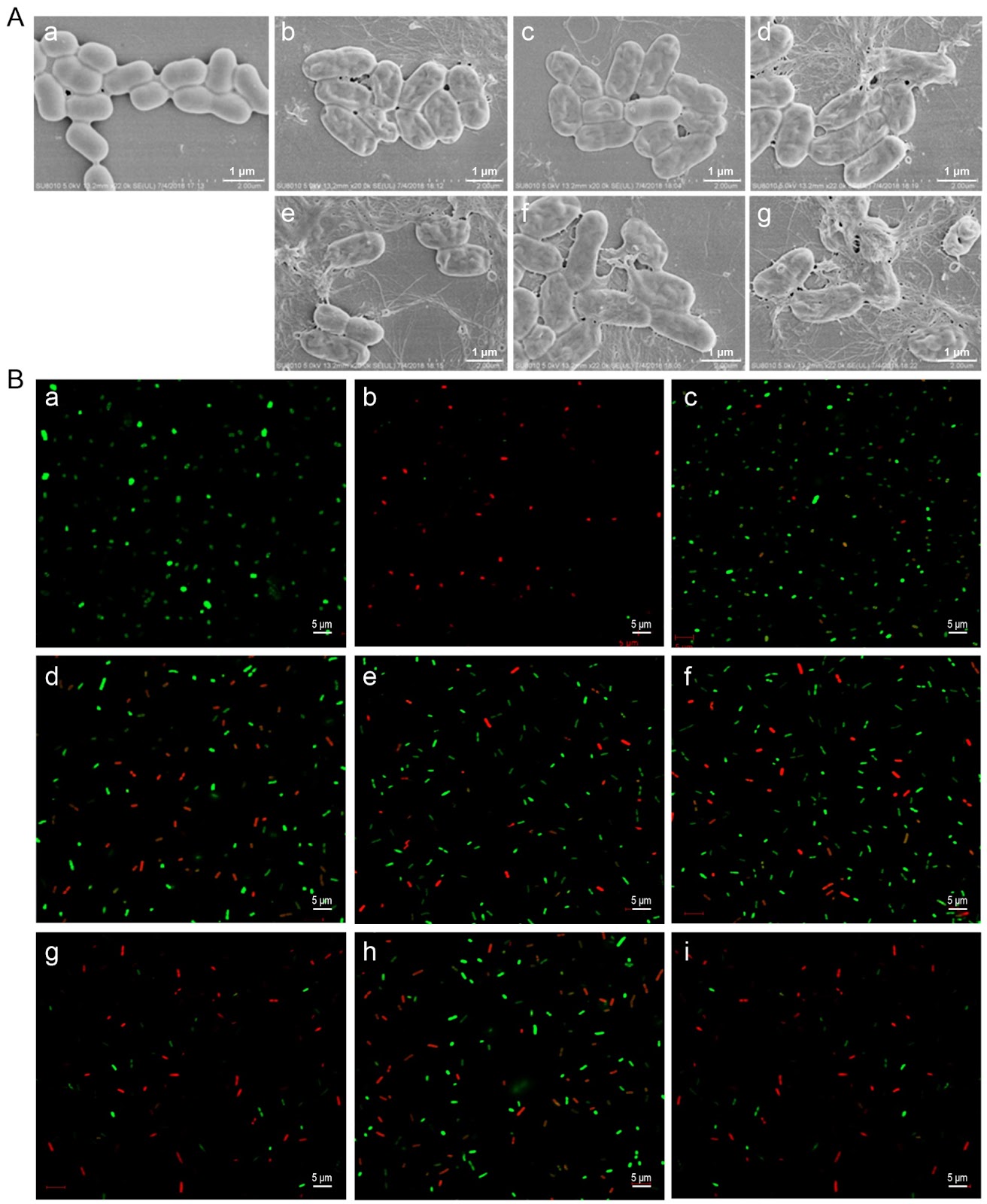
Tryptophan (Trp)-substituted antimicrobial peptides (AMPs) exhibit enhanced interactions with bacterial cell membranes, potentially improving their antimicrobial efficacy. Klebsiella pneumoniae (20.59% of 2054 ICU isolates) is resistant to multiple clinically used antibiotics and presents significant treatment challenges. In the present study, three Trp-modified peptides (I4W, L12W, and I1WL5W) are generated by substituting Ile or Leu residues in temporin-1CEb, a peptide derived from frog skin, with Trp at various sites to assess their antibacterial effects and mechanisms against K. pneumoniae. Compared with L12W, both I4W and I1WL5W display superior antimicrobial activity and lower cytotoxicity. Mechanistic studies reveal that AMPs exert antibacterial and bactericidal effects through bacterial surface charge neutralization, insertion into bacterial cell membranes, increase permeability of both the inner and outer membranes, and disruption of membrane integrity. Notably, I1WL5W exhibit the most potent membrane-disrupting activity. Assessment of the impact of Trp-containing peptides on bacterial biofilms reveals that these peptides not only inhibit exopolysaccharide production and biofilm formation but also degrade preformed biofilms. A murine lung infection model is established to investigate the therapeutic efficacy of I1WL5W against MDRKP 1203-induced lung infection in mice. Compared with the control treatment, treatment with I1WL5W results in reduced bacterial counts and levels of IL-6 and TNF-α in both the blood and lung tissues of MDRKP 1203-infected mice, and treatment with I1WL5W improves lung tissue structure. The present study provides valuable insights for designing Trp-containing peptides with potent antimicrobial properties by facilitating their penetration across bacterial membranes.

Tryptophan-substituted antimicrobial peptide temporin-1CEb: in vitro and in vivo antibacterial activity against clinically isolated multidrug-resistant Klebsiella pneumonia
Graphical Abstract
Original ResearchVol. xx, Issue xx • pp. xx-xxDOI: 10.3724/abbs.2026065

Ginsenoside Rh2 alleviates osteoporosis by attenuating oxidative stress-induced osteoblast dysfunction via the FoxO1/β-catenin pathway

Authors: Wenqi Jin, Yujing Tan, Xuenan Chen, Zehan Guo, Jixiang Ren, Liwei Sun

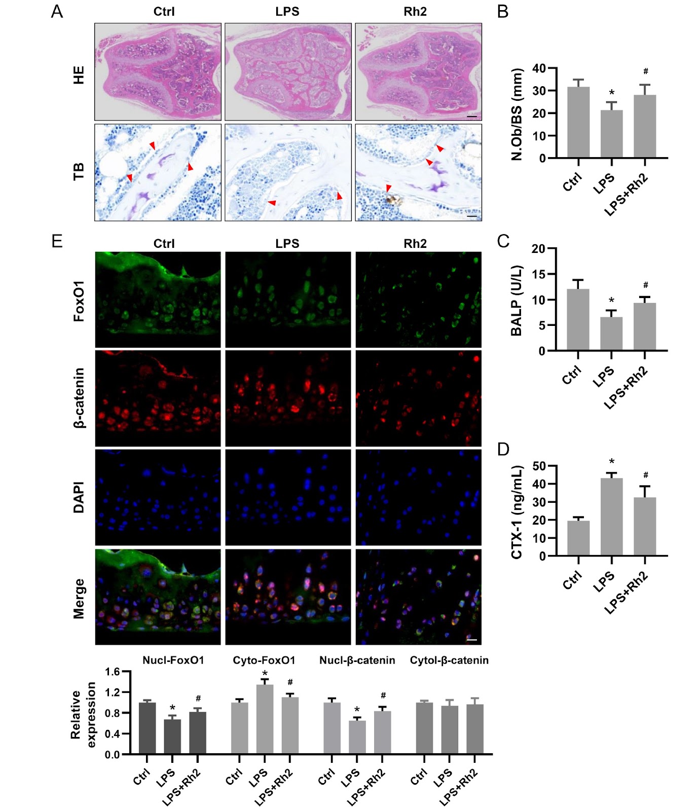
The degree of oxidative stress decreases osteoblast function with age, which leads to a decline in bone compressive capacity. Ginsenoside Rh2 is a known clinical or adjuvant therapy for various tissues. In this study, we investigate the pharmacological effects of Rh2 against oxidative stress-induced osteoblasts. Osteoblasts are pretreated with Rh2 for 48 h and then exposed to hydrogen peroxide (H2O2), which results in significantly decreased ROS levels, increased antioxidant enzyme activity, and enhanced mitochondrial function. Functionally, Rh2 increases alkaline phosphatase (ALP) expression, together with enhanced mineralization and expression of osteogenesis-associated genes. Rh2 also promotes the nuclear translocation of FoxO1 and β-catenin, whereas it does not reverse reduced mineralization caused by decreased FoxO1 or β-catenin activity, indicating that its effect is mediated through the functional interaction between FoxO1 and β-catenin. In a mouse model of lipopolysaccharide (LPS)-induced bone loss, Rh2 administration improves trabecular microstructure, increases osteoblast numbers, and upregulates serum metabolites associated with bone formation. Immunofluorescence analysis further reveals that Rh2 promotes the nuclear co-localization of FoxO1 and β-catenin in femurs, indicating their coordinated action within this signaling axis. These findings indicate that Rh2 mitigates oxidative stress-induced osteoblast dysfunction via the FoxO1/β-catenin pathway, highlighting the pivotal role of redox balance in bone remodeling and suggesting a promising therapeutic strategy for osteoporosis.

Ginsenoside Rh2 alleviates osteoporosis by attenuating oxidative stress-induced osteoblast dysfunction via the FoxO1/β-catenin pathway
Graphical Abstract