🧬 SinoBioData Academic Portal
🏛️ Indexed Academic JournalOriginal: 生物化学与生物物理学报

Acta Biochimica et Biophysica Sinica

Premier Chinese Biomedical Journal indexed in SinoBioData: Acta Biochimica et Biophysica Sinica (生物化学与生物物理学报).

Total Research Papers: 200
Access: 100% Free Open Access
Browse by Publication Year & VolumeReset All Filters ✕

Published Research PapersFiltered: Year 2024 • 56 • 9

Showing 11 of 200 peer-reviewed papers with full Graphical Abstracts.

Original ResearchVol. 56, Issue 9 • pp. 1256-1266DOI: 10.3724/abbs.2024105

Molecular insight into the potential functional role of pseudoenzyme GFOD1 via interaction with NKIRAS2

Authors: Jiawen Shi, Xinyi Guo, Chan Liu, Yilun Wang, Xiaobao Chen, Guihua Wu, Jianping Ding, Tianlong Zhang

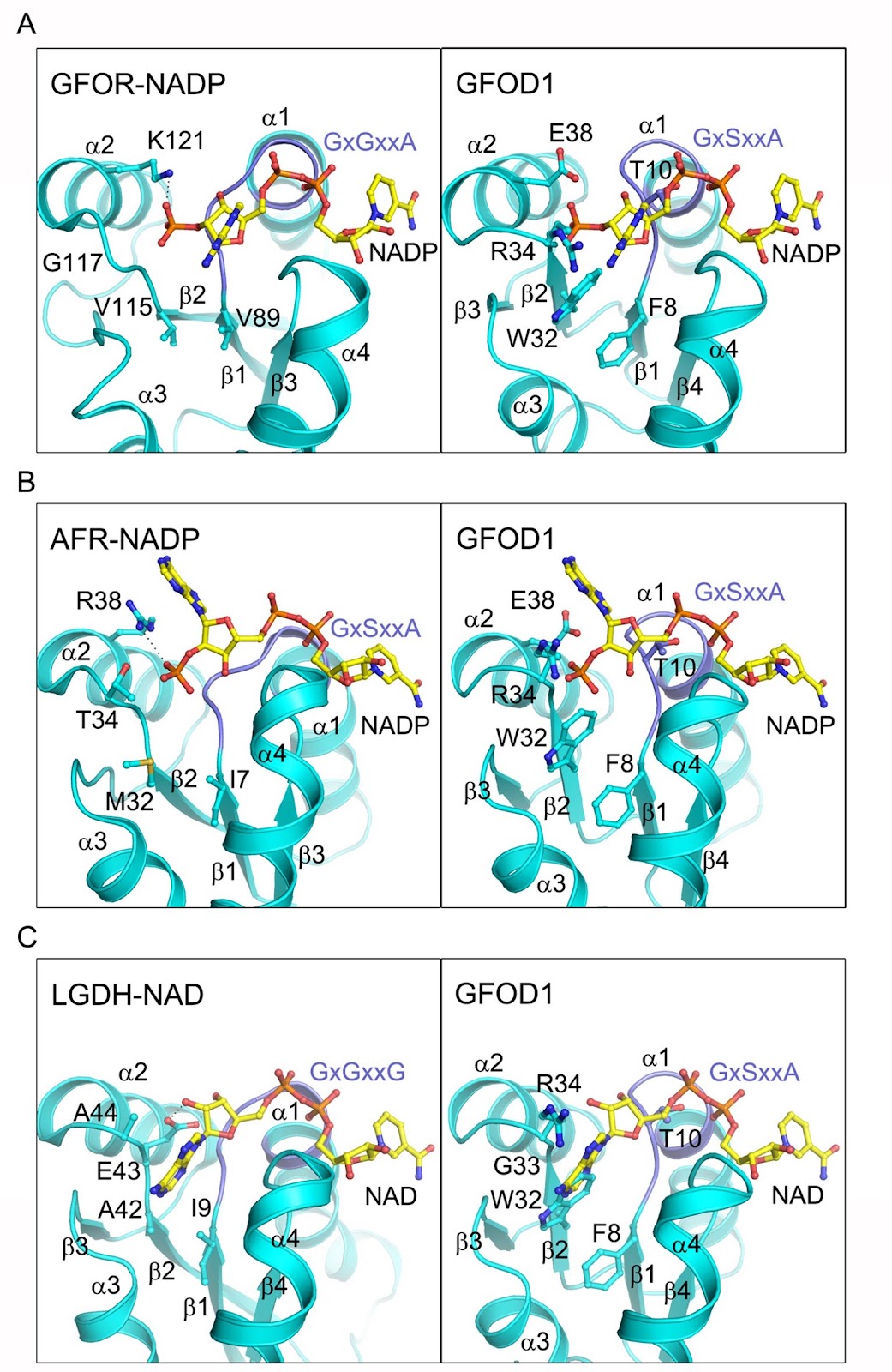
The glucose-fructose oxidoreductase/inositol dehydrogenase/rhizopine catabolism protein (Gfo/Idh/MocA) family includes a variety of oxidoreductases with a wide range of substrates that utilize NAD or NADP as redox cofactor. Human contains two members of this family, namely glucose-fructose oxidoreductase domain-containing protein 1 and 2 (GFOD1 and GFOD2). While GFOD1 exhibits low tissue specificity, it is notably expressed in the brain, potentially linked to psychiatric disorders and severe diseases. Nevertheless, the specific function, cofactor preference, and enzymatic activity of GFOD1 remain largely unknown. In this work, we find that GFOD1 does not bind to either NAD or NADP. Crystal structure analysis unveils that GFOD1 exists as a typical homodimer resembling other family members, but lacks essential residues required for cofactor binding, suggesting that it may function as a pseudoenzyme. Exploration of GFOD1-interacting partners in proteomic database identifies NF-κB inhibitor-interacting Ras-like 2 (NKIRAS2) as one potential candidate. Co-immunoprecipitation (co-IP) analysis indicates that GFOD1 interacts with both GTP- and GDP-bound forms of NKIRAS2. The predicted structural model of the GFOD1-NKIRAS2 complex is validated in cells using point mutants and shows that GFOD1 selectively recognizes the interswitch region of NKIRAS2. These findings reveal the distinct structural properties of GFOD1 and shed light on its potential functional role in cellular processes.

Molecular insight into the potential functional role of pseudoenzyme GFOD1 via interaction with NKIRAS2
Graphical Abstract
Original ResearchVol. 56, Issue 9 • pp. 1373-1386DOI: 10.3724/abbs.2024111

Lactate activates CCL18 expression via H3K18 lactylation in macrophages to promote tumorigenesis of ovarian cancer

Authors: Jinrui Sun, Qinmei Feng, Yue He, Ming Wang, Yumei Wu

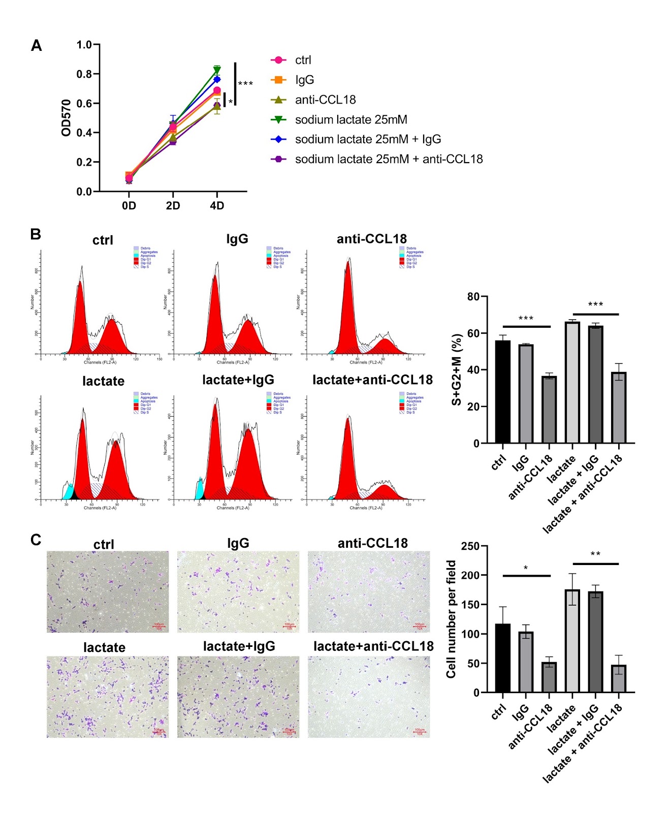
This study investigates the role of lactate in the genesis and progression of ovarian cancer (OV) and explores the underlying mechanisms. Serum lactate levels show a positive correlation with tumor grade and poor prognosis in patients with OV. Bioinformatics analysis identifies CCL18 as a lactate-related gene in OV. CCL18 is up-regulated in cancerous tissues and positively related to serum lactate levels in OV patients. THP-1 cells are exposed to phorbol-12-myristate-13-acetate for M0 macrophage induction. The results of RT-qPCR and ELISA for M1/M2 macrophage-related markers and inflammatory cytokines show that the exposure of lactate to macrophages induces M2 polarization. Based on the coculture of OV cells with macrophages, lactate-treated macrophages induces a significant increase in the proliferation and migration of OV cells. However, these effects can be reversed by silencing of Gpr132 in macrophages or treatment with anti-CCL18 antibody. Experiments using the xenograft model verify that the oncogenic role of lactate in tumor growth and metastasis relies on Gpr132 and CCL18. ChIP-qPCR and luciferase reporter assays reveal that lactate regulates CCL18 expression via H3K18 lactylation. In conclusion, lactate is a potential therapeutic target for OV. It is involved in tumorigenesis by activating CCL18 expression via H3K18 lactylation in macrophages.

Lactate activates CCL18 expression via H3K18 lactylation in macrophages to promote tumorigenesis of ovarian cancer
Graphical Abstract
Original ResearchVol. 56, Issue 9 • pp. 1289-1299DOI: 10.3724/abbs.2024084

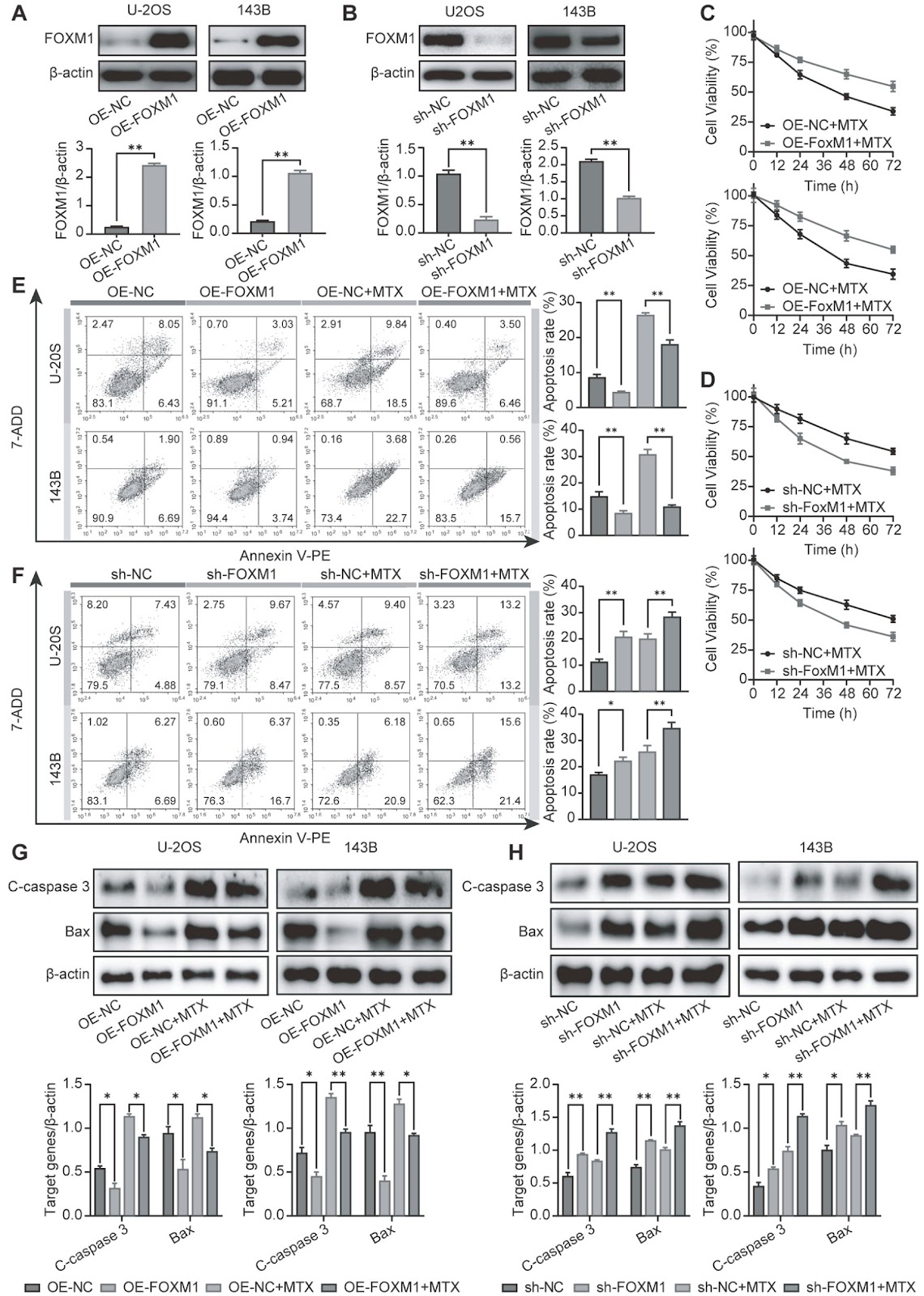
FOXM1 mediates methotrexate resistance in osteosarcoma cells by promoting autophagy

Authors: Luoyang Wang, Dongchang Zhai, Lei Tang, Hui Zhang, Xinlong Wang, Ning Ma, Xiaoyue Zhang, Mingguo Cheng, Ruowu Shen

Osteosarcoma (OS) is a primary bone cancer mostly found in adolescents and elderly individuals. The treatment of OS is still largely dependent on traditional chemotherapy. However, the high incidence of drug resistance remains one of the greatest impediments to limiting improvements in OS treatment. Recent findings have indicated that the transcription factor FOXM1 plays an important role in various cancer-related events, especially drug resistance. However, the possible role of FOXM1 in the resistance of OS to methotrexate (MTX) remains to be explored. Here, we find that FOXM1, which confers resistance to MTX, is highly expressed in OS tissues and MTX-resistant cells. FOXM1 overexpression promotes MTX resistance by enhancing autophagy in an HMMR/ATG7-dependent manner. Importantly, silencing of FOXM1 or inhibiting autophagy reverses drug resistance. These findings demonstrate a new mechanism for FOXM1-induced MTX resistance and provide a promising target for improving OS chemotherapy outcomes.

FOXM1 mediates methotrexate resistance in osteosarcoma cells by promoting autophagy
Graphical Abstract
Original ResearchVol. 56, Issue 9 • pp. 1311-1322DOI: 10.3724/abbs.2024049

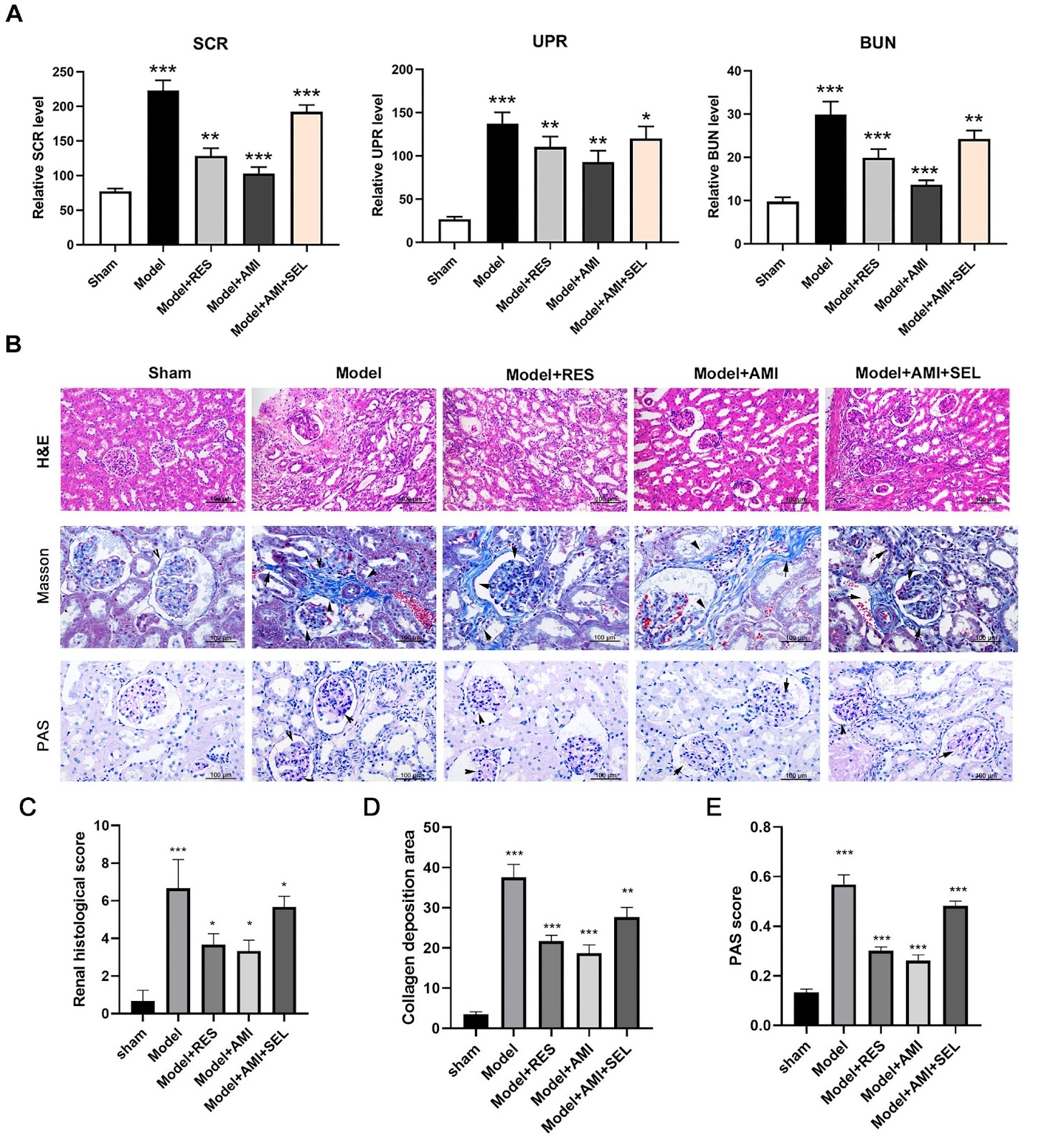
Aminophylline suppresses chronic renal failure progression by activating SIRT1/AMPK/mTOR-dependent autophagy

Authors: Xin Liao, Jieyi Lu, Zhifeng Huang, Jinai Lin, Miao Zhang, Huanru Chen, Xiaoqing Lin, Xia Gao, Sitang Gong

Chronic renal failure (CRF) is a severe syndrome affecting the urinary system for which there are no effective therapeutics. In this study, we investigate the effects and mechanisms of aminophylline in preventing CRF development. A rat model of chronic renal failure is established by 5/6 nephrectomy. The levels of serum creatinine (SCR), urinary protein (UPR), and blood urea nitrogen (BUN) are detected by ELISA. Histological evaluations of renal tissues are performed by H&E, Masson staining, and PAS staining. Functional protein expression is detected by western blot analysis or immunofluorescence microscopy. Glomerular cell apoptosis is determined using the TUNEL method. Results show that Aminophylline significantly reduces the levels of SCR, UPR, and BUN in the CRF model rats. Histological analyses show that aminophylline effectively alleviates renal tissue injuries in CRF rats. The protein expression levels of nephrin, podocin, SIRT1, p-AMPK, and p-ULK1 are greatly increased, while p-mTOR protein expression is markedly decreased by aminophylline treatment. Additionally, the protein level of LC3B in CRF rats is significantly increased by aminophylline. Moreover, aminophylline alleviates apoptosis in the glomerular tissues of CRF rats. Furthermore, resveratrol promotes SIRT1, p-AMPK, and p-ULK1 protein expressions and reduces p-mTOR and LC3B protein expressions in CRF rats. Selisistat (a SIRT1 inhibitor) mitigates the changes in SIRT1, p-AMPK, p-ULK1, p-mTOR, and LC3B expressions induced by aminophylline. Finally, RAPA alleviates renal injury and apoptosis in CRF rats, and 3-MA eliminates the aminophylline-induced inhibition of renal injury and apoptosis in CRF rats. Aminophylline suppresses chronic renal failure progression by modulating the SIRT1/AMPK/mTOR-mediated autophagy process.

Aminophylline suppresses chronic renal failure progression by activating SIRT1/AMPK/mTOR-dependent autophagy
Graphical Abstract
Original ResearchVol. 56, Issue 9 • pp. 1365-1372DOI: 10.3724/abbs.2024091

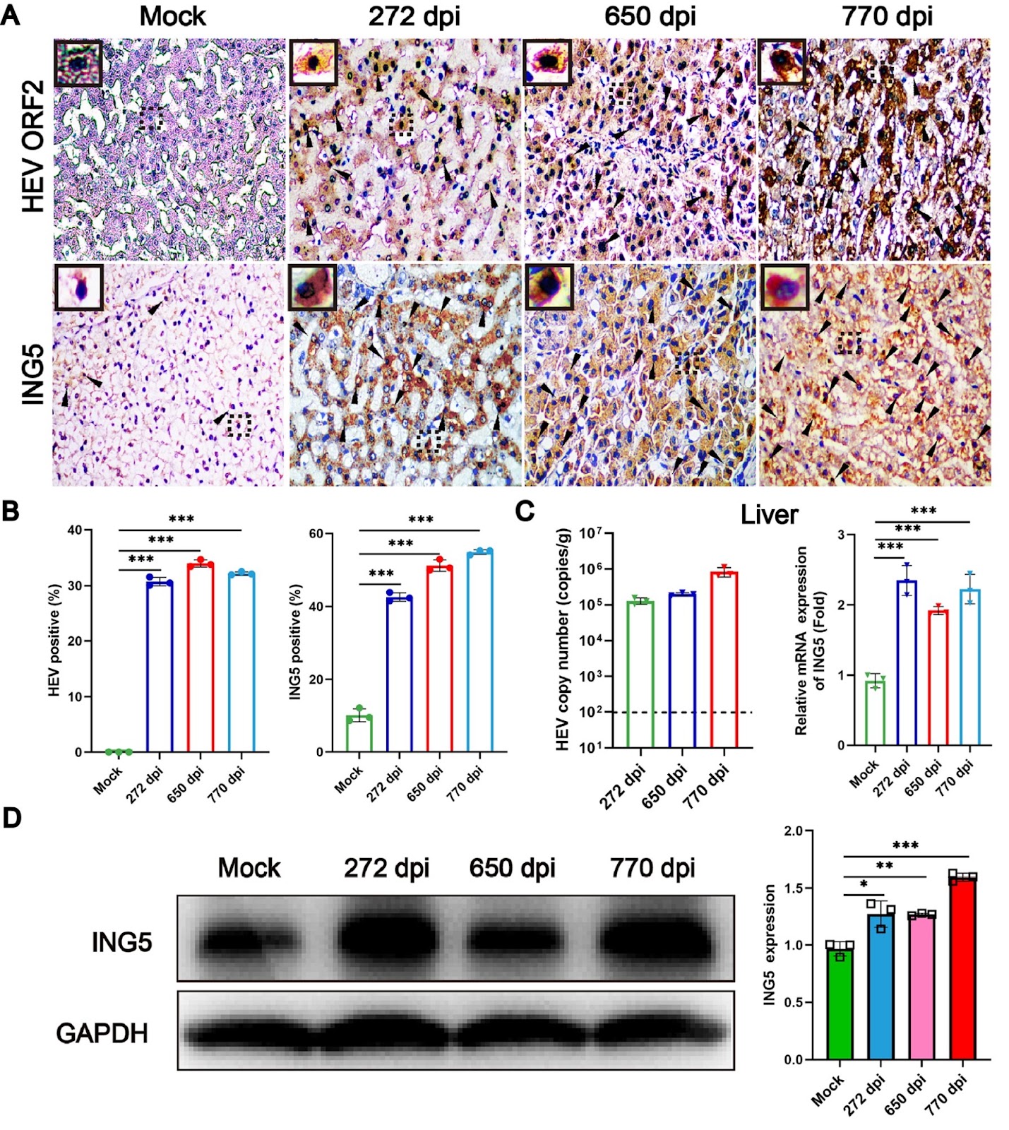
Hepatitis E virus infection upregulates ING5 expression in vitro and in vivo

Authors: Wanqiu Zhao, Yueping Xia, Tengyuan Li, Huichan Liu, Guo Zhong, Dongxue Chen, Wenhai Yu, Yunlong Li, Fen Huang

Hepatitis E virus (HEV) is the major pathogen of viral hepatitis. Immunocompromised individuals infected by HEV are prone to chronic hepatitis and increase the risk of hepato-cellular carcinoma (HCC). Inhibitor of growth family member 5 (ING5) is a tumor suppressor that is expressed at low levels in cancer tumors or cells. However, the underlying relationship between ING5 and HEV infection is unclear. In the present study, acute and chronic HEV animal models are used to explore the interaction between ING5 and HEV. Notably, the expression of ING5 is significantly increased in both the livers of acute HEV-infected BALB/c mice and chronic HEV-infected rhesus macaques. In addition, the relationship between HEV infection and ING5 expression is further identified in human hepatoma (HepG-2) cells. In conclusion, HEV infection strongly upregulates ING5 expression both in vivo and in vitro, which has significant implications for further understanding the pathogenic mechanism of HEV infection.

Hepatitis E virus infection upregulates ING5 expression in vitro and in vivo
Graphical Abstract
Original ResearchVol. 56, Issue 9 • pp. 1340-1351DOI: 10.3724/abbs.2024050

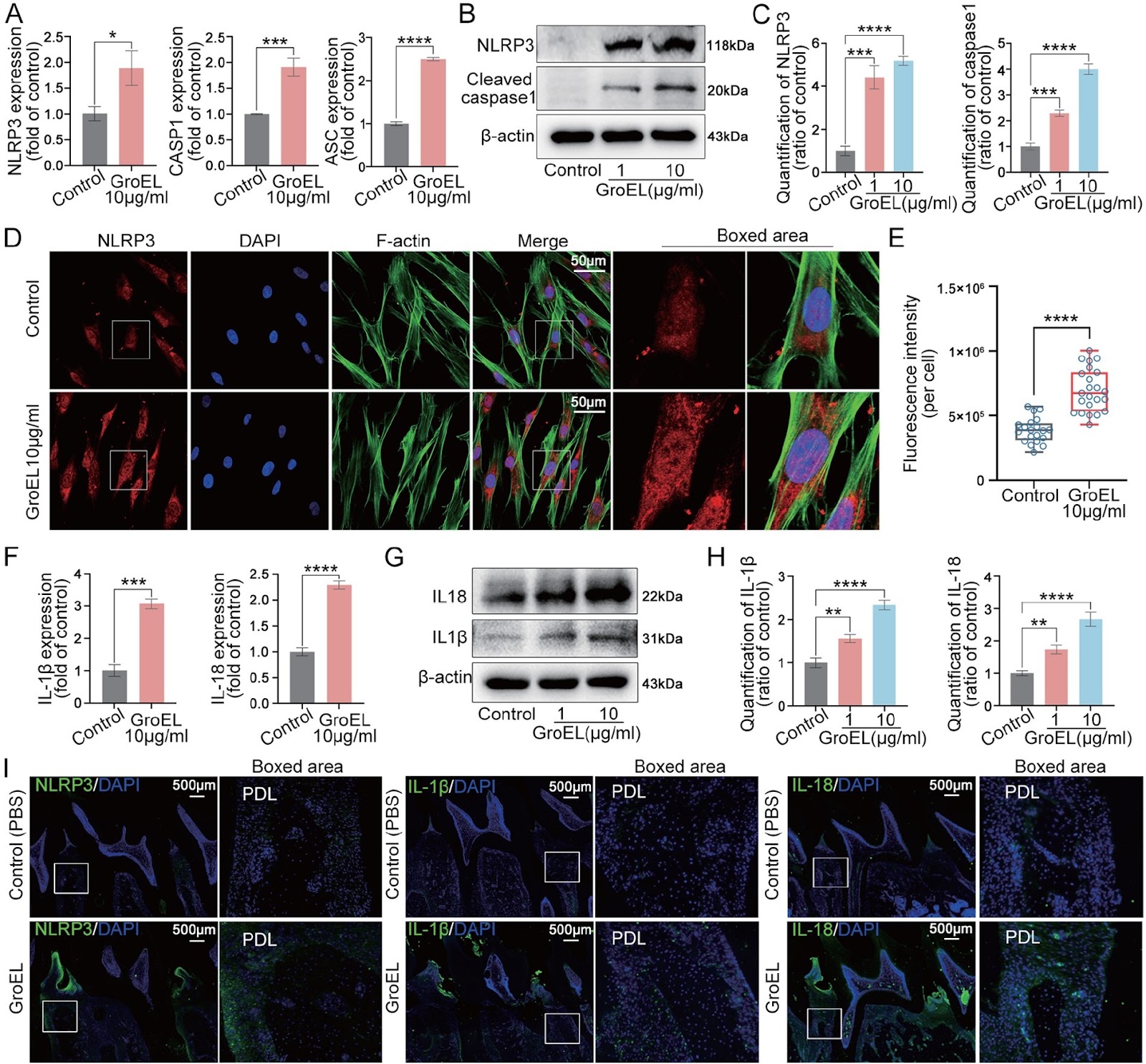
GroEL triggers NLRP3 inflammasome activation through the TLR/NF-κB p-p65 axis in human periodontal ligament stem cells

Authors: Li Zhang, Mengmeng Duan, Xiaohua Pu, Huiling Zheng, Xinjie Ning, Ying Tu, Chunming Xu, Demao Zhang, Chengcheng Liu, Jing Xie

The interaction between bacteria and the host plays a vital role in the initiation and progression of systemic diseases, including gastrointestinal and oral diseases, due to the secretion of various virulence factors from these pathogens. GroEL, a potent virulence factor secreted by multiple oral pathogenic bacteria, is implicated in the damage of gingival epithelium, periodontal ligament, alveolar bone and other peripheral tissues. However, the underlying biomechanism is still largely unknown. In the present study, we verify that GroEL can trigger the activation of NLRP3 inflammasome and its downstream effector molecules, IL-1β and IL-18, in human periodontal ligament stem cells (hPDLSCs) and resultantly induce high activation of gelatinases (MMP-2 and MMP-9) to promote the degradation of extracellular matrix (ECM). GroEL-mediated activation of the NLRP3 inflammasome requires the participation of Toll-like receptors (TLR2 and TLR4). High upregulation of TLR2 and TLR4 induces the enhancement of NF-κB (p-p65) signaling and promotes its nuclear accumulation, thus activating the NLRP3 inflammasome. These results are verified in a rat model with direct injection of GroEL. Collectively, this study provides insight into the role of virulence factors in bacteria-induced host immune response and may also provide a new clue for the prevention of periodontitis.

GroEL triggers NLRP3 inflammasome activation through the TLR/NF-κB p-p65 axis in human periodontal ligament stem cells
Graphical Abstract
Original ResearchVol. 56, Issue 9 • pp. 1406-1409DOI: 10.3724/abbs.2024115

Identification and validation of disease severity-related circular RNA in acute pancreatitis

Authors: Jiarong Li, Zefang Sun, Caihong Ning, Chiayen Lin, Dingcheng Shen, Gengwen Huang, Shuai Zhu, Lu Chen

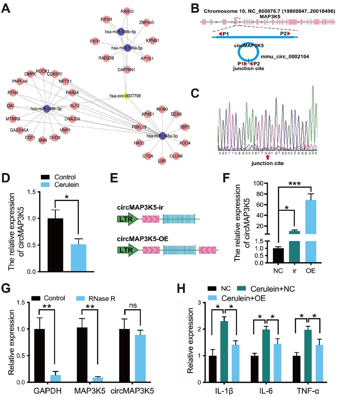
Acute pancreatitis arises from the activation of digestive enzymes in pancreatic acinar cells, leading to autodigestion of the pancreas and surrounding tissues. It is a common digestive tract emergency which requires hospitalization, and its incidence is increasing worldwide. In the past decade, several advances have been made in the treatment of acute pancreatitis. However, there is still a lack of efficacious drugs for clinical practice, and the limited value of existing biomarkers for early warning of the severity of acute pancreatitis is a major obstacle. Thus, there is an urgent need to gain a better understanding of the molecular mechanisms of acute pancreatitis. Circular RNAs (circRNAs) are a unique class of RNA molecules that are covalently closed. Ongoing investigations have provided evidence that circRNAs govern downstream target expression by acting as miRNA sponges, functioning as transcription factors, interacting with RNA-binding proteins, and regulating alternative splicing. These mechanisms support the pivotal role of circRNAs in a wide variety of physiological and pathological conditions, such as innate immunity, inflammation, neuronal function, and tumorigenesis. To explore the role of circular RNA in acute pancreatitis, we employed circRNA microarray technology (Arraystar Human circRNA Array V2) to examine the circRNA expression profile in the blood of three acute pancreatitis patients and three healthy controls. Clinical acute pancreatitis samples were obtained from Xiangya Hospital, Central South University. This study was approved by the Ethics Committee of Xiangya hospital (No. 2019010008). Normal control patients were recruited from among individuals who had visited Xiangya Hospital for a routine checkup. Written informed consent was obtained from all participants or their legal representatives for publication of data. The diagnosis and severity classification of acute pancreatitis were performed according to the American Gastroenterological Association guidelines and the Revised Atlanta Classification (RAC). circRNAs with a fold change ≥1.5 and a P value<0.05 were considered to be differentially expressed. As shown in Figure 1A, the two groups presented different expression profiles. We found that 91 circRNAs were significantly differentially expressed in the blood of acute pancreatitis patients, with 10 circRNAs exhibiting increased expression and 81 exhibiting decreased expression (Figure 1B,C). Among the differentially expressed circRNAs in acute pancreatitis, downregulated circRNAs are more prevalent than upregulated circRNAs, and the differential expression is more significant. Therefore, the present study focused on downregulated circRNAs. We selected circRNAs that are downregulated at least 2.5-fold and excluded those with fewer than 1000 bases to ensure the accuracy of qPCR. Based on these criteria, we identified nine circRNAs (circ_0006554, circ_0007798, circRNA_405815, circ_0001847, circ_0069748, circ_0001850, circ_0008417, circ_0002560, and circ_0000008) for validation by qPCR (Applied Biosystems, Foster City, USA) in blood samples from 30 acute pancreatitis patients (10 patients each with mild acute pancreatitis, moderate severe acute pancreatitis, and severe acute pancreatitis) and 15 healthy individuals. The levels of circ_0007798, circ_0001847, and circ_0069748 were significantly lower in acute pancreatitis patients than in normal controls, while the remaining circRNAs were not significantly differentially expressed (Figure 1D). In addition, the levels of circ_0007798 increased gradually with the severity of acute pancreatitis, suggesting that circ_0007798 is associated with the clinical severity of the disease (Figure 1E). Differential circRNAs have been studied for the diagnosis of pancreatic diseases. The expression level of circ_0007798 can be used to grade the severity of acute pancreatitis and provide individualized treatment. Furthermore, homology analysis (NCBI blast) revealed that circ_0007798 has a high degree of conservation between rats and humans according to the basic local alignment search tool. In conclusion, according to the
Identification and validation of disease severity-related circular RNA in acute pancreatitis
Graphical Abstract
Original ResearchVol. 56, Issue 9 • pp. 1352-1364DOI: 10.3724/abbs.2024024

The mechanism by which piR-000699 targets SLC39A14 regulates ferroptosis in aging myocardial ischemia/reperfusion injury

Authors: Hongyang Chi, Yue’e Chai, Lingju Ma, Yichen Wang, Qianqian Wu, Lexin Wang, Junjie Zhai, Fufun Ma, Yancheng Tian, Ning Qi, Jianhong Peng, Youjuan Fu, Xiaoling Yang, Hui Huang, Shengchao Ma

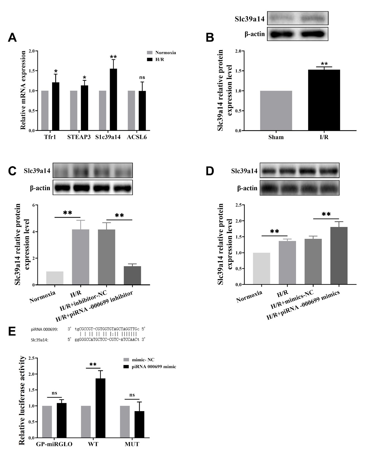
Myocardial ischemia/reperfusion (I/R) injury is a classic type of cardiovascular disease characterized by injury to cardiomyocytes leading to different types of cell death. The degree of irreversible myocardial damage is closely related to age, and ferroptosis is involved in cardiomyocyte damage. However, the mechanisms underlying ferroptosis regulation in aging myocardial I/R injury are still unclear. The present study aims to explore the underlying mechanism of piRNA regulation in ferroptosis. Using left anterior descending coronary artery ligation in an aging rat model and a D-galactose-induced rat cardiomyocyte line (H9C2) to construct an aging cardiomyocyte model, we investigate whether ferroptosis occurs after reperfusion injury in vitro and in vivo. This study focuses on the upregulation of piR-000699 after hypoxia/reoxygenation treatment in aging cardiomyocytes by observing hypoxia/reoxygenation (H/R) injury indicators and ferroptosis-related indicators and clarifying the role of piR-000699 in H/R injury caused by ferroptosis in aging cardiomyocytes. Bioinformatics analysis reveals that SLC39A14 is a gene that binds to piR-000699. Our data show that ferroptosis plays an important role in I/R injury both in vivo and in vitro. Furthermore, the results show the potential role of piR-000699 in regulating SLC39A14 in ferroptosis in aging cardiomyocytes under hypoxia/reoxygenation conditions. Together, our results reveal that the mechanism by which piR-000699 binds to SLC39A14 regulates ferroptosis in aging myocardial I/R injury.

The mechanism by which piR-000699 targets SLC39A14 regulates ferroptosis in aging myocardial ischemia/reperfusion injury
Graphical Abstract
Original ResearchVol. 56, Issue 9 • pp. 1267-1277DOI: 10.3724/abbs.2024010

TRMT13 inhibits the growth of papillary thyroid cancer by targeting ANAPC4

Authors: Lianyong Liu, Yan Wang, Mei Zou, Shiwei Chen, Fengying Wu, Xiangqi Li

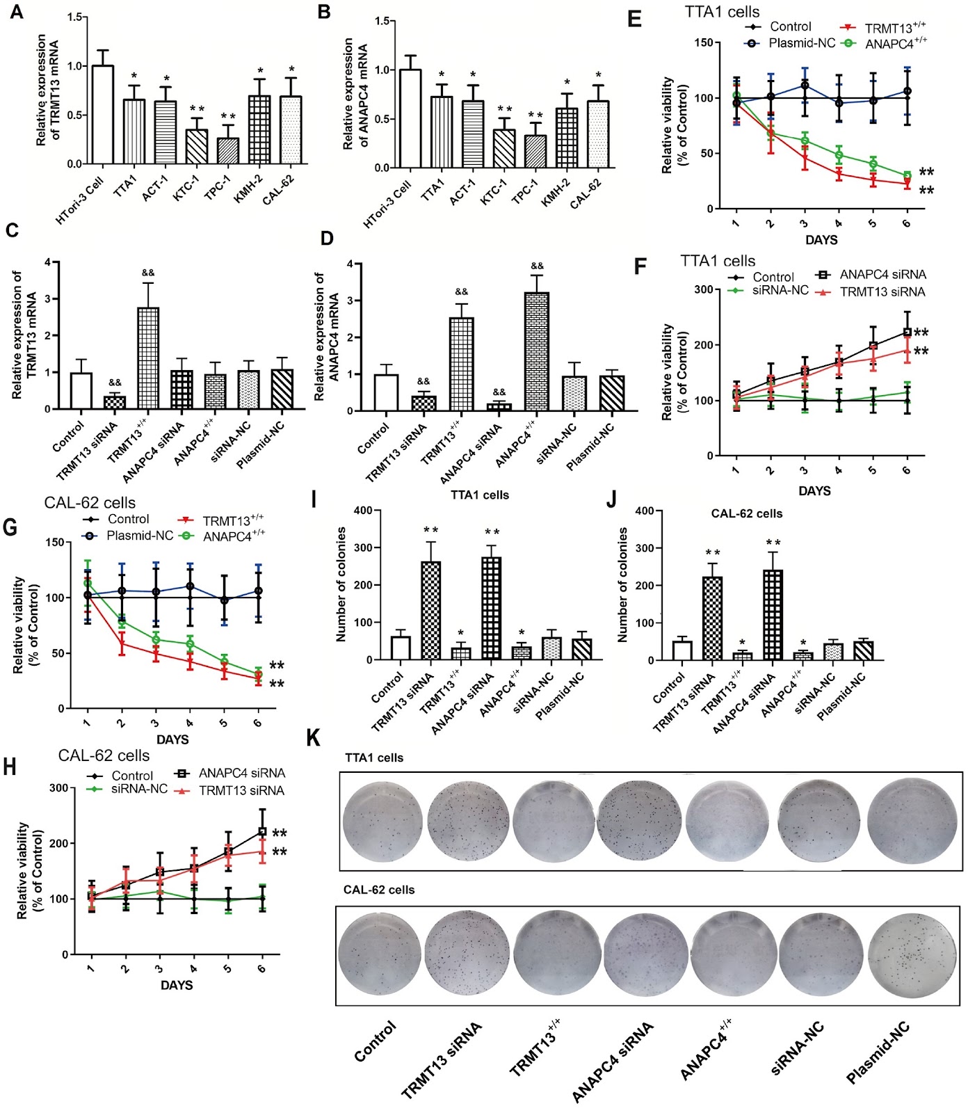
The recently discovered gene TRMT13 encodes a type of RNA methylase and is a member of the CCDC family (also called CCDC76). Here, we delineate its role in papillary thyroid cancer (PTC). Bioinformatics analysis shows significant TRMT13 and ANAPC4 downregulation in PTC and reveals that the expression levels of both genes are linearly correlated. Subsequent analyses confirm that both TRMT13 and ANAPC4 expressions are downregulated in PTC tissues and that this change in expression has a significant impact on cancer diagnosis. We conduct assays on PTC cells subjected to TRMT13 and ANAPC4 silencing or overexpression to assess the biological effects of these genes. We also perform rescue experiments to validate the regulatory effects of TRMT13 on ANAPC4. A nude mouse tumor model is used to evaluate the effects of TRMT13 and ANAPC4 on PTC tumorigenesis. TRMT13 expression is decreased in PTC tissues and cell lines and is positively correlated with that of ANAPC4. Cell assays reveal that TRMT13/ANAPC4 attenuates the malignancy of PTC cells by restraining cell proliferation, migration and invasion, while rescue experiments corroborate that ANAPC4 is a downstream target of TRMT13. In the nude mouse xenograft model, both TRMT13 and ANAPC4 inhibit tumor growth, and TRMT13 and ANAPC4 expression levels are significantly associated with survival. Taken together, these findings lead to the conclusion that TRMT13 inhibits PTC growth via ANAPC4, indicating a new role of TRMT13 and providing insights into the tRNA methyltransferase and coiled-coil domain-containing protein families.

TRMT13 inhibits the growth of papillary thyroid cancer by targeting ANAPC4
Graphical Abstract
Original ResearchVol. 56, Issue 9 • pp. 1300-1310DOI: 10.3724/abbs.2024042

Proanthocyanidins isolated from lotus seed skin mitigate glycolipid metabolism disorder through the p38/Nrf2/NF-κB signaling pathway

Authors: Yuhang Yi, Tao Li, Chenghao lv, Wenjiang He, Wenzhi Li, Xixin Zhou, Si Qin

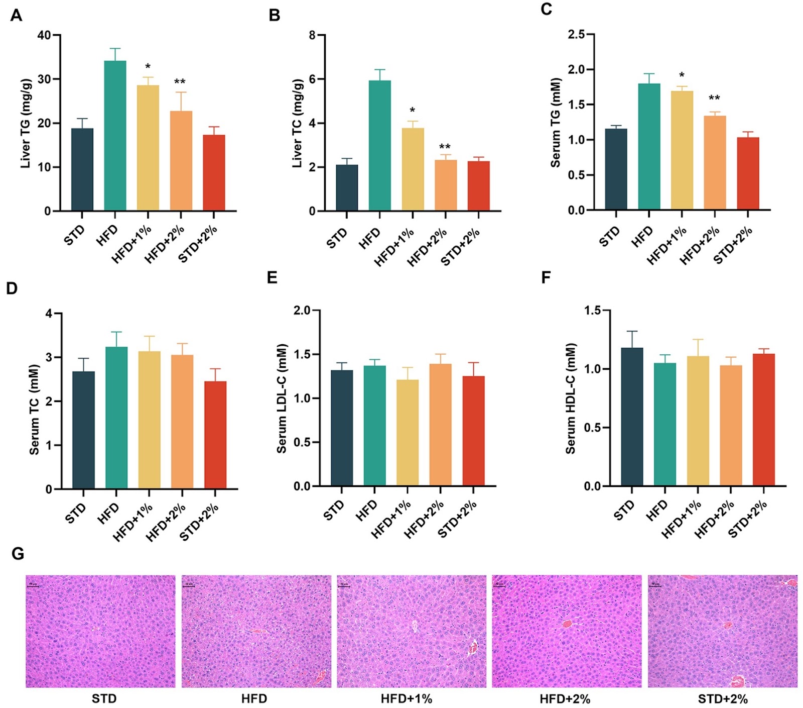
Lotus seed skin extract is rich in flavonoids, making it a promising candidate for developing health products. In a previous study, we found that proanthocyanidins from lotus seed skin, particularly proanthocyanidin B1 (PB1), can indirectly activate the Nrf2 signaling pathway, exerting an antioxidant effect. In this study, we isolate proanthocyanidins from lotus seed skin (PLS) using ethanol extraction and RP-HPLC identification, and investigate its effects on glycolipid metabolism both in vivo and in vitro. Our results demonstrate that PLS reduces body weight in high-fat diet (HFD) mice by decreasing feed efficiency. PLS also normalizes serum glucose, insulin secretion, glycosylated hemoglobin (HbA1c), and intraperitoneal glucose tolerance (IPGTT). Furthermore, PLS significantly improves blood lipid parameters and inhibits the expressions of six proinflammatory factors, including IL-1α, IL-1β, IL-3, IL-6, IFN-γ and TNF-α in HFD mice. Additionally, analysis of fresh liver tissues reveals that PLS and PB1 induce the expressions of antioxidant proteins such as HO-1 and NQO1 by activating the p38-Nrf2 signaling pathway and inhibiting the NF-κB signaling pathway. In conclusion, proanthocyanidins from lotus seed skin regulate glycolipid metabolism disorders by targeting the p38/Nrf2/NF-κB signaling pathway. Our study offers a new approach for the high-value comprehensive utilization of lotus seed skin by-products and precise dietary intervention for metabolic syndrome.

Proanthocyanidins isolated from lotus seed skin mitigate glycolipid metabolism disorder through the p38/Nrf2/NF-κB signaling pathway
Graphical Abstract
Original ResearchVol. 56, Issue 9 • pp. 1278-1288DOI: 10.3724/abbs.2024066

The rod cell, a small form of Candida albicans, possesses superior fitness to the host gut and adaptation to commensalism

Authors: Yinxing Xu, Wencheng Zhu, Baodi Dai, Hui Xiao, Jiangye Chen

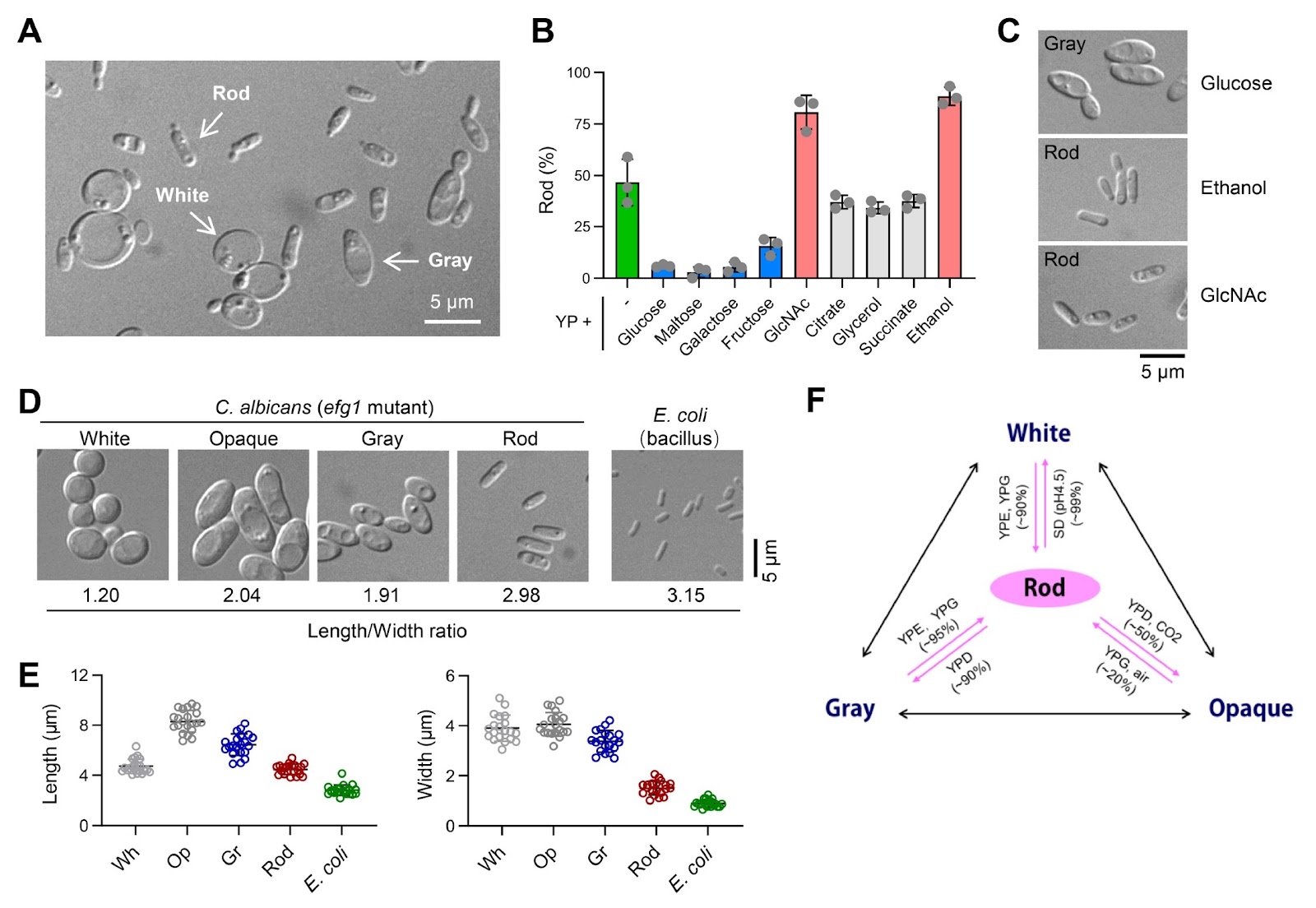
Candida albicans deploys various morphological forms through complex switching mechanisms, ensuring its survival and thriving as a commensal or pathogen in vastly different human niches. In this study, we demonstrate that a novel ''rod'' morphological form of C. albicans coexists and is interchangeable with previously reported white, gray, and opaque forms, constituting a tetra-stable phenotypic switching system. Rod cells arise from the efg1 mutant of SC5314 cells or from the clinical BJ1097 strain cultured under glucose-free conditions. They are characterized by a distinct gene expression profile and can be stably maintained through in vitro passaging or in vivo inhabitation of the gastrointestinal (GI) tract of mice. Remarkably, the majority of the efg1 mutant cells become rod cells in N-acetylglucosamine (GlcNAc)-containing medium, and the GlcNAc sensor Ngs1 is instrumental in converting the white or gray cells to the rod cells. Conversely, glucose inhibits rod cells through Cph1; consequently, the loss of Cph1 in the efg1 mutant cells permits their conversion to rod cells in glucose-replete media. Notably, rod cells of the efg1/cph1 mutant display superior adaptation and longer persistence in the murine GI environment than wild-type white cells. Taken together, these findings establish rod cells as a previously unappreciated form that is not only morphologically and transcriptionally distinguishable but also defined by specific genetic and environmental determinants, shedding light on complex fungus-host interactions.

The rod cell, a small form of Candida albicans, possesses superior fitness to the host gut and adaptation to commensalism
Graphical Abstract