🧬 SinoBioData Academic Portal
🏛️ Indexed Academic JournalOriginal: 生物化学与生物物理学报

Acta Biochimica et Biophysica Sinica

Premier Chinese Biomedical Journal indexed in SinoBioData: Acta Biochimica et Biophysica Sinica (生物化学与生物物理学报).

Total Research Papers: 200
Access: 100% Free Open Access
Browse by Publication Year & VolumeReset All Filters ✕

Published Research PapersFiltered: Year 2026 • 58 • 6

Showing 12 of 200 peer-reviewed papers with full Graphical Abstracts.

Original ResearchVol. 58, Issue 6 • pp. 1402-1412DOI: 10.3724/abbs.2025224

Bioinformatics classification of the MgtE Mg2+ channel and de novo protein design for the stabilization of its novel subclass

Authors: Zhixuan Zhao, Kimiho Omae, Wataru Iwasaki, Ziyi Zhang, Fazhi Pan, Eun-Jin Lee, Koichi Ito, Motoyuki Hattori

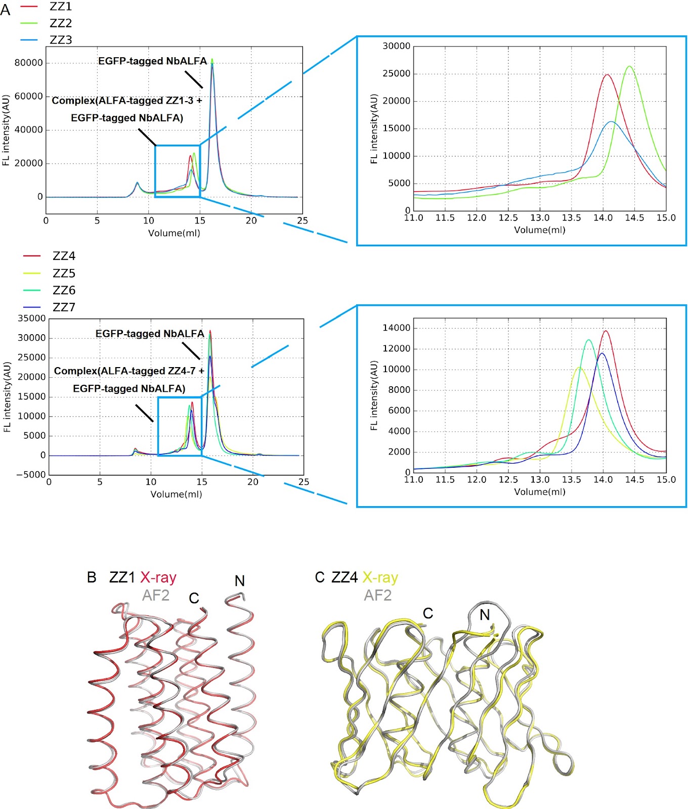
MgtE channels play crucial roles in Mg2+ homeostasis and are implicated in bacterial survival under antibiotic exposure. Previous structural and biophysical studies have focused predominantly on Thermus thermophilus MgtE, leaving the structural and mechanistic diversity of MgtE family proteins largely unexplored. In this study, via a genome mining approach, we identify diverse MgtE homologs, including a novel subclass termed the “mini-N type”, which lacks the canonical cytoplasmic N and CBS domains but possesses a unique small N-like domain. Despite extensive expression screening, mini-N-type homologs cannot be stably purified. To address this issue, we design a series of de novo proteins and determine their crystal structures. A selected de novo protein is fused to a mini-N-type MgtE, enabling successful purification and preliminary cryo-EM imaging. Our findings demonstrate that de novo-designed protein fusions serve as powerful tools for stabilizing and purifying otherwise unstable membrane proteins, opening new avenues for structural and functional studies of otherwise inaccessible membrane proteins.

Bioinformatics classification of the MgtE Mg2+ channel and de novo protein design for the stabilization of its novel subclass
Graphical Abstract
Original ResearchVol. 58, Issue 6 • pp. 1281-1290DOI: 10.3724/abbs.2026042

Structural and functional insights into the distinct DNA recognition mechanisms of the terminase small subunit TerS from cyanophages

Authors: De-Qin Dong, Feng Yang, Kang Du, Kang Xu, Wen-Bin Cheng, Yuxing Chen, Cong-Zhao Zhou, Yong-Liang Jiang

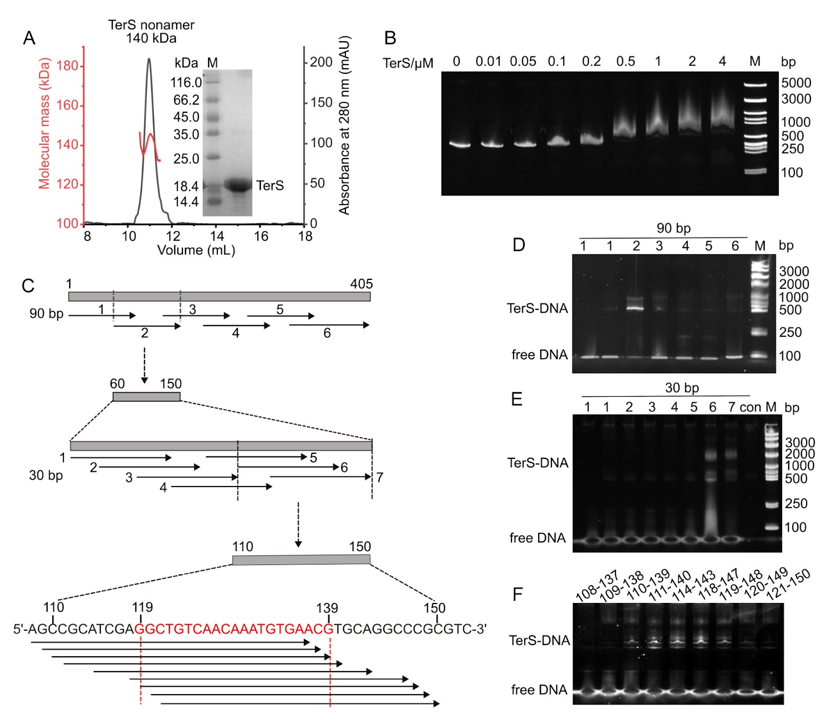
Efficient genome packaging is a critical step in the phage life cycle, directly influencing the viral maturation and infectivity. In tailed phages, this process is driven by a packaging motor composed of a portal protein and a terminase complex. The terminase complex usually consists of a large subunit (TerL) and a small subunit (TerS), which cooperate to recognize, cleave, and translocate genomic DNA into the capsid. However, due to the remarkable diversity and complexity of phage packaging systems, the molecular mechanisms governing TerS-mediated DNA recognition remain poorly understood. Here, we report the 3.51 Å cryo-electron microscopy structure of the TerS from the short-tailed cyanophage Pam5, which infects the host Pseudanabaena mucicola Chao 1806. Pam5 TerS assembles into a nonameric ring with a radially symmetric spiral architecture. Biochemical assays show that Pam5 TerS recognizes the genomic DNA via a specific interaction between the N-terminal helix-turn-helix (HTH) domain of TerS and a 21-bp DNA sequence within the terS gene. In contrast, the TerS from another short-tailed cyanophage, Pam1, which infects the same host, binds to DNA in a sequence-independent manner. These findings reveal that cyanophages, even infecting the same host, could adopt two distinct DNA recognition strategies: HTH-mediated sequence-dependent or sequence-independent modes. This work provides structural and mechanistic insights into the diverse DNA-recognition strategies of TerS and advances our understanding of the evolutionary plasticity of viral genome packaging mechanisms.

Structural and functional insights into the distinct DNA recognition mechanisms of the terminase small subunit TerS from cyanophages
Graphical Abstract
Original ResearchVol. 58, Issue 6 • pp. 1431-1432DOI: 10.3724/abbs.2026006

Caught the ‘Catch’ of midnolin: structural basis for broad substrate specificity in ubiquitin-independent proteasomal degradation

Authors: Chuanyin Li, Ronggui Hu

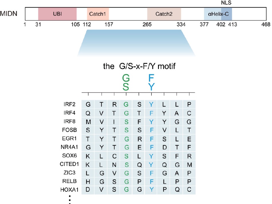
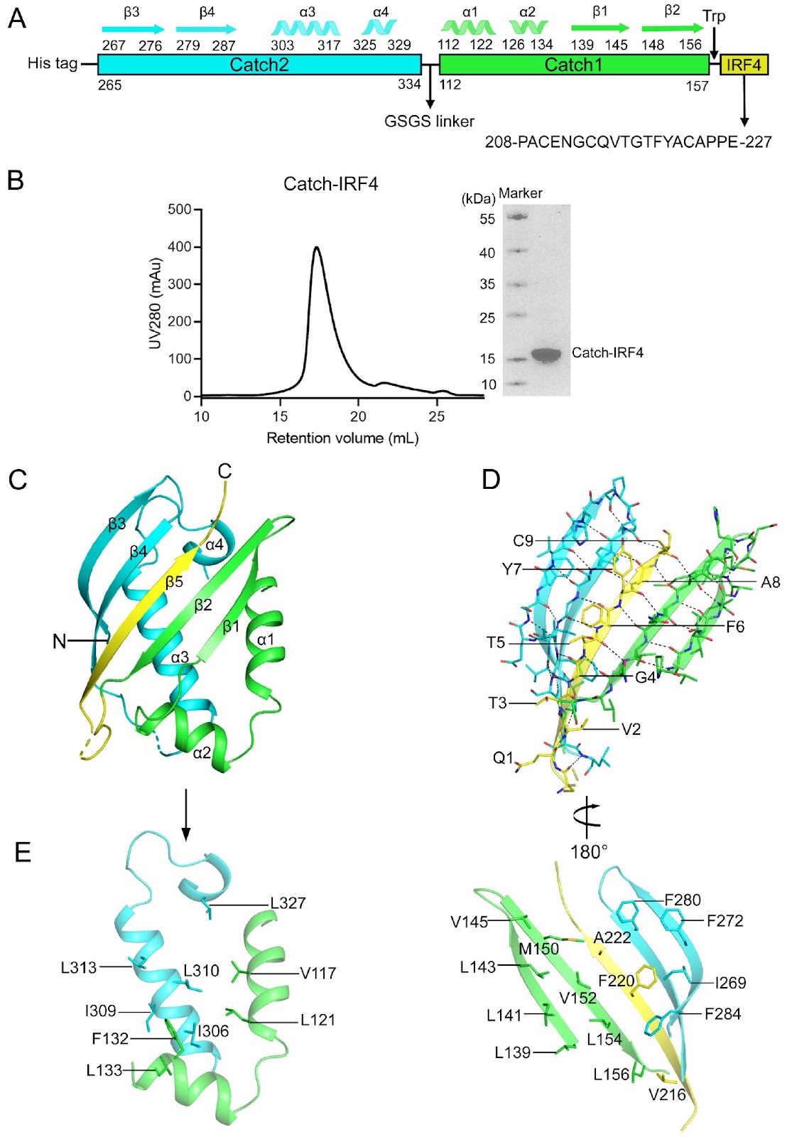
Protein homeostasis serves as the foundation for every cellular decision—division, differentiation, stress adaptation, or death—by precisely balancing the proteome across abundance, quality, spatial distribution, and temporal dynamics; its dysregulation drives numerous human pathologies, including cancers and neurological disorders. In the traditional ubiquitin-dependent degradation cascade, target proteins are marked by covalent attachment of polyubiquitin chains, a process requiring E1 activating enzymes, E2 conjugating enzymes, and E3 ligases that confer substrate specificity. This ubiquitin signal is then recognized by the 19S regulatory particle of the proteasome, which unfolds and translocates the tagged protein into the 20S core for proteolytic destruction. The ubiquitin-independent proteasomal degradation pathway mediated by midnolin (MIDN) has recently emerged as a distinct and biologically important mechanism for regulating nuclear protein turnover. While earlier genetic, biochemical, and cryo-electron microscopy studies established MIDN as a proteasome-associated adaptor for immediate-early gene (IEG) products, the molecular logic underlying its broad yet selective substrate recognition remains unresolved. In the current study, Zhong et al. provide a comprehensive structural and biochemical analysis of the MIDN Catch domain bound to IRF4-derived peptides, substantially advancing our understanding of MIDN substrate recognition. By solving multiple crystal structures of the Catch-IRF4 complex, including wild-type and systematically engineered mutants, the authors demonstrate that MIDN recognizes substrates through a conserved β-strand insertion mechanism at the interface of the Catch1 and Catch2 subdomains. This investigation generalizes and expands prior structural observations of MIDN-IRF4 to diverse substrates, demonstrating that β-strand complementation constitutes a universal recognition mechanism utilized by MIDN. A major conceptual advance of this study is the identification of a minimal and generalizable recognition principle centered on two highly conserved positions within the substrate motif. The authors show that a reciprocal phenylalanine-glycine interaction between the substrate and Catch2—forming an “F-G zipper”—constitutes the dominant energetic determinant for binding. Disruption of this zipper severely compromises protein stability and binding, explaining prior functional observations that single-point mutations in IRF4 or EGR1 abolish MIDN-mediated degradation. In contrast, flanking residues within the binding motif display remarkable tolerance to substitution. Through combined mutagenesis, thermostability analysis, AlphaFold3 modeling, and structural determination, the study demonstrates that these positions occupy large and plastic hydrophobic pockets in the Catch domain. This architectural flexibility provides a direct molecular explanation for how MIDN can accommodate a wide spectrum of substrates while preserving selectivity. From these data, the authors derive a concise consensus recognition motif (G/S-x-F/Y) embedded within an unstructured or loop region, offering a predictive framework for identifying additional MIDN targets. Importantly, this work bridges a critical gap between previous cryo-EM studies describing MIDN-proteasome engagement and earlier substrate-specific structural analyses. Whereas prior studies clarified how MIDN delivers captured substrates to the proteasome, the present study elucidates how MIDN initially selects and binds those substrates. Together, these findings unify MIDN biology across structural, biochemical and functional dimensions. Overall, this study represents a significant advance in the field of proteostasis by revealing how a ubiquitin-independent adaptor achieves broad substrate specificity through a simple yet robust structural principle. Beyond MIDN, the work provides a paradigm for short-linear-motif-based proteasomal targeting and has important implications for immune regulation, neurodegeneration and cancer biology.

Caught the ‘Catch’ of midnolin: structural basis for broad substrate specificity in ubiquitin-independent proteasomal degradation
Graphical Abstract
Original ResearchVol. 58, Issue 6 • pp. 1235-1249DOI: 10.3724/abbs.2026002

Biochemical and structural studies of the midnolin Catch domain bound with both wild-type and mutant IRF4 peptides reveal the molecular basis for its broad substrate specificity

Authors: Yanling Zhong, Ziyue Chen, Guanchao Wang, Jianping Ding

The newly discovered midnolin-proteasome pathway is a unique ubiquitin-independent mechanism for degrading nuclear proteins, which is crucial for maintaining cellular protein homeostasis. The Catch domain of midnolin is essential for substrate recognition and binding, yet the underlying mechanism for its broad substrate specificity remains elusive. Transcription factor IRF4, essential for the functions of B and T cells, is a substrate of midnolin. This study presents comprehensive biochemical and structural analyses of the human midnolin Catch domain in complexes with both wild-type and mutant IRF4 peptides. The crystal structure of the Catch-IRF4 complex reveals that the Catch domain creates a substrate-binding groove at the interface of the Catch1 and Catch2 subdomains, recognizing and binding to the 215-QVTGTFYAC-223 sequence motif of IRF4. The binding motif of IRF4 forms a β-strand that is embedded into the substrate-binding groove, resulting in an antiparallel five-stranded β-sheet. The interactions between the IRF4 peptide and the Catch domain are predominantly hydrophobic and exhibit high spatial complementarity. Additionally, the biochemical, modeling and structural data indicate that the V2 and A8 positions of the IRF4 sequence motif can be substituted with other hydrophobic or small polar residues (G/A/V/L/I/M/P/F/Y/C/S/T), but not with large polar and charged residues (D/N/E/Q/H/K/R). The G4 position can be replaced by Ser, while the F6 position can be substituted with Tyr. These results suggest that the Catch domain can recognize and bind to a variety of substrates containing the sequence motif x[G/A/V/L/I/M/P/F/Y/C/S/T]x[G/S]x[F/Y]x[G/A/V/L/I/M/P/F/Y/C/S/T]x or briefly the G/SxF/Y motif (where x represents polar residues) located in an unstructured or loop region on the protein surface, and the hydrophobic interactions and spatial complementarity between the binding motifs of substrates and the Catch domain govern the substrate specificity. Collectively, these findings elucidate the molecular basis for midnolin’s broad substrate specificity.

Biochemical and structural studies of the midnolin Catch domain bound with both wild-type and mutant IRF4 peptides reveal the molecular basis for its broad substrate specificity
Graphical Abstract
Original ResearchVol. 58, Issue 6 • pp. 1423-1426DOI: 10.3724/abbs.2025222

Mixed fungal polysaccharides enhance intestinal health, antioxidant capacity, and microbiota diversity in broiler chickens

Authors: Bingyu Zhu, Enze Zhang, Min Yang, Ye Zhang, Can Liu, Runxin Jiao, Mengling Peng, Jie Zhou, Jianbo Cheng, Juhua Wang

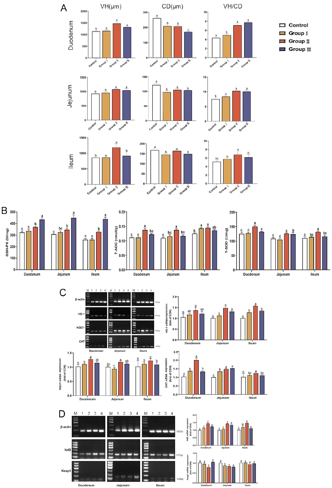
Poultry production faces escalating challenges from intensive farming practices, where stressors, including high stocking density, pathogen exposure, and dietary fluctuations, disrupt intestinal integrity, microbiota balance, and antioxidant defenses. These disruptions impair nutrient absorption, growth performance, and immune function, leading to significant economic losses. Although antibiotics have historically mitigated such issues, growing restrictions due to antimicrobial resistance necessitate natural alternatives. Fungal polysaccharides (FP)—notably lentinan (LNT) from Lentinula edodes and polysaccharide from Ganoderma lucidum (GLP), are promising candidates owing to their immunomodulatory, antioxidant, and prebiotic properties. However, existing research focuses predominantly on individual FP, neglecting potential synergies in blended formulations. Structurally, LNT (β-(1→3)-D-glucan backbone) enhances rumen volatile fatty acid production and fiber degradation, whereas GLP (heterogeneous α/β-glycans) potently activates the Nrf2/HO-1 antioxidant pathway and modulates Th1/Th2 immunity. These divergent mechanisms imply complementary effects when combined. In our previous experiments on broiler feeding, we reported that a combination of GLP (68.32% polysaccharide content, composed of mannose, glucose, arabinose, rhamnose, and galactose at a molar ratio of 1.00:16.37:18.82:1.42:17.42) and LNT (76.52% polysaccharide content, composed of mannose, galacturonic acid, arabinose, galactose, glucose, and rhamnose at a molar ratio of 1.00:15.22:8.23:2.05:1.78:4.26) at a 1:1 ratio maximally promoted broiler growth (unpublished data), but their impacts on intestinal morphology, antioxidant signaling, and the microbiota remain uncharacterized. We therefore hypothesize that mixed FP synergistically may enhance intestinal health by simultaneously improving nutrient absorption, activating antioxidant pathways, and stabilizing microbial ecosystems. To investigate the effects of mixed FP on intestinal development, 240 one-day-old Arbor Acres male broilers were randomly assigned to the 0 mg/kg FP (Control), 200 mg/kg FP (Group I), 400 mg/kg FP (Group II), and 600 mg/kg FP (Group III) groups. Broilers were housed in three-tier battery cages (0.7 m × 0.7 m × 0.4 m; 12 broilers/cage), with five replicate cages per experimental group maintained under identical conditions. The experiments were approved by the College of Animal Science and Technology in Anhui Agricultural University (approval number: SYXK 2016-007). All the cages were subjected to a 16 h light: 8 h dark cycle with ad libitum access to water and twice-daily feeding (09:00/16:00) of basal diets (Supplementary Table S1). On day 42, the duodenum, jejunum, and ileum segments were collected, fixed in 4% paraformaldehyde, sectioned at 5 μm, and stained with hematoxylin-eosin. Villus height (VH), crypt depth (CD), and VH/CD ratios were measured via Case Viewer software. The results revealed that Group II significantly increased VH and VH/CD across all the intestinal segments while reducing CD (Figure 1A; P < 0.05 vs the control); these findings suggest enhanced nutrient absorption capacity and intestinal health. To evaluate antioxidant capacity and signaling pathway activation, intestinal tissues were homogenized in PBS (1:9, w/v). The total antioxidant capacity (T-AOC), total superoxide dismutase (T-SOD), and glutathione peroxidase (GSH-Px) activities were determined via commercial kits (Nanjing Jiancheng Bioengineering Institute, Nanjing, China)). For gene expression analysis, total RNA was extracted and reverse-transcribed. The qPCR was performed via specific primers for HO-1, NQO1, CAT, Nrf2, and Keap1, with β-actin used as the reference gene (primer sequences and product sizes are listed in Supplementary Table S2). The results demonstrated that Group II significantly elevated antioxidant enzyme activities (P < 0.05), upregulated HO-1, NQO1, CAT, and Nrf2, and
Mixed fungal polysaccharides enhance intestinal health, antioxidant capacity, and microbiota diversity in broiler chickens
Graphical Abstract
Original ResearchVol. 58, Issue 6 • pp. 1329-1341DOI: 10.3724/abbs.2025178

circDCBLD2 regulates the Th1/Th2 immune balance via the miR-26a-5p/PTEN axis

Authors: Yue Zhao, Guangling Liu, Rui Li, Shuting Yu, Beibei Zhu, Xunzhou Liu, Hanyi Jiang, Jinya Wang

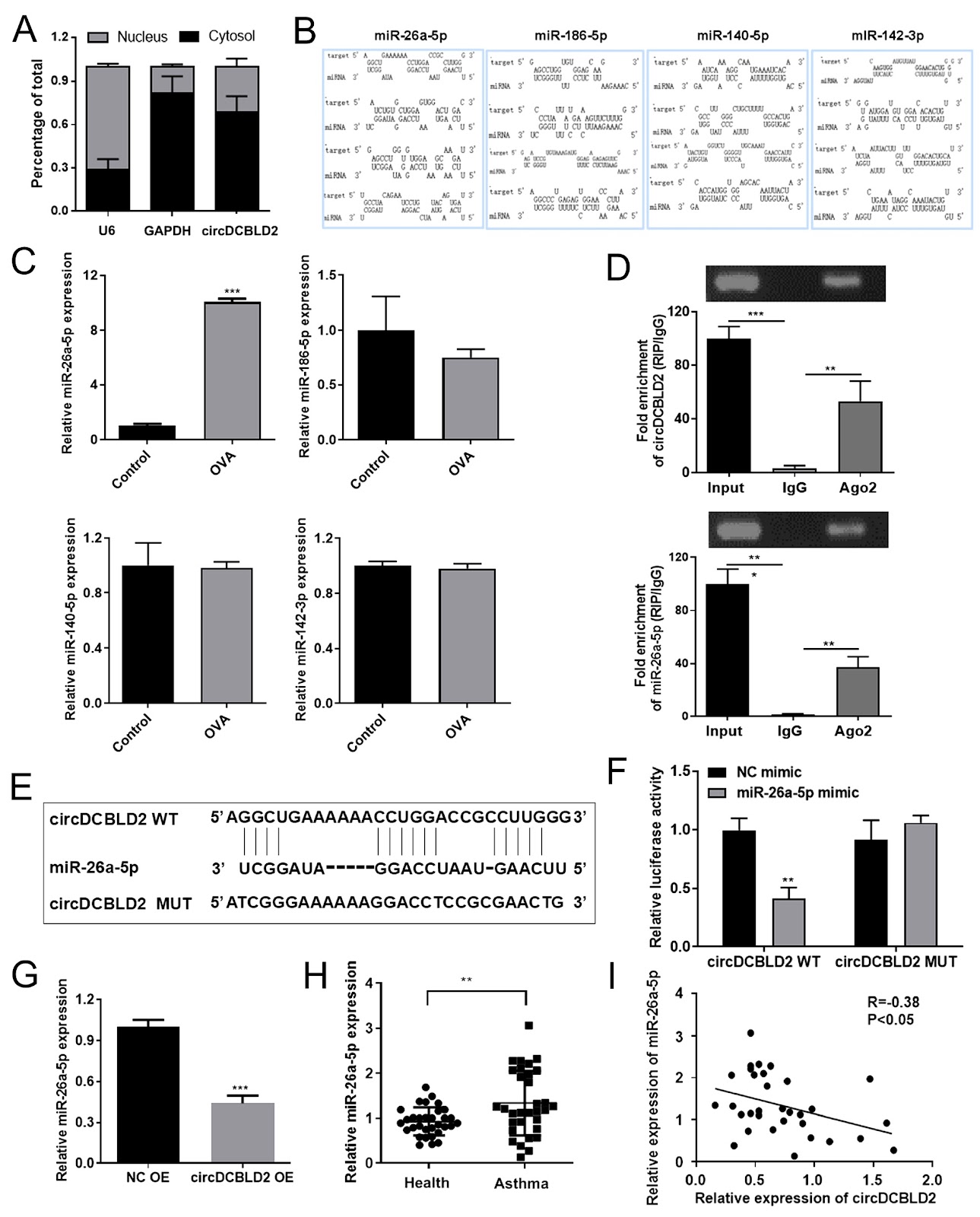
Asthma is a prevalent chronic respiratory disease in children. Recently, adjusting the Th1/Th2 imbalance has become a significant focus in asthma immunotherapy. The present study aims to investigate the roles and mechanisms of circDCBLD2 in maintaining the Th1/Th2 immune balance. CircDCBLD2 is downregulated in CD4+ T cells from asthmatic patients and in CD4+ T cells from an OVA-induced asthmatic mouse model. Additionally, circDCBLD2 levels are significantly decreased in the PBMCs of asthmatic mice. The expression of circDCBLD2 is positively correlated with the Th1 cytokines IFN-γ and IL-2 but negatively correlated with the Th2 cytokines IL-4 and IL-13. Flow cytometry and ELISA analyses demonstrate that circDCBLD2 overexpression increases the proportion of Th1 cells (CD4+IFN-γ+) and the levels of Th1 cytokines while decreasing the proportion of Th2 cells (CD4+IL-4+) and the levels of Th2 cytokines. Furthermore, circDCBLD2 overexpression alleviates the asthma phenotype in OVA-induced mice, reduces the infiltration of inflammatory cells in the lungs, and corrects the Th1/Th2 imbalance. Mechanistically, circDCBLD2 is found to target miR-26a-5p. Rescue experiments indicate that circDCBLD2 regulates the Th1/Th2 immune balance by targeting miR-26a-5p. Additionally, PTEN has been identified as a direct target of miR-26a-5p. The overexpression of PTEN partially reverses the effects of miR-26a-5p on the Th1/Th2 immune balance. These findings indicate that circDCBLD2 increases the proportion of Th1 cells and decreases the proportion of Th2 cells via the miR-26a-5p/PTEN axis, providing a promising target for asthma treatment.

circDCBLD2 regulates the Th1/Th2 immune balance via the miR-26a-5p/PTEN axis
Graphical Abstract
Original ResearchVol. 58, Issue 6 • pp. 1374-1386DOI: 10.3724/abbs.2025152

A Mycobacterium tuberculosis multi-epitope DNA vaccine encoding adaptive immune antigens provokes IFNγ/Th1 immunity and confers potential protection

Authors: Jingyao Xue, Yumeng Li, Chi Li, Yu Zhang, Chiuan Yee Leow, Gaoqian Feng, Minjun Ji, Qiao Liu, Zhipeng Xu

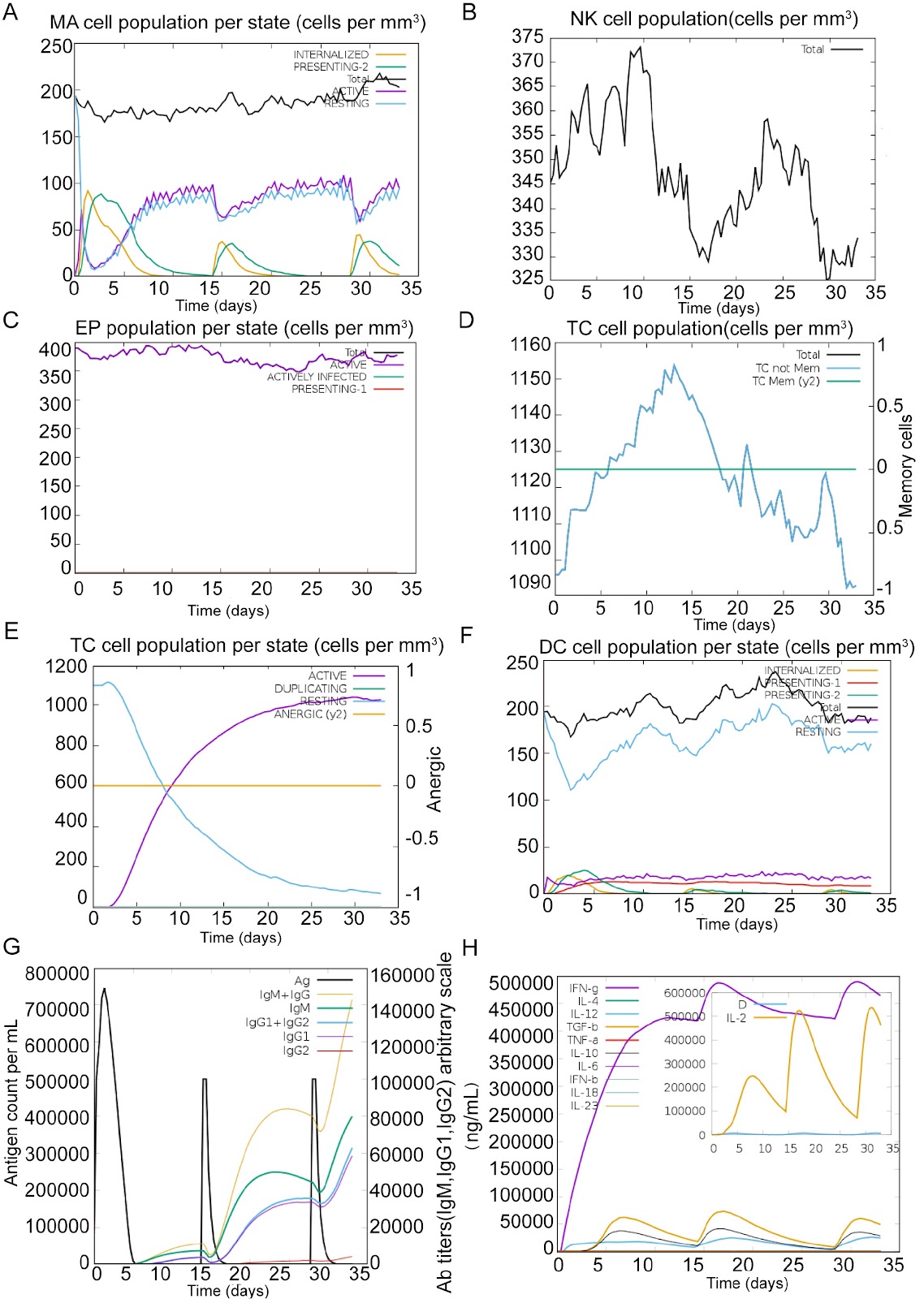
Tuberculosis (TB), caused by Mycobacterium tuberculosis (MTB), remains a significant global health threat. However, the licensed Bacille Calmette-Guérin (BCG) vaccine provides only limited protection in adults, underscoring the urgent need for more effective preventive strategies. Recent studies have shown that multi-epitope DNA vaccines are superior to traditional vaccines in terms of immunogenicity, safety and stability. In this study, we develop a multi-epitope DNA vaccine that contains CD8+ T-cell epitopes, CD4+ T-cell epitopes, and B-cell epitopes using bioinformatics tools. These epitopes are derived from three genome-encoded proteins, ESAT-6, Rv2660c, and RpfB, which exhibit stage-specific immunodominance in the early, resting, and convalescent stages of MTB infection. Using reverse vaccinology and computational immunomodulation, we demonstrate that the multiepitope vaccine increases antigen-specific antibody titres, activates CD8+ T and CD4+ T cells, and enhances IFN-γ secretion. In vitro validation studies in HEK293T cells confirm high-yield expression of multi-epitope-encoded antigens, whereas in vivo immunization experiments reveal significant expansion of NK cells and Th1-polarized lymphocytes, with concomitant upregulation of pro-inflammatory mediators. Collectively, these results highlight the potent activation of adaptive immunity through Th1-driven mechanisms and IFN-γ-mediated mycobacterial clearance, which are crucial for defending against MTB.

A Mycobacterium tuberculosis multi-epitope DNA vaccine encoding adaptive immune antigens provokes IFNγ/Th1 immunity and confers potential protection
Graphical Abstract
Original ResearchVol. 58, Issue 6 • pp. 1413-1422DOI: 10.3724/abbs.2025176

The D826V point mutation in IREB2 causes early-onset neurodegeneration in mice

Authors: Zhenglong Guo, Yibing Lv, Jianmei Huang, Yingying Shao, Yuwei Zhang, Yibin Hao, Bingtao Hao, Zhenbo Cheng, Shixiu Liao

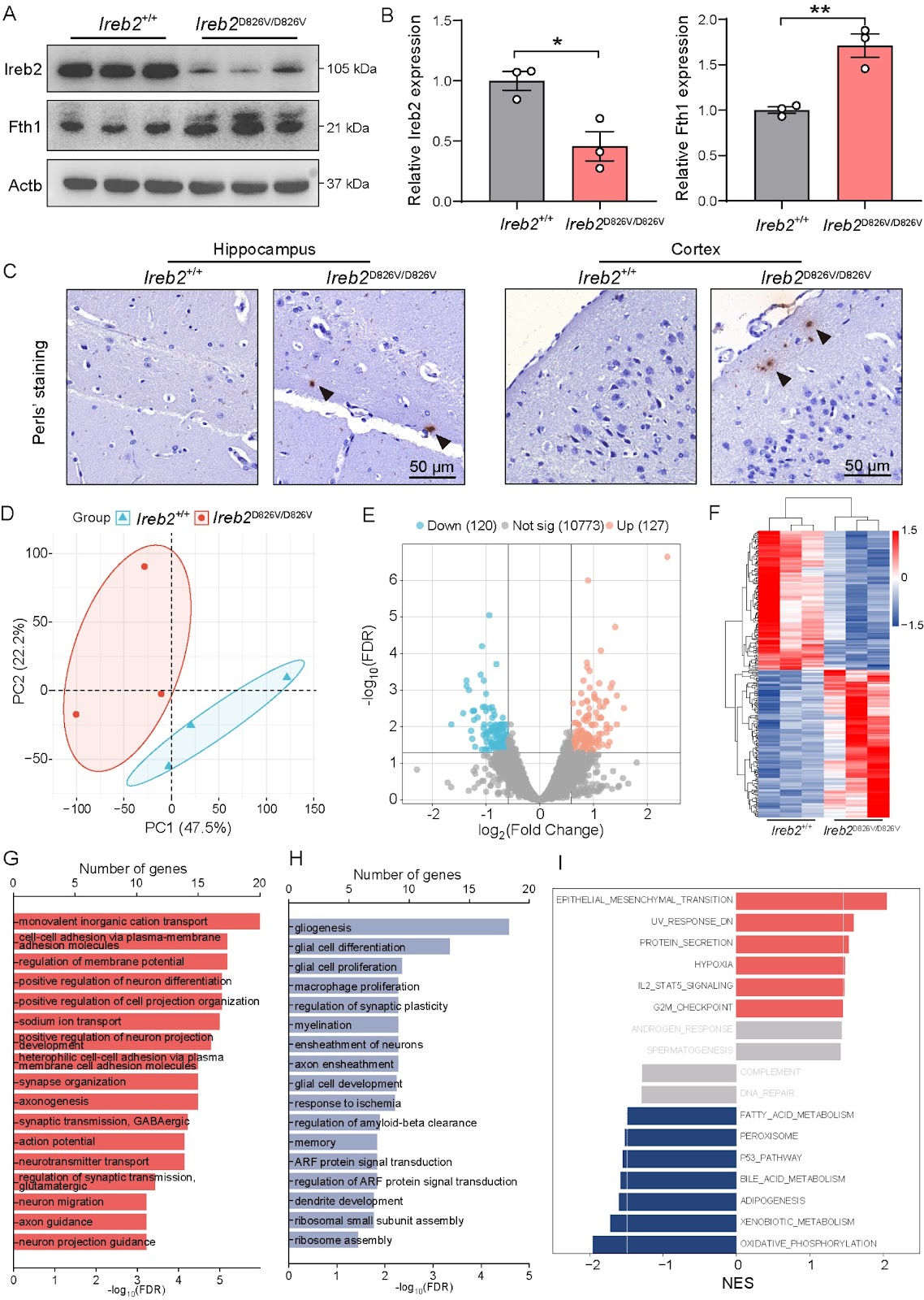
The iron regulatory protein IREB2 (Iron Responsive Element Binding Protein 2) plays a crucial role in maintaining cellular iron homeostasis through the posttranscriptional regulation of genes involved in iron metabolism. Mutations in the IREB2 gene have been linked to NDCAMA (OMIM#618451), a rare genetic neurological disorder characterized by early-onset neurodegeneration, choreoathetoid movements, and microcytic anemia. However, the absence of an IREB2-mutated animal model has left the underlying pathogenic mechanisms poorly understood. To investigate this, we establish a CRISPR-Cas9-mediated Ireb2D826V/D826V mouse model, which carries the c.2477A>T (p.D826V) pathogenic variant in IREB2 identified in a Chinese pedigree with NDCAMA. Behavioral studies, including the Morris water maze (MWM), open field test (OFT), and Y-maze, reveal significant neurobehavioral deficits, such as impaired spatial learning and memory and reduced motor activity, in Ireb2D826V/D826V mice. Furthermore, we observe increased microglial activation and decreased dendritic spine density in the hippocampus, along with impaired long-term potentiation (LTP) and elevated paired-pulse facilitation (PPF), indicating synaptic dysfunction. Mechanistically, Ireb2D826V/D826V mice present reduced Ireb2 protein levels, dysregulated iron metabolism, and an altered expression profile associated with neurological function. This study elucidates the molecular mechanisms underlying NDCAMA and establishes Ireb2D826V/D826V mice as a model for iron metabolism-driven neurodegeneration. This finding links the instability of IREB2 to synaptic failure and neuroinflammation, highlighting potential therapeutic implications for neurodegenerative diseases.

The D826V point mutation in IREB2 causes early-onset neurodegeneration in mice
Graphical Abstract
Original ResearchVol. 58, Issue 6 • pp. 1250-1264DOI: 10.3724/abbs.2025236

Biochemical and structural studies of NFIA and NFIC reveal a conserved mechanism for specific DNA recognition and provide insight into potential pathogenicity of disease-associated mutations

Authors: Shuli Pan, Wenjie Pei, Jin Zhang, Jinrong Min, Ke Liu

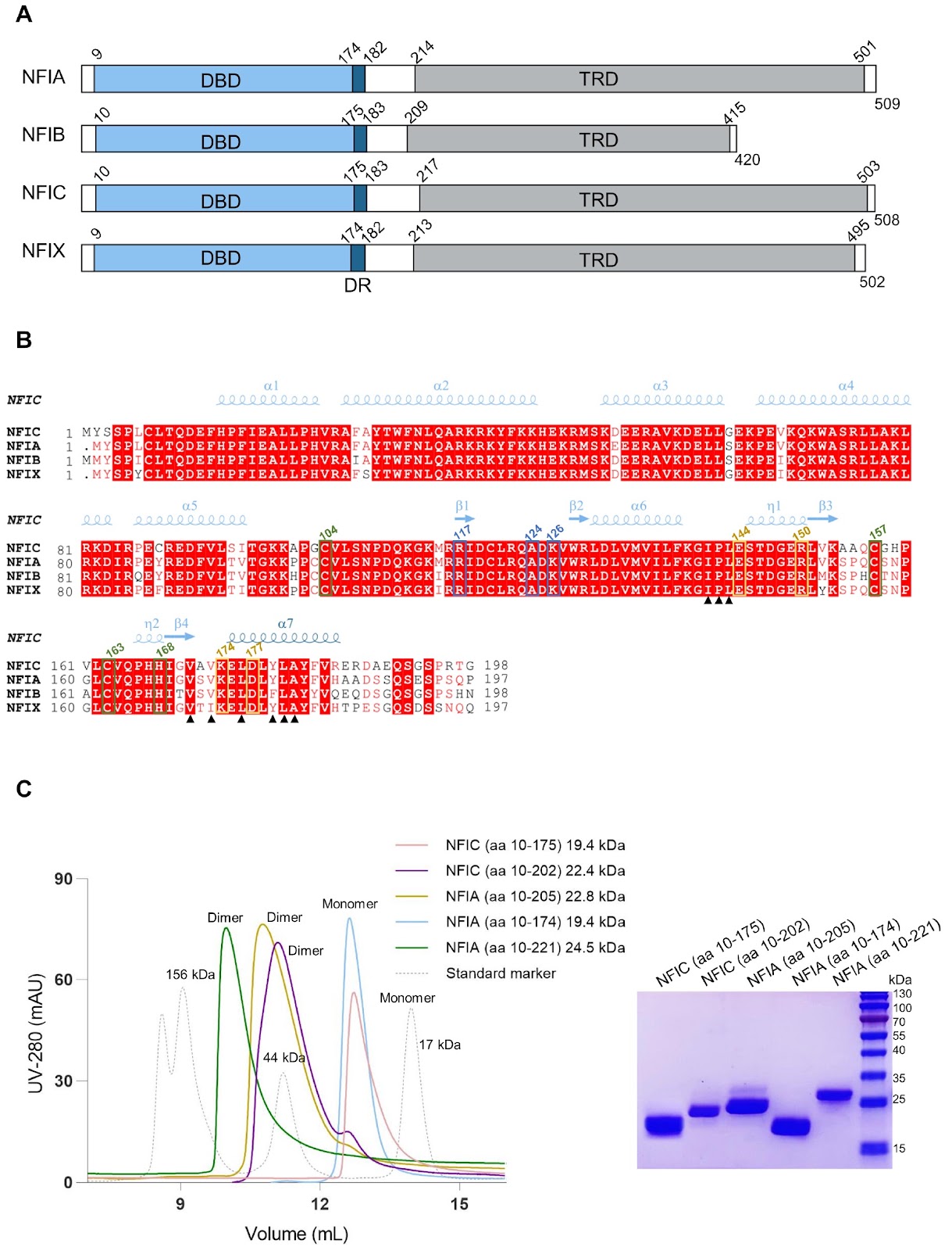
Nuclear factor I (NFI) transcription factors play essential roles in multiple aspects of nervous system development, including radial glia maturation, neurogenesis, gliogenesis, and brain morphogenesis. Numerous NFI variants have been identified in individuals with neurodevelopmental disorders, yet the molecular basis of their pathogenicity remains unclear. The absence of resolved NFI-DNA complex structures continues to impede mechanistic insights and therapeutic exploration. Here, we define the oligomeric states of NFIA and NFIC, and determine the crystal structures of the NFIC homodimer, as well as the NFIA and NFIC monomers lacking their dimerization region, in complexes with double-stranded DNAs. Structural analysis reveals the molecular mechanism underlying NFI dimerization and recognition of a dyad-symmetric TGGCA(N3)TGCCA sequence motif, and demonstrates that dimerization enhances both DNA-binding affinity and specificity of NFI proteins. The functional importance of key NFI residues and DNA bases involved in the protein-DNA interaction is further validated by mutagenesis and binding assays. Additionally, we systematically evaluate the effects of the neurodevelopmental disorders-associated NFI mutations on DNA binding of NFIA, providing insights into their potential pathogenic mechanisms. Together, our findings elucidate the structural basis of NFI dimerization and dyad-symmetric DNA recognition and highlight pathogenic variants for further mechanistic studies in neurodevelopmental disorders.

Biochemical and structural studies of NFIA and NFIC reveal a conserved mechanism for specific DNA recognition and provide insight into potential pathogenicity of disease-associated mutations
Graphical Abstract
Original ResearchVol. 58, Issue 6 • pp. 1313-1328DOI: 10.3724/abbs.2025151

Gut-brain axis and exosome-mediated communication in postoperative cognitive dysfunction associated with colorectal cancer

Authors: Ying Gao, Biao Xi, Yang Zhang, Mei Mei, Ming Zhai, Yunzhi Ling, Weiwei Chu

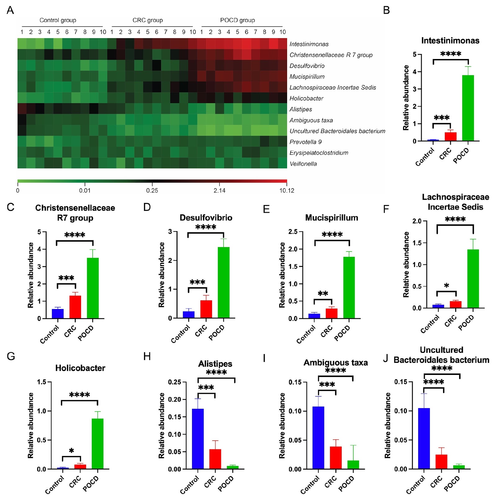
Postoperative cognitive dysfunction (POCD) is a serious complication in patients undergoing colorectal cancer (CRC) surgery. It is characterized by significant impairments in memory, information processing and attention, and may also result in mood and personality changes, thereby increasing the risk of postoperative mortality. Currently, there are no effective interventions available, highlighting the need for further investigation into its pathogenesis. While the current literature has identified an association between gut microbiota dysregulation and cognitive deficits, the precise mechanisms involved remain insufficiently understood. This study hypothesizes that exosome-like (Exos-like) nanoparticles derived from the gut microbiota contribute to POCD by modulating autophagy-dependent ferroptosis in hippocampal neurons. In a rat model of CRC, significant alterations in the gut microbiota composition, including reduced microbial diversity and changes in the abundance of key taxa, are observed. Exosomes derived from these microbiota enhance neuronal uptake and trigger markers of ferroptosis, as evidenced by increased expressions of ATG5 and COX2, along with decreased levels of GPX4 and FTH1. These findings establish a mechanistic link between microbial dysbiosis, ferroptosis, and cognitive decline in POCD, providing new insights into potential therapeutic targets for CRC-associated POCD.

Gut-brain axis and exosome-mediated communication in postoperative cognitive dysfunction associated with colorectal cancer
Graphical Abstract
Original ResearchVol. 58, Issue 6 • pp. 1342-1355DOI: 10.3724/abbs.2025172

Long noncoding RNA LINC02432 inhibits papillary thyroid cancer via promoting ferroptosis

Authors: Yilin Zhang, Chuimian Zeng, Junxin Chen, Weijian Ke, Yi Zhao, Niandong Yi, Xueying Chen, Jinmei Deng, Xianying Zhu, Yanbing Li, Hongyu Guan

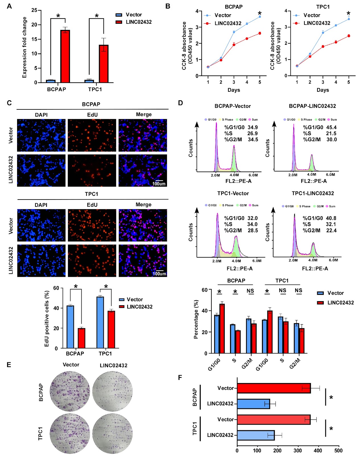
Long noncoding RNAs (lncRNAs) are essential in regulating the development and progression of different types of cancer. However, our insights into their impact and mechanisms in papillary thyroid carcinoma (PTC) are still insufficient. In this study, we investigate the effects of the ferroptosis-associated long noncoding RNA LINC02432 on PTC, which recognizes ferroptosis as a critical mechanism in cancer biology and lncRNAs as significant factors in various malignancies. To identify lncRNAs associated with ferroptosis in PTC, we conduct bioinformatics analyses and perform functional assays to evaluate the biological impact of LINC02432 on PTC cells, as well as its relationship with ferroptosis. Mechanistic studies employ methods such as western blot analysis, flow cytometry, and real-time PCR. Our analysis of transcriptome data from TCGA reveals that LINC02432 is the only lncRNA consistently identified by all 10 machine learning methods used, and its expression is significantly downregulated in PTC. Overexpression of LINC02432 in PTC cells inhibits cell proliferation and migration while promoting ferroptosis through inactivation of the NRF2 pathway. LINC02432 knockdown in PTC cells yields the opposite result. These findings highlight the potential of LINC02432 as a tumor suppressor in PTC progression, offering new insights into the mechanisms underlying the development and progression of this malignancy.

Long noncoding RNA LINC02432 inhibits papillary thyroid cancer via promoting ferroptosis
Graphical Abstract
Original ResearchVol. 58, Issue 6 • pp. 1427-1430DOI: 10.3724/abbs.2025180

Yaf9 conditionally contributes to cell size control in Candida albicans

Authors: Wencheng Zhu, Baodi Dai, Yinxing Xu, Jiangye Chen

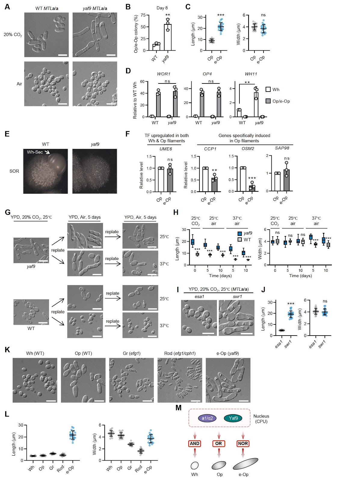
Candida albicans is an opportunistic fungal pathogen renowned for its ability to transition between distinct phenotypic states, such as the yeast-hyphae transition and the white-opaque switching. This morphological plasticity allows the organism to adapt to various host environments and evade immune responses. The white state is characterized by yeast-like cells with high proliferative capacity, whereas the opaque state features elongated cells with enhanced mating ability. The regulation of white-opaque switching is primarily controlled by a complex network of transcription factors. White-Opaque Regulator 1 (Wor1) serves as a master regulator crucial for the establishment and maintenance of the opaque state by activating the expression of genes required for opaque cell formation [1–3]. Conversely, the Mating-Type Like (MTL) locus in C. albicans acts as a critical barrier to white-opaque switching. The genes present at this locus strictly repress the white-to-opaque transition by the formation of a1/α2 complex; therefore, only MTLa/a or MTLα/α strains frequently switch to the opaque state [4,5]. Although the MTLa/α lab strain CAI4 is typically locked in the white state, some MTLa/α clinical isolates can switch to opaque [6]. Several genes were found to modulate this repression. For example, loss of HBR1, which is an activator of MTLALPHA1 and MTLALPHA2 gene expression, enables switching in MTLa/α cells [7]. Deletion of transcriptional repressors of the opaque state such as TUP1 also facilitates white-to-opaque switching [8]. The SWR1 complex incorporates H2A.Z into chromatin, and loss of Swr1 enhances switching and stabilizes the opaque state in MTL homozygous cells [9]. Our previous work revealed that the NuA4 histone acetyltransferase complex and the SWR1 complex merge into a supercomplex via Yaf9 in white-state yeast cells in C. albicans [10]. Here, we first tested whether Yaf9 is involved in white-to-opaque switching in MTLa/α heterozygous cells. The knockout of the YAF9 gene was validated by genotyping and qRT-PCR, confirming its loss at both the genomic and transcriptional levels (Supplementary Figure S1). The yaf9 null mutant cells were spread onto YPD plates and incubated in 20% CO2 at 25°C. After eight days of growth, sectors containing opaque cells were observed (Figure 1A). The frequency of opaque cell formation in the yaf9 mutant exceeded that in wild-type (WT) cells overexpressing WOR1 (Figure 1B). qRT-PCR analysis confirmed significant upregulation of opaque cell-specific markers, including WOR1 and OP4, in yaf9 mutant opaque cells, whereas the white cell marker WH11 was downregulated (Figure 1C). To examine whether YAF9 deletion affects the expression of MTL genes, we performed qRT-PCR for MTLA1 and MTLALPHA2 in white WT cells and in both white and opaque yaf9 mutant cells. The expression of both genes remained unchanged in yaf9 mutant cells (Figure 1D), suggesting that Yaf9-mediated repression of white-to-opaque switching occurs independently of MTL gene regulation. As Yaf9 is a component of the NuA4 and SWR1 complexes, we next investigated the roles of the NuA4 core enzyme Esa1 and the SWR1 core enzyme Swr1 in white-to-opaque switching in MTLa/α heterozygous cells. As shown in Figure 1E (upper panel), esa1 cells failed to switch to the opaque form under 20% CO2 stimulation, indicating that Esa1 activity is essential for opaque cell formation under the tested conditions. In contrast, swr1 cells readily underwent white-to-opaque switching (Figure 1E, lower panel), similar to the yaf9 mutant. These results indicate that Yaf9 functions as a repressor of white-to-opaque switching and that its deletion bypasses the repression imposed by the MTLa/α configuration. We then examined the role of YAF9 in white-to-opaque switching in MTLa/a cells, where MTL repression is removed. In air, yaf9 cells remained white; however, when exposed to 20% CO2, they frequently (> 50%) switched to the opaque form, which occurred at a significantly higher frequency than WT cells (Figure 2A,B). Notably, yaf9 cells exhibited a novel elongated opaque morphology, which we term e-Op cells. Quantification revealed that e-Op cells had similar width but were two to three times longer than WT opaque cells (Figure 2C). At the transcriptional level, e-Op cells displayed comparable upregulation of WOR1 and OP4 and downregulation of WH11 (Figure 2D). Notably, WH11 expression in yaf9 white cells was slightly higher than that in WT white cells. Like white cells, opaque cells are also capable of forming filaments under specific conditions [11]. To determine whether e-Op cells represent a filamentous form of opaque cells, we examined their gene expression and morphological stability. Multiple lines of evidence indicate that e-Op cells are distinct from these filamentous forms. First, when cultured on SOR medium, which promotes filamentous g

Yaf9 conditionally contributes to cell size control in Candida albicans
Graphical Abstract