🧬 SinoBioData Academic Portal
🏛️ Indexed Academic JournalOriginal: 生物化学与生物物理学报

Acta Biochimica et Biophysica Sinica

Premier Chinese Biomedical Journal indexed in SinoBioData: Acta Biochimica et Biophysica Sinica (生物化学与生物物理学报).

Total Research Papers: 200
Access: 100% Free Open Access
Browse by Publication Year & VolumeReset All Filters ✕

Published Research PapersFiltered: Year 2025 • 57 • 10

Showing 11 of 200 peer-reviewed papers with full Graphical Abstracts.

Original ResearchVol. 57, Issue 10 • pp. 1660-1669DOI: 10.3724/abbs.2025036

HDAC11 in ovarian granulosa cells coordinates LH in the maturation of oocytes in Tan sheep

Authors: Jiaqi Shi, Donghuan Lv, Yaxiu Xu, Xiangyan Wang, Zhipeng Qi, Yujie Yan, Jinghua Wang, Hongyuan Song, Hui Yang, Luguo Jin, Zhengyi Yang, Xiaoning Yang, Xiumei Kang, Xinfeng Liu, Zhuming Zhang, Chao Wang

Oocyte maturation plays an important role in supporting mammalian reproduction. Histone deacetylase 11 (HDAC11), the only member of the class IV histone deacetylase family and the smallest histone deacetylases (HDACs), has been shown to regulate oocyte maturation in mice and pigs. However, the epigenetic effects of HDACs in follicular granulosa cells in response to LH induction remain elusive in sheep. In this study, the effects of follicular somatic cell-derived HDAC11 on oocyte maturation in Tan sheep are evaluated. The expression changes of HDAC11 and related proteins are detected by means of immunofluorescence, immunohistochemistry, western blot analysis and enzyme-linked immunosorbent assay. Our results indicate that the level of HDAC11 in follicular granulosa cells as well as oocytes in Tan sheep increases with the growth and maturation of the follicles. Specific inhibition of HDAC11 by SIS17 remarkably reduces the oocyte maturation rate under LH supplementation in vitro. Accordingly, the acetylation level of H3K9 in granulosa cells is increased, while the EGF-like growth factor AREG is remarkably decreased. Furthermore, inhibition of HDAC11 markedly decreases the level of YAP1, which is a negative regulator of AREG in granulosa cells. Conclusively, HDAC11 in the granulosa cells of Tan sheep contributes to the LH induced production of AREG during oocyte in vitro maturation by decreasing the level of H3K9 acetylation and increasing the level of YAP1.

HDAC11 in ovarian granulosa cells coordinates LH in the maturation of oocytes in Tan sheep
Graphical Abstract
Original ResearchVol. 57, Issue 10 • pp. 1611-1624DOI: 10.3724/abbs.2025056

Triptonide facilitates autophagy-mediated apoptosis in esophageal squamous cell carcinoma by targeting the AMPK-mTOR-ULK1 axis

Authors: Jiujun Ju, Nuo Xu, Bohan Li, Dan Shi, Jiahui Cai, Qiusheng Zheng, Lei Ye, Shaosen Zhang, Caixia Wang

Triptonide (TN) is a small-molecule compound initially derived from Tripterygium wilfordii Hook. f used in traditional Chinese medicine. However, its potential antitumor mechanisms are still far from adequately understood. The purpose of this research is to elucidate the antitumor and pharmacological effects of TN on esophageal squamous cell carcinoma (ESCC). Functional assays, such as CCK-8 and colony formation assays, are used to evaluate the effects of TN on KYSE450 and KYSE510 cells. Subsequently, western blot analysis, Hoechst 33258 staining, flow cytometric analysis, autophagic flux detection, and transmission electron microscopy (TEM) are used to determine the effects of TN on apoptosis and autophagy in ESCC cells. Additionally, the autophagy inhibitor 3-methyladenine (3-MA) and the AMPK inhibitor dorsomorphin (Compound C, CC) are administered to explore the molecular mechanisms and crucial pathways in ESCC cells. Our findings provide strong evidence that TN induces autophagy-dependent apoptosis by targeting the AMPK-mTOR-ULK1 axis in ESCC cells. Collectively, this study sheds light on the anticancer mechanisms of TN in esophageal squamous cell carcinoma and suggests that TN is a promising candidate for the antitumor phytomedicine.

Triptonide facilitates autophagy-mediated apoptosis in esophageal squamous cell carcinoma by targeting the AMPK-mTOR-ULK1 axis
Graphical Abstract
Original ResearchVol. 57, Issue 10 • pp. 1589-1600DOI: 10.3724/abbs.2025034

Melatonin attenuates kidney injury by alleviating lysosomal damage in diabetic kidney disease

Authors: Jiaqi Chen, Shuting Zhang, Xiaoquan Xue, Xiaoqin Ma, Aomiao Chen, Yichuan Wu, Geningyue Wang, Qian Zhang, Yaoming Xue, Yijie Jia, Zongji Zheng

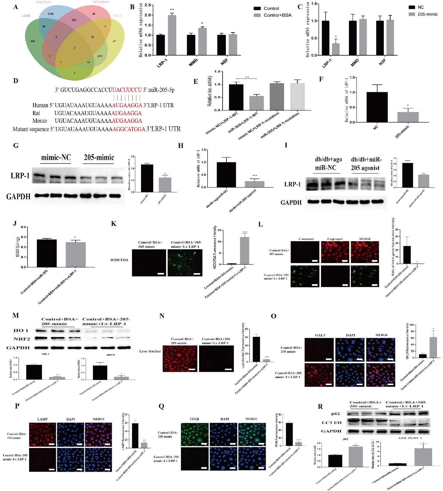
Proteinuria-induced damage to renal tubular epithelial cells is one of the main causes of diabetic kidney disease (DKD), and the clearance of overloaded albumin by lysosomes is crucial for maintaining the homeostasis of renal tubular epithelial cells. Therefore, lysosomal damage is closely related to the pathogenesis of DKD, but effective prevention and treatment measures are still lacking. Melatonin (MLT) is secreted by the pineal gland and can not only regulate circadian rhythms but also maintain lysosomal homeostasis. In this study, we demonstrate the presence of significant lysosomal damage in the renal tubules of DKD patients, which causes autophagy impairment and a concomitant oxidative stress imbalance; however, MLT can upregulate transcription factor EB (TFEB) to improve lysosomal damage and restore the biosynthesis of this organelle. Mechanistically, MLT may protect lysosomes via the upregulation of TFEB and the miR-205-5p-LRP-1 pathway in renal tubules, thus improving autophagy dysfunction and oxidative imbalance in DKD.

Melatonin attenuates kidney injury by alleviating lysosomal damage in diabetic kidney disease
Graphical Abstract
Original ResearchVol. 57, Issue 10 • pp. 1574-1588DOI: 10.3724/abbs.2025102

Nanchangmycin suppresses influenza A virus infection by blocking endosomal acidification

Authors: Hao Zhang, Changhai Liu, Shuo Cai, Yuting Wu, Lu Shang, Fayu Yang, Jing Liu, Nan Wei, Yingchun Liu, Mi Wang, Fei Gao, Qinfang Liu, Hongjun Chen, Guangzhi Tong, Yin Chen, Feng Gu

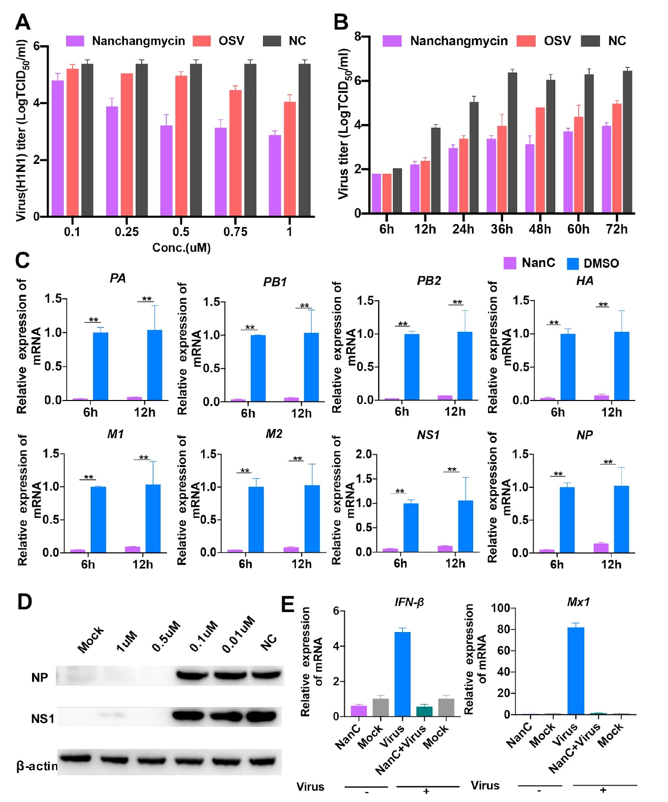
Influenza A viruses (IAVs) constitute a major threat to human and animal health. Currently, M2 ion-channel inhibitors, neuraminidase (NA) inhibitors, RNA polymerase inhibitors, and cap-dependent endonuclease inhibitors have been applied clinically as therapeutics against IAVs. However, IAVs possess adaptive mutations to these inhibitors, especially M2 ion channel and NA inhibitors. Thus, novel antiviral agents should be developed. In the present study, we screen approximately 5500 compounds and identify an IAV inhibitor, nanchangmycin, which possesses a robust antiviral activity both in vitro and in vivo. In addition, it exhibits broad-spectrum antiviral activity for additional virus infections, including pseudorabies virus, herpes virus, porcine epidemic diarrhea virus, porcine reproductive and respiratory syndrome virus. Most importantly, it has antiviral activity against oseltamivir-resistant strains in sub-μM ranges and promotes the survival of MDCK cells infected with the oseltamivir-resistant influenza A virus strain. Further studies reveal that it blocks the nuclei migration of viral nuclear proteins (NPs), resulting in NP accumulation in the cytoplasm, particularly within perinuclear endosomes. Also, it inhibits IAVs by blocking endosomal acidification. Overall, nanchangmycin has the potential to be developed as an anti-influenza agent.

Nanchangmycin suppresses influenza A virus infection by blocking endosomal acidification
Graphical Abstract
Original ResearchVol. 57, Issue 10 • pp. 1625-1635DOI: 10.3724/abbs.2025042

PDGFC secreted by cancer-associated fibroblasts promotes epithelial-mesenchymal transition and immunosuppression in lung adenocarcinoma

Authors: Meimei Cui, Xiaodi Ding, Yu Jiang, Liying Zhang, Wangkai Cao, Yongming Wang, Zhimei Sheng, Wei Sun, Ai Guo, Lihui Gu, Xiurong Zhang, Wanli Duan, Lihong Shi, Baogang Zhang

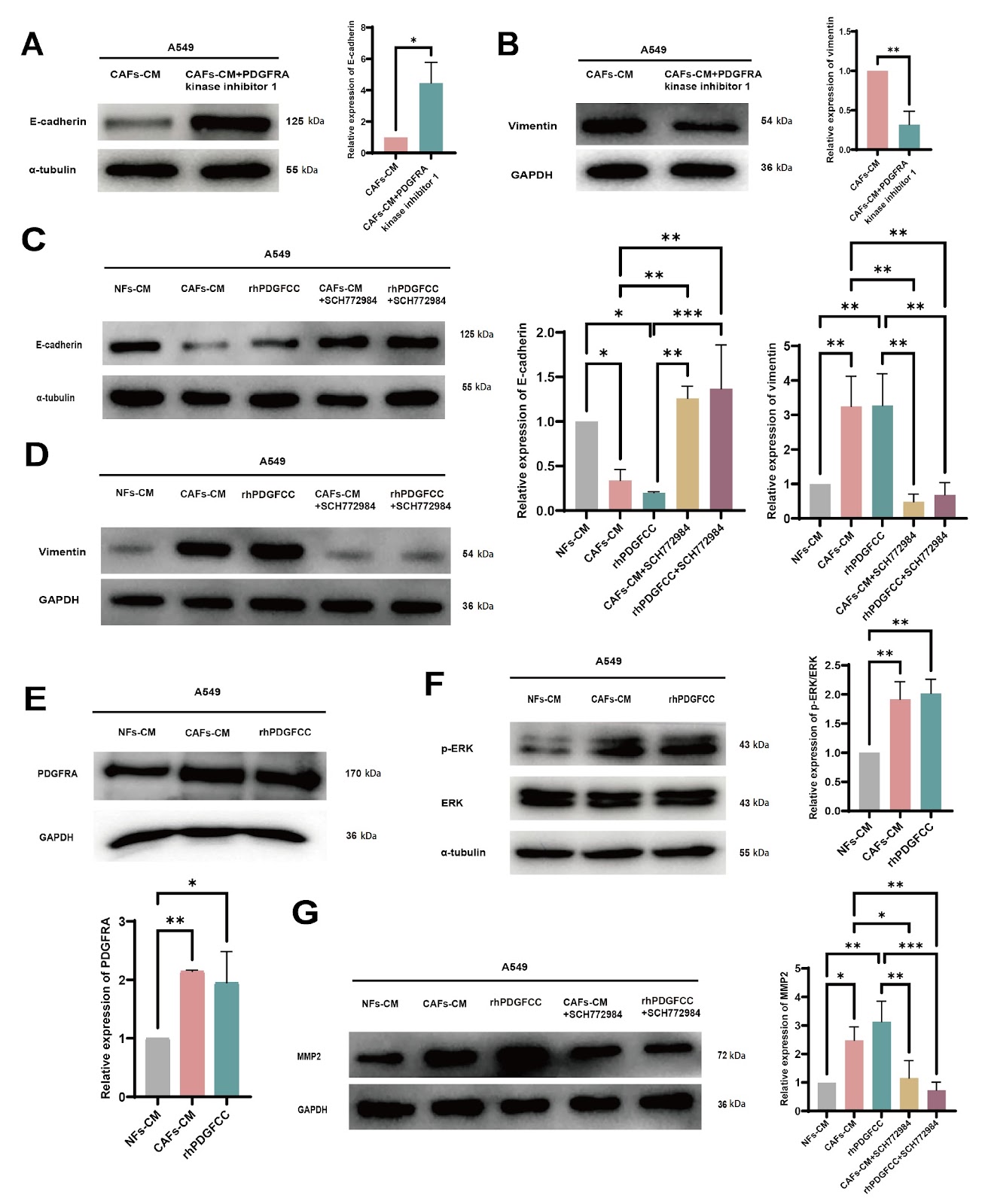
This study elucidates the mechanisms by which cancer-associated fibroblast (CAF)-derived platelet-derived growth factor C (PDGFC) promotes the progression of lung adenocarcinoma (LUAD) and explores the impact of PDGFC on immune regulation within the tumor microenvironment (TME). Our results show that there is higher expression of PDGFC in CAFs than in nontumor tissue fibroblasts (NFs) and that higher expression of PDGFC is correlated with poor prognosis in LUAD patients. Furthermore, CAF-derived PDGFC promotes epithelial-mesenchymal transition (EMT) in cancer cells as well as matrix metalloproteinase 2 (MMP2) expression through the PDGF receptor A (PDGFRA)-mitogen-activated protein kinase/extracellular signal-regulated kinase (MAPK/ERK) pathway. Moreover, our study demonstrates that CAF-derived PDGFC is essential for the activation and infiltration of fibroblasts in the TME, as well as the inflammatory infiltration of different immune cell types and the immunosuppressive conditions within the TME. In particular, PDGFC induces increased PDGFRA expression in both tumor cells and fibroblasts, which can lead to reciprocally positive feedback to accelerate malignant tumor progression. This discovery provides a novel TME-targeted strategy for LUAD treatment.

PDGFC secreted by cancer-associated fibroblasts promotes epithelial-mesenchymal transition and immunosuppression in lung adenocarcinoma
Graphical Abstract
Original ResearchVol. 57, Issue 10 • pp. 1684-1695DOI: 10.3724/abbs.2025045

TLR4 mediates lipotoxic β-cell dysfunction by inhibiting the TMEM24/PI3K/AKT pathway

Authors: Chao Lan, Yan Li, Zhiyan Weng, Wei Pan, Wanxin Lin, Zhen Jiang, Liyong Yang, Ximei Shen

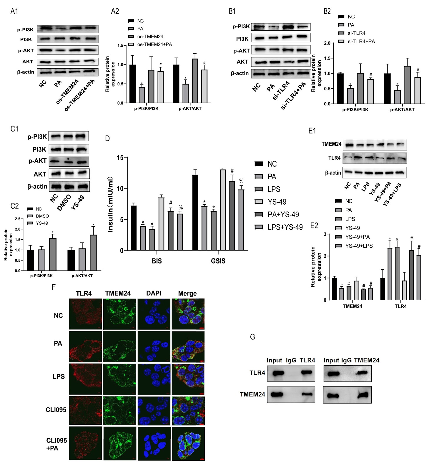
Immune imbalance is the core pathophysiological mechanism of the deterioration of β-cell function driven by lipid metabolism disorders. Toll-like receptor 4 (TLR4) inflammatory signaling is a key pathway that mediates lipotoxic injury in β-cells, but the underlying mechanism needs to be further elucidated. Transmembrane protein 24 (TMEM24) is a key transporter that regulates pulsatile insulin secretion, but its pathophysiology in lipotoxicity remains unclear. In this study, we investigate whether TLR4-mediated lipotoxicity is affected by the inhibition of TMEM24 expression. The PPI network shows that TLR4 is associated with both insulin secretion and ER stress proteins in islets from obese rats. Using in vitro lipotoxic β-cell models, we found that TMEM24 is the target signal of palmitic acid (PA)-induced insulin secretion impairment in islet β-cells, and TLR4 plays a mediating role in this process. Mechanistically, TLR4 mediates lipotoxicity by binding to TMEM24 and downregulating its protein expression to suppress PI3K/AKT signaling, leading to β-cell dysfunction. TLR4 knockout ameliorates islet function impairment through TMEM24/PI3K/AKT signaling in HFD-induced obese rats. Taken together, our results show that TLR4 mediates lipotoxicity in islet β-cells by inhibiting the TMEM24/PI3K/AKT pathway, and the mechanism of TLR4-mediated lipotoxicity is elucidated from the perspective of insulin vesicular secretion.

TLR4 mediates lipotoxic β-cell dysfunction by inhibiting the TMEM24/PI3K/AKT pathway
Graphical Abstract
Original ResearchVol. 57, Issue 10 • pp. 1557-1573DOI: 10.3724/abbs.2025071

CD47-mediated tumor microenvironment remodeling: a central mechanism in immune evasion

Authors: Hemei Yuan, Lingling Zhu, Longhuan Yang, Yong Yi, Tao Lv

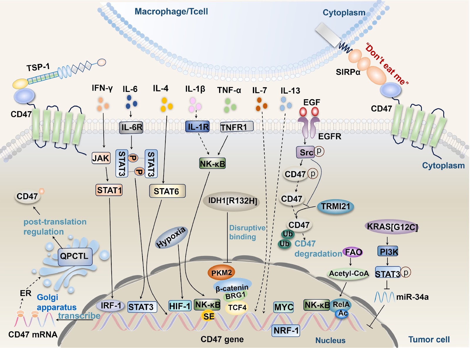
Immune evasion is a crucial strategy for tumor growth and survival, with the tumor microenvironment facilitating tumor immune evasion and cancer progression. CD47, a transmembrane protein highly expressed in various cancer cell types, interacts with its ligands SIRPα and TSP-1 to induce immune tolerance, enabling tumor cells to evade immune surveillance and phagocytosis by immune cells. Understanding the pathways driving CD47 signaling and related activation factors is essential. In this review, we discuss the interactions between CD47 and its ligands SIRPα and TSP-1; their roles in inhibiting the functions of immune cells (macrophages, dendritic cells (DCs), glial cells, T cells, NK cells, etc.); and the mechanisms involved. Furthermore, we also explore the influence of factors within the tumor microenvironment, including TNF-α, IFN-γ, ILs, HIF-1, oncogenes, isocitrate dehydrogenase 1, metabolic enzymes, and exosomes, on CD47-mediated immune evasion. Recent monoclonal antibody drugs targeting CD47 for cancer treatment have shown side effects and cause economic losses. Researchers can explore alternative approaches, such as designing targeted drugs with minimal side effects or investigating other related molecules or pathways. Combination therapy and further research into the molecular mechanisms of CD47 could offer new directions for antitumor drug development.

CD47-mediated tumor microenvironment remodeling: a central mechanism in immune evasion
Graphical Abstract
Original ResearchVol. 57, Issue 10 • pp. 1601-1610DOI: 10.3724/abbs.2025063

FOXD3 promotes homologous recombination repair and genomic stability by facilitating MRE11-mediated DNA end resection

Authors: Shibin Xu, Jingyu Zhang, Congwen Gao, Ziyi Xiong, Yamin Gong, Bao Chai, Hongxiang Chen, Xingzhi Xu

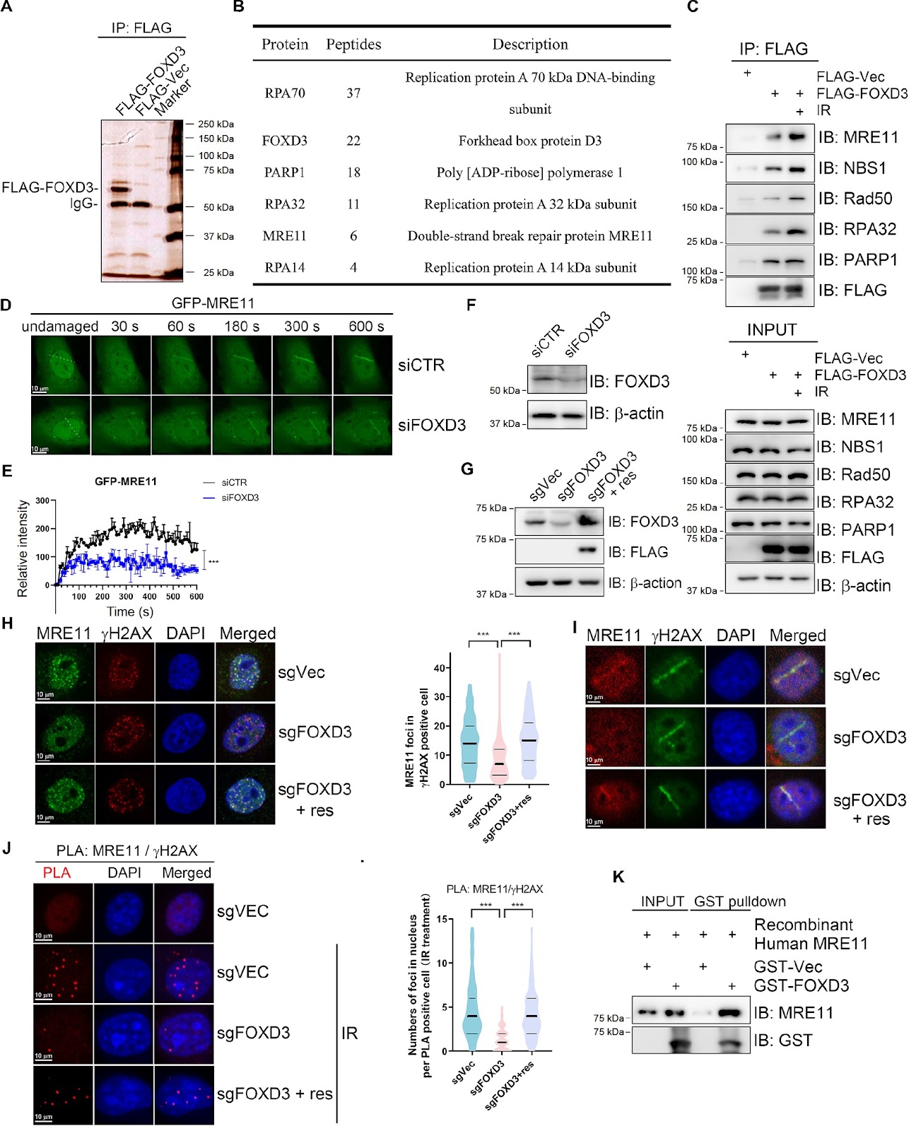
Homologous recombination (HR) is crucial for the high-fidelity repair of DNA double-strand breaks (DSBs), ensuring the maintenance of genome stability. In this study, we show that FOXD3 interacts with poly (ADP-ribose) polymerase 1 (PARP1) and is recruited to DSBs in a PARP1-dependent manner. FOXD3 directly binds to the DSB repair protein MRE11 and promotes its recruitment to DSB sites, ensuring proper end resection. Inhibition of FOXD3 expression compromises HR-mediated DSB repair and chromosome stability and sensitizes cancer cells to ionizing radiation. Collectively, our findings demonstrate that FOXD3 promotes HR-mediated DSB repair and genome stability.

FOXD3 promotes homologous recombination repair and genomic stability by facilitating MRE11-mediated DNA end resection
Graphical Abstract
Original ResearchVol. 57, Issue 10 • pp. 1709-1717DOI: 10.3724/abbs.2025005

ATF3 triggers M2 macrophage polarization to protect against pulp inflammation through WNT4 regulation

Authors: Liu Liu, Jie Wang, Jie Yu, Jing Wang, Jinhua Yu

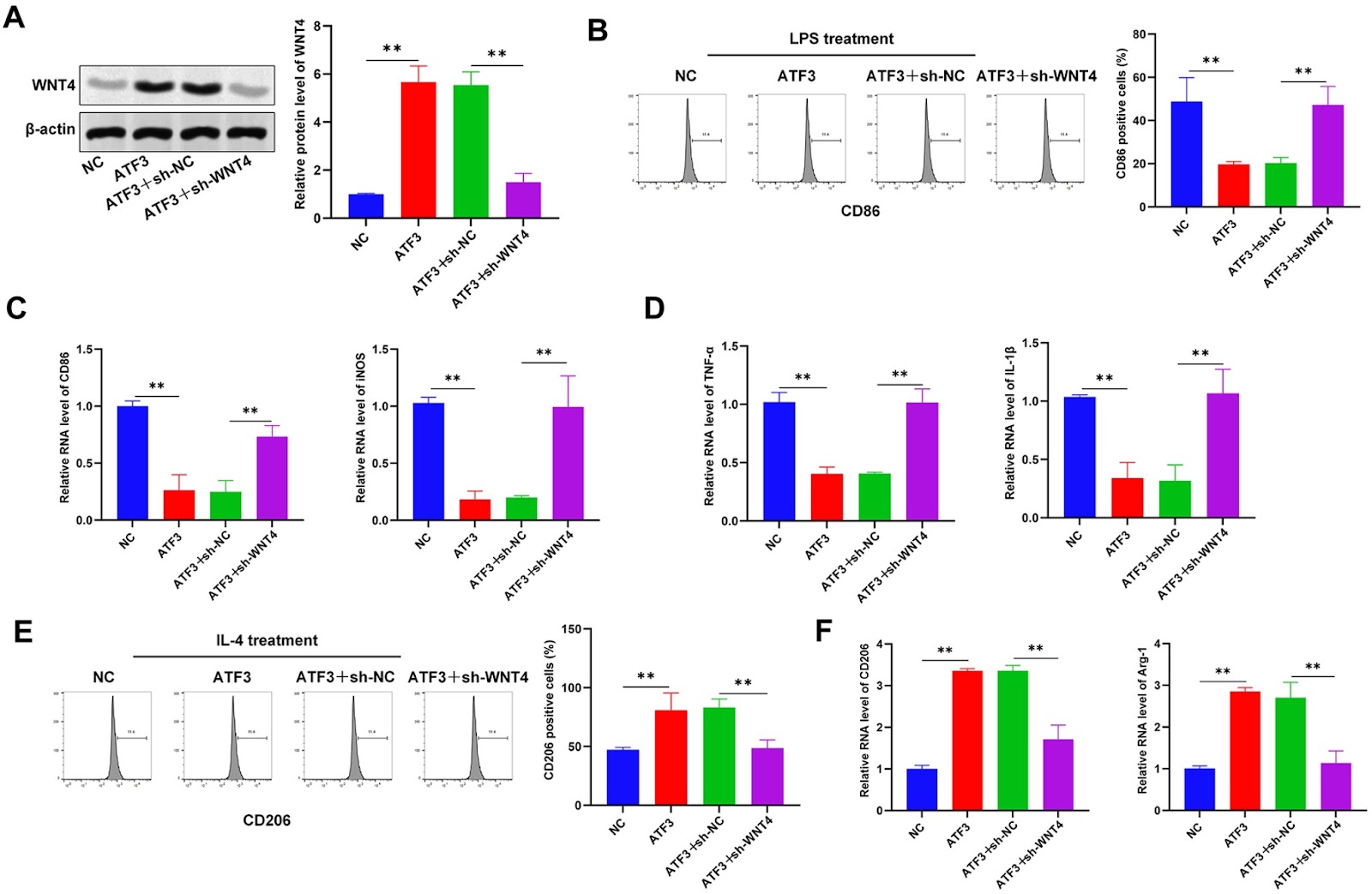
Pulpitis is a common inflammatory oral disease that can lead to pulp necrosis. The aim of this study is to investigate the expression and regulatory mechanisms of ATF3, a potential therapeutic marker, in pulpitis. A mouse pulpitis model with different degrees of inflammation is established, and the expression of ATF3 in pulpitis is explored. The histological features of healthy pulp and pulpitis are analyzed by HE staining, and classical inflammatory factors are detected by immunohistochemistry (IHC). In an in vitro study, we investigate the role of ATF3 in the regulation of WNT4 transcription and explore the effects of the ATF3/WNT4 axis on the polarization of RAW264.7 macrophages, the inflammatory response and the osteogenic differentiation of human dental pulp stem/stromal cells (hDPSCs). Our results show that ATF3 is expressed at low levels in inflamed pulp tissues; overexpression of ATF3 reduces the area of pulp necrosis, decreases the level of pro-inflammatory factors, and promotes macrophage polarization toward the M2 type. Furthermore, we reveal that ATF3 binds to the WNT4 promoter region and positively regulates the expression of WNT4 and that ATF3 downregulates M1 markers and increases the expression of M2 markers by regulating WNT4 expression. In addition, ATF3 promotes the osteogenic differentiation of dental pulp stem cells. In summary, this study reveals that ATF3 promotes M2 macrophage polarization by regulating WNT4, which in turn inhibits pulpal inflammatory responses and promotes the osteogenic differentiation of dental pulp stem cells. These findings suggest that ATF3 may be a potential target for pulpitis treatment.

ATF3 triggers M2 macrophage polarization to protect against pulp inflammation through WNT4 regulation
Graphical Abstract
Original ResearchVol. 57, Issue 10 • pp. 1647-1659DOI: 10.3724/abbs.2025070

Schisandrin A ameliorates the diabetes-associated memory impairment by alleviating inflammation and ferroptosis

Authors: Guandi Ma, Min Lei, Shuang Guo, Yuqing Zhang, Yixuan Sun, Huimin Ji, Changhan Ouyang, Xiaosong Yang, Youzhi Zhang, Xiufen Liu, Baoqing Zhao, Xiying Guo

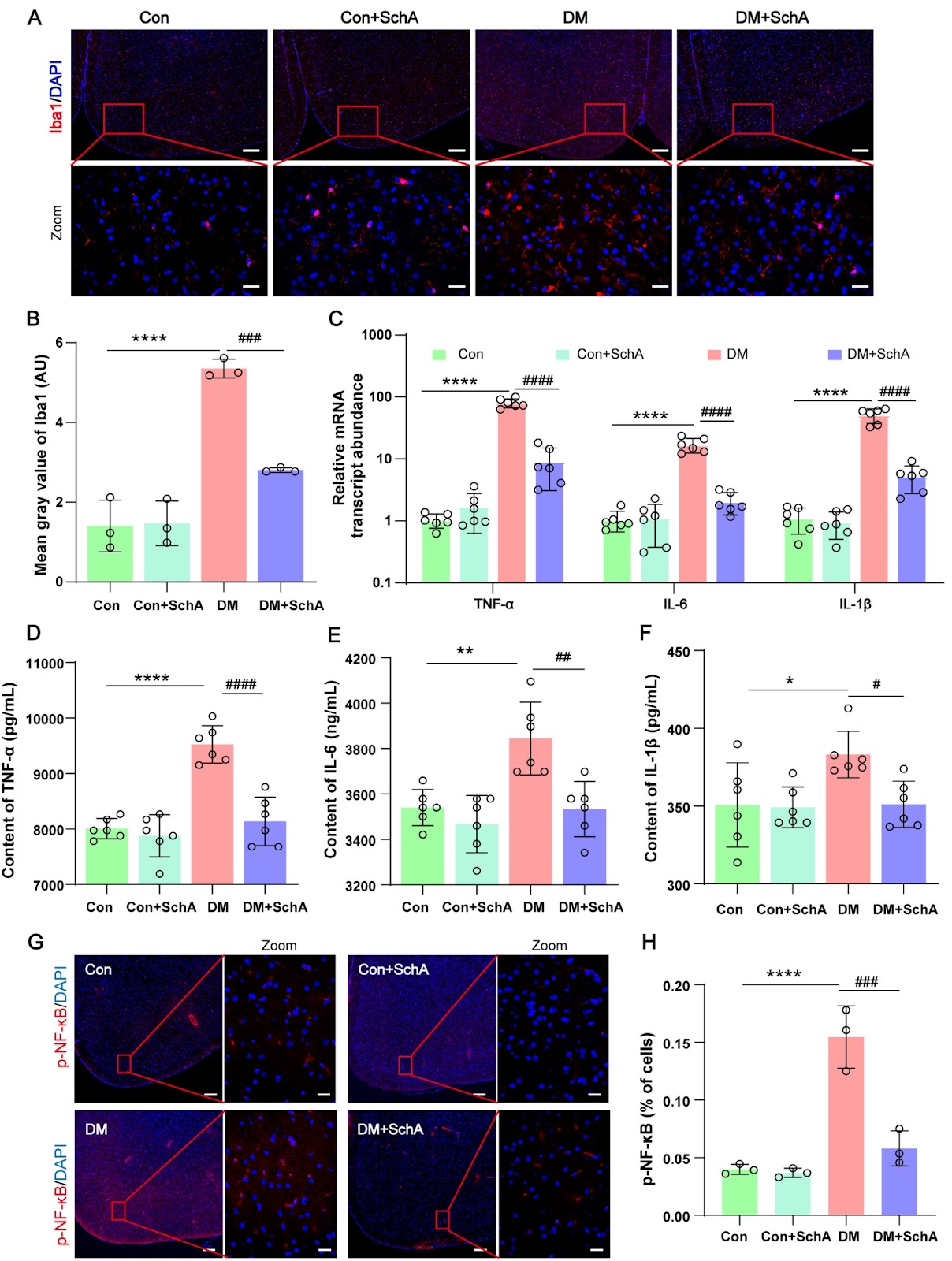
Schisandrin A (SchA), a bioactive lignan that was isolated from the dried fruit of Schisandra chinensis, has attracted much attention because of its diverse spectrum of pharmacological effects. The aim of this study is to clarify the function of SchA in diabetes-related fear memory impairment and its molecular mechanisms. Rats are randomly assigned to 4 groups: the control group (Con group), the DM group, the DM + SchA group, and the Con + SchA group. The results demonstrate that SchA treatment improves insulin sensitivity, reduces blood glucose, and significantly reduces memory impairment. SchA treatment also prevents histological damage, enhances synaptic protein production, and significantly decreases Aβ42 formation in the diabetic prefrontal cortex. Further research reveals that SchA therapy decreases microglial activation and the expression levels of variables linked to inflammation while increasing the phosphorylation of proteins implicated in the insulin resistance signaling pathway. Furthermore, in the prefrontal cortex of diabetic rats, SchA decreases ferroptosis by increasing the protein expressions of GPX4, SLC7A11, Nrf2, HO-1, and SIRT1. Overall, our findings suggest that SchA may lessen diabetes-associated fear memory impairment symptoms by, most likely, lowering ferroptosis and inflammatory responses in the prefrontal brain of diabetic rats. SchA may be a useful therapy for diabetes, including memory impairment.

Schisandrin A ameliorates the diabetes-associated memory impairment by alleviating inflammation and ferroptosis
Graphical Abstract
Original ResearchVol. 57, Issue 10 • pp. 1740-1742DOI: 10.3724/abbs.2025107

ArfGAP2 deficiency ameliorates autoinflammation by regulating STING signaling and proton channel activity

Authors: Min Zhang, Yufei Wang, Zhenwang Zhao, Xiaobo Hu

The cGAS-STING pathway is a critical regulator of innate immunity. When cyclic GMP-AMP synthase (cGAS) detects aberrant cytosolic DNA, it synthesizes the second messenger 2′3′-cGAMP, which binds and activates stimulator of interferon genes (STING) on the endoplasmic reticulum (ER). Activated STING then translocates to the Golgi apparatus, where it recruits and mutually phosphorylates TANK-binding kinase 1 (TBK1) and interferon regulatory factor 3 (IRF3). Phosphorylated IRF3 dimerizes and translocates to the nucleus, where it drives the production of type 1 interferons (IFN-1). In addition to being activated by IFN-1, STING also engages the nuclear factor kappa-B (NF-κB) pathway to induce the production of proinflammatory cytokines and chemokines. Moreover, IFN-1 signaling upregulates the expressions of interferon-stimulated genes (ISGs) through the IFN-α receptor (IFNAR). STING-associated vasculopathy with onset in infancy (SAVI) is a severe pediatric autoimmune disease caused by heterozygous gain-of-function mutations in STING, such as the N154S variant in humans and N153S in mice. Initially, classified as an interferonopathy due to constitutive activation of the STING pathway, SAVI is characterized by systemic inflammation, vasculopathy, interstitial lung disease, T-cell cytopenia, skin ulcerations, and premature death. However, recent studies challenge this paradigm, demonstrating that SAVI pathology develops independently of IFNAR-1 and IRFs (IRF3 and IRF7) [1,2], suggesting that alternative mechanisms drive disease progression. Emerging evidence indicates that STING restricts microbial infection through noncanonical autophagy and cell death pathways [3,4]. These functions may depend on its recently identified role as a proton channel in the Golgi apparatus [4,5]. Xun et al. [4] demonstrated that ligand-bound STING forms an ion channel in its transmembrane domain, facilitating proton efflux from post-Golgi vesicles and inducing Golgi deacidification. This raises a critical question: could STING-mediated Golgi deacidification be a potential mechanism underlying SAVI pathogenesis? A recent study by Poddar et al. [6] identified ADP ribosylation factor GTPase-activating protein 2 (ArfGAP2), which is involved in coatomer protein-1 (COP-1) coating in Golgi vesicles, as a key regulator of both STING signaling and proton channel activity, offering novel therapeutic insights for SAVI. First, to elucidate the role of STING in SAVI pathogenesis, they conducted a genome-wide CRISPR-Cas9 screen in T cells resistant to chronic STING activation and identified ArfGAP2 as a critical STING modulator among multiple Golgi-related proteins. Further investigation revealed that genetic ablation of ArfGAP2 in Jurkat T cells significantly attenuated STING-mediated ISG induction. Compelling evidence indicates that STING activation is associated with its subcellular location [7]. While ArfGAP family proteins typically regulate Golgi membrane trafficking, vesicle transport, and cargo sorting [8], Poddar et al. [6] surprisingly reported that ArfGAP2 enhances STING-mediated ISG induction and promotes LC3 lipidation without altering STING palmitoylation or its Golgi localization. Further experiments revealed that ArfGAP2 enhances STING signaling and IFN-β secretion in mouse bone marrow-derived macrophages (BMDMs). In addition to promoting IFN-1 induction, ArfGAP2 promotes the secretion of NF-κB-dependent proinflammatory cytokines activated by STING in THP-1 monocytes [6]. In addition to inducing ISGs and NF-κB signaling, activated STING acts as a proton channel triggering Golgi deacidification [4]. Given the well-established importance of the Golgi pH in regulating enzyme activity, protein modification, and membrane trafficking [9], researchers have further explored how STING and ArfGAP2 modulate cargo transport and secretion. They reported that the loss of ArfGAP2 impairs STING-mediated proton channel activity in the Golgi, leading to a lower luminal pH. Moreover, ArfGAP2-deficient cells presented significant alterations in the cell surface proteome upon STING activation, accompanied by altered sorting, secretion and trafficking rates of specific protein cargos in the Golgi [6].
ArfGAP2 deficiency ameliorates autoinflammation by regulating STING signaling and proton channel activity
Graphical Abstract