🧬 SinoBioData Academic Portal
🏛️ Indexed Academic JournalOriginal: 生物化学与生物物理学报

Acta Biochimica et Biophysica Sinica

Premier Chinese Biomedical Journal indexed in SinoBioData: Acta Biochimica et Biophysica Sinica (生物化学与生物物理学报).

Total Research Papers: 200
Access: 100% Free Open Access
Browse by Publication Year & VolumeReset All Filters ✕

Published Research PapersFiltered: Year 2025 • 57 • 8

Showing 11 of 200 peer-reviewed papers with full Graphical Abstracts.

Original ResearchVol. 57, Issue 8 • pp. 1222-1233DOI: 10.3724/abbs.2025041

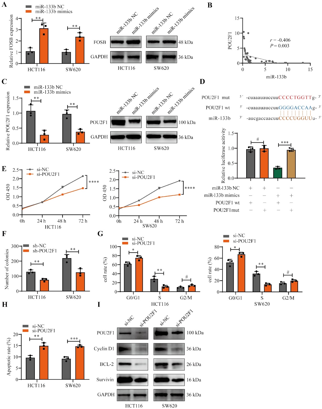
A positive feedback loop between FOSB and miR-133b controls colon cancer cell proliferation

Authors: Wanwan Li, Qionggui Hu, Changwei Lin, Xiaorong Li, Yang Bai, Min Ma

FOSB, a member of the FOS gene family, forms heterodimers with JUN family proteins to engage in diverse cellular processes. Its biological impacts vary among different types of tumors, yet its specific function in colon cancer (CC) remains ambiguous. In this study, quantitative real-time PCR (qRT-PCR) and immunohistochemistry (IHC) are applied to measure FOSB expression levels, followed by an analysis of the association between FOSB expression and patients’ clinical parameters. In vitro experiments are performed to assess cell proliferation, including growth rate, cell cycle distribution, and apoptosis. A subcutaneous xenograft model in nude mice is utilized to monitor tumor growth in vivo. Additionally, chromatin immunoprecipitation (ChIP) and luciferase reporter assays are conducted to dissect the interactions among FOSB, miR-133b, and POU2F1. The results indicate that FOSB expression is downregulated in CC tissues relative to normal controls. Overexpression of FOSB suppresses proliferation and promotes apoptosis in CC cells. Mechanistically, FOSB binds to the promoter region of miR-133b, enhancing its transcription and subsequently repressing POU2F1 expression. Notably, decreased POU2F1 expression also alleviates the transcriptional repression of the FOSB promoter region, establishing a FOSB-miR-133b-POU2F1 feedback loop that inhibits CC proliferation. In summary, our findings suggest that FOSB acts as a tumor suppressor gene in CC and may exert its inhibitory effects on CC growth via the FOSB-miR-133b-POU2F1 feedback loop.

A positive feedback loop between FOSB and miR-133b controls colon cancer cell proliferation
Graphical Abstract
Original ResearchVol. 57, Issue 8 • pp. 1270-1280DOI: 10.3724/abbs.2025043

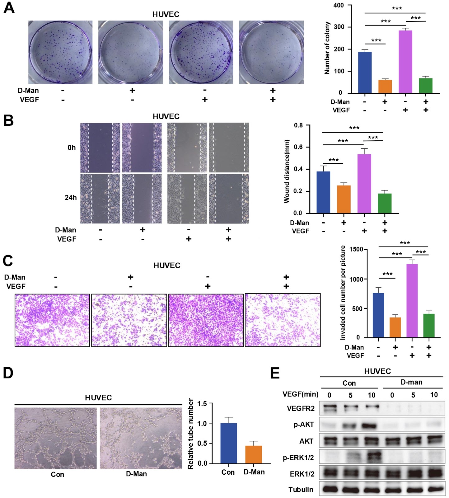
D-mannose suppresses the angiogenesis and progression of colorectal cancer

Authors: Yu Du, Xinchao Zhang, Yixin Xu, Yuefan Zhou, Yanping Xu

Angiogenesis is an important factor influencing the development of solid tumors, and vascular endothelial growth factor receptor-2 (VEGFR2) is a central regulator of angiogenesis. Antibodies and inhibitors against VEGFR2 have been widely used in various malignancies. However, the regulatory mechanism of VEGFR2 has not been fully clarified. Here, we show that D-mannose can significantly inhibit angiogenesis and tumor growth by degrading VEGFR2. Specifically, D-mannose inactivates GSK3β by promoting the phosphorylation of GSK3β at Ser9, enhances the nuclear translocation of TFE3, and promotes lysosomal biogenesis, thereby increasing the lysosome-mediated degradation of VEGFR2. Thus, D-mannose significantly inhibits the proliferation, migration, and capillary formation of human umbilical vein endothelial cells (HUVECs) in vitro. Oral administration of D-mannose dramatically inhibits angiogenesis and tumor growth in mice. Our findings reveal a previously unrecognized anti-tumor mechanism of D-mannose by destabilizing VEGFR2 and provide a new strategy for the clinical treatment of colorectal cancer (CRC).

D-mannose suppresses the angiogenesis and progression of colorectal cancer
Graphical Abstract
Original ResearchVol. 57, Issue 8 • pp. 1260-1269DOI: 10.3724/abbs.2024221

NCOA6 knockdown enhances RSL3-induced ferroptosis in pancreatic cancer cells and increases the sensitivity to gemcitabine

Authors: Yuming Jia, Zeng Ye, Xin Wang, Yanli Deng, Chao Wang, Zhilei Zhang, Guixiong Fan, Wuhan Yang, Xiaowu Xu, Yi Qin, Li Peng

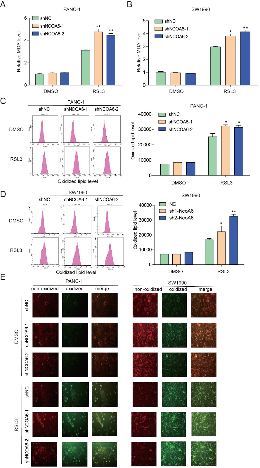
Ferroptosis is a type of programmed death characterized by iron-dependent lipid peroxidation, and targeting ferroptosis has been shown to efficiently kill highly aggressive cancer cells. Previously, we confirmed that nuclear receptors regulate ferroptosis in pancreatic cancer. However, whether nuclear receptor co-activators regulate ferroptosis is unclear. Here, we show that knocking down the nuclear receptor co-activator, NCOA6, enhances the sensitivity of pancreatic cancer cells to ferroptosis. Mechanistically, NCOA6 knockdown promotes the expression of ACSL4 while inhibiting the expression of SCD1, resulting in changes in lipid metabolism, sensitivity to RSL3-induced ferroptosis, and sensitivity to gemcitabine in pancreatic cancer. The relationships between NCOA6 and ACSL4 or SCD1 are further explored in clinical specimens. This study reveals that targeting NCOA6 might alleviate gemcitabine resistance in pancreatic cancer.

NCOA6 knockdown enhances RSL3-induced ferroptosis in pancreatic cancer cells and increases the sensitivity to gemcitabine
Graphical Abstract
Original ResearchVol. 57, Issue 8 • pp. 1281-1291DOI: 10.3724/abbs.2025013

GWAS study of myelosuppression among NSCLC patients receiving platinum-based combination chemotherapy

Authors: Hanxue Huang, Junyan Liu, Qi Xiao, Chenxue Mao, Lei She, Lulu Yu, Bing Yu, Mengrong Lei, Ying Gao, Baimei He, Pinhua Pan, Xi Li, Jiye Yin, Zhaoqian Liu

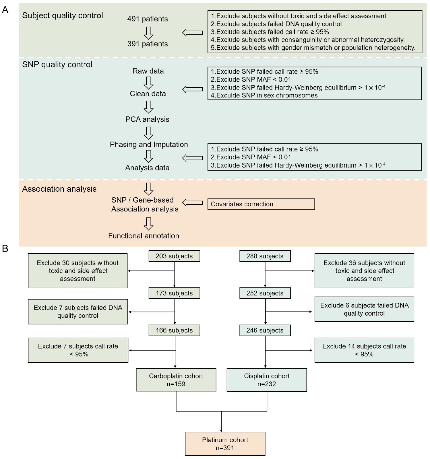
Platinum-based chemotherapy remains the mainstay for non-small cell lung cancer (NSCLC), but it frequently causes dose-limiting myelosuppression, with significant individual variability in susceptibility. However, the genetic basis of myelosuppression side effects remains elusive, greatly hindering personalized therapeutic approaches. In this study, we perform a comprehensive genome-wide association analysis on 491 NSCLC patients receiving platinum-based chemotherapy, examining 4,690,998 single-nucleotide polymorphisms (SNPs) to identify relevant genetic variants. LDBlockShow, FUMA, and MAGMA are utilized to explore linkage disequilibrium, expression quantitative trait loci (eQTLs), chromatin interaction, and conduct gene-based and gene set-based analysis of candidate SNPs. The GWAS results reveal that rs6856089 and its linked SNPs are significantly associated with platinum-based chemotherapy-induced myelosuppression. Specifically, patients with the A allele of rs6856089 have a significantly lower risk of myelosuppression [odds ratio (OR) = 0.1300, P = 7.59 × 10⁻⁸]. Furthermore, gene-based analysis reveals that EMCN (P = 2.47 × 10⁻⁵), which encodes endomucin, a marker for hematopoietic stem cells, might mediate myelosuppression. This study provides a scientific basis for the individual differences in platinum-based chemotherapy-induced myelosuppression.

GWAS study of myelosuppression among NSCLC patients receiving platinum-based combination chemotherapy
Graphical Abstract
Original ResearchVol. 57, Issue 8 • pp. 1350-1362DOI: 10.3724/abbs.2025014

Knockdown of lncRNA XR_877193.1 suppresses ferroptosis and promotes osteogenic differentiation via the PI3K/AKT signaling pathway in SONFH

Authors: Huixia Yang, Ning Ding, Shi Qing, Yinju Hao, Cilin Zhao, Kai Wu, Guizhong Li, Huiping Zhang, Shengchao Ma, Zhigang Bai, Yideng Jiang

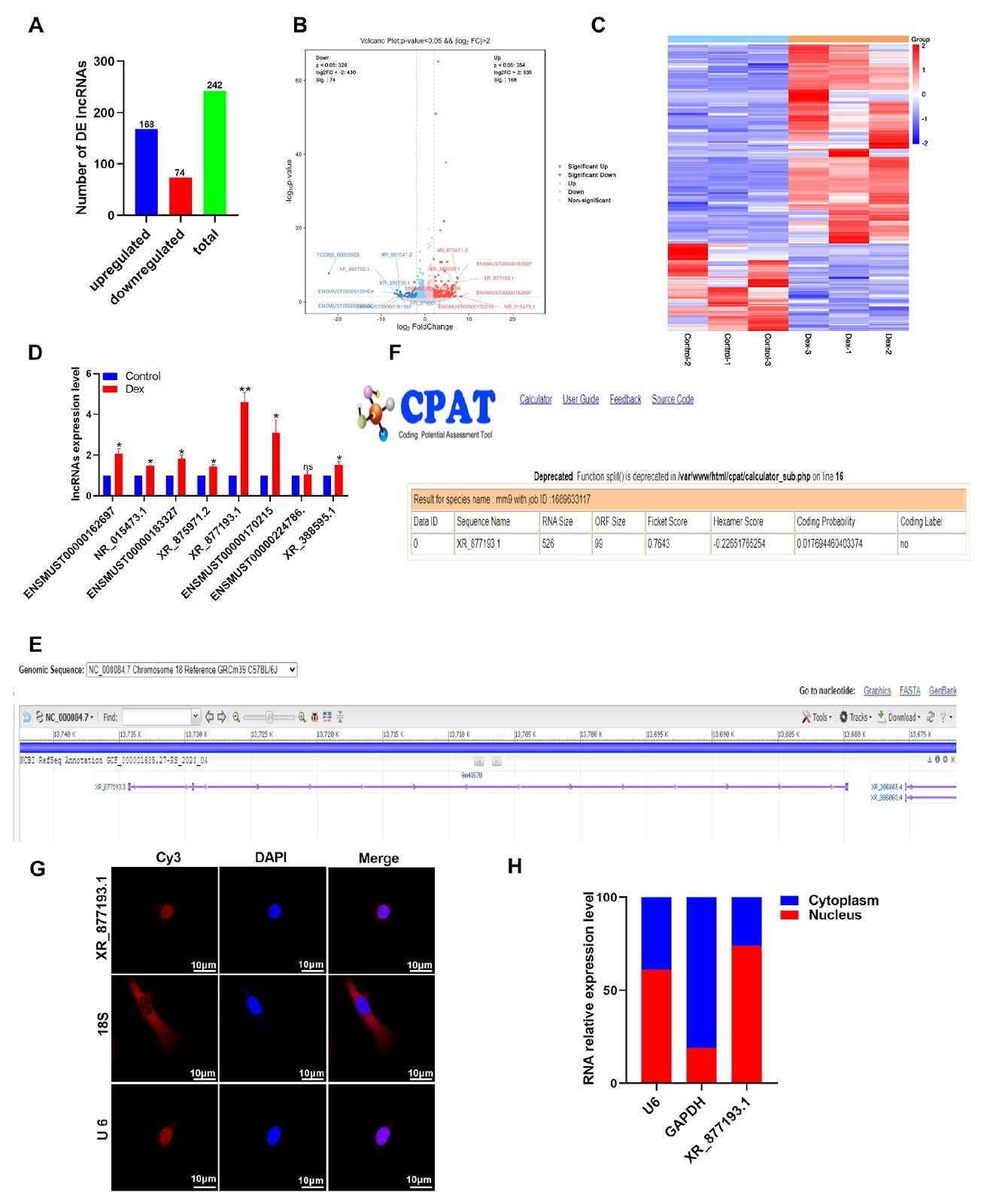
Ferroptosis is a novel form of regulated cell death characterized by the iron-dependent accumulation of lipid peroxides. Recent research has suggested that ferroptosis in osteoblasts contributes to steroid-induced osteonecrosis of the femoral head (SONFH). However, the relationship between ferroptosis and SONFH remains unclear. In this study, in vitro experiments show that dexamethasone (Dex) treatment reduces the expressions of key ferroptosis regulators, SLC7A11 and GPX4, in MC3T3-E1 cells. This reduction leads to a decrease in intracellular glutathione (GSH) levels, accompanied by elevated levels of total iron, malondialdehyde (MDA), and reactive oxygen species (ROS). Importantly, the ferroptosis inhibitor ferrostatin-1 (Fer-1) effectively reverses Dex-induced ferroptosis in MC3T3-E1 cells. Furthermore, RNA-seq analysis reveals that the long noncoding RNA (lncRNA) XR_877193.1 is significantly upregulated in Dex-treated MC3T3-E1 cells. Functional studies demonstrate that the knockdown of lncRNA XR_877193.1 promotes osteogenic differentiation by inhibiting Dex-induced ferroptosis in MC3T3-E1 cells, whereas its overexpression exacerbates cell death via ferroptosis. Kyoto Encyclopedia of Genes and Genomes (KEGG) enrichment analysis reveals that the differentially expressed lncRNA XR_877193.1 is enriched in ferroptosis-related pathways, including the PI3K/AKT signaling pathway. Moreover, PI3K/AKT inhibitors reverse ferroptosis in MC3T3-E1 cells inhibited by lncRNA XR_877193.1 knockdown. Collectively, our findings indicate that lncRNA XR_877193.1 knockdown exerts anti-ferroptosis effects by stimulating the PI3K/AKT signaling pathway, suggesting a promising therapeutic strategy for attenuating SONFH.

Knockdown of lncRNA XR_877193.1 suppresses ferroptosis and promotes osteogenic differentiation via the PI3K/AKT signaling pathway in SONFH
Graphical Abstract
Original ResearchVol. 57, Issue 8 • pp. 1371-1375DOI: 10.3724/abbs.2025016

CRATS: a one-pot CRISPR-Cas12b and RPA combined assay with a temperature switch for highly sensitive detection of monkeypox virus

Authors: Guangxi Yu, Yue Wang, Yukang Chen, Jiangyuan Liu, Hongtao Kang, Xiaodong Luan, Song Gao, Pei Wang

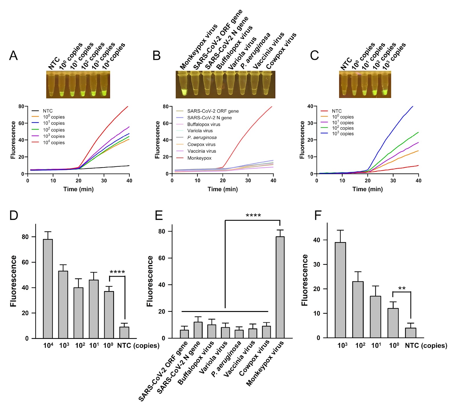
CRISPR-Cas nucleases have been extensively used in molecular detections, especially highly sensitive nucleic acid detections. In these detections, Cas nucleases are programmed by the guide RNA to respond to the detection targets and cleave the chemically labelled molecular beacons by the trans-cleavage activity to produce the detection signal. To improve sensitivity, nucleic acid amplification technologies are usually introduced to give a pre-amplification of the nucleic acid targets, increasing the detection sensitivity extraordinarily. Polymerase chain reaction (PCR) technology has been used for pre-amplification in laboratories, and isothermal amplification technologies are applied to meet point-of-care testing (POCT) needs because they avoid the use of sophisticated thermal cycling devices. The recombinase polymerase amplification (RPA) technology that amplifies nucleic acid targets isothermally at 37–42°C has been combined with CRISPR-Cas nucleases to establish advantageous nucleic acid detection assays, e.g., the SHERLOCK, which combines with Cas13a, and the DETECTR, which combines with Cas12a. It has been challenging to integrate Cas nucleases and RPA in a one-pot reaction system because the cleavage activity of Cas stimulated by even small amounts of the targets can interfere with amplification by digesting the primers or the newly amplified fragments. Thus, many assays based on Cas nucleases and RPA are in a two-step setting, with pre-amplification and Cas cleavage being isolated as two independent procedures. The two-step setting ensures that amplification and cleavage occur under favorable conditions but sacrifices operational convenience and introduces the risk of cross-contamination. In efforts to establish one-pot RPA-Cas assays, many strategies have been applied, including the use of photocontrolled guide RNA to activate the Cas nuclease at a preferred timepoint, the use of a suboptimal protospacer adjacent motif (PAM) to suppress Cas activity, the generation of dynamic aqueous multiphase with sucrose or glycerol to partially separate the two reactions, and extensive optimization of the RPA-CRISPR reaction system to achieve a subtle balance between the two reactions. In this study, a one-pot CRISPR-Cas12b and RPA combined assay with a temperature switch (CRATS) was established utilizing the reaction temperature difference between RPA and CRISPR-Cas12b cleavage. The Cas12b used in this study, AaCas12b, is a type V-B CRISPR‒Cas nuclease from Alicyclobacillus acidiphilus. It has a bi-lobed architecture consisting of an α-helical recognition lobe containing the REC domains and a nuclease lobe containing the WED, RuvC and Nuc domains. As a dual-RNA-guided DNA endonuclease, Cas12b can be guided by a chimeric single-guide (sg) RNA, and its trans-cleavage activity is specifically activated by the DNA target and results in nonspecific cleavage of single-stranded (ss) DNA molecules, which can be used to produce detection signals if the ssDNA is appropriately labelled as the molecular beacon. As the temperature for RPA is 37°C and the trans-cleavage of Cas12b is active at 60°C, CRATS uses temperature switching to adjust the major on-going reaction in the one-pot system and realizes sequential amplification of the target and cleavage reactions for signal detection. The detection target of this study, monkeypox virus, is an infectious pathogen that has caused the announcement of the Public Health Emergency of International Concern (PHEIC) by the World Health Organization (WHO) twice in recent years. In this one-pot CRATS assay, the reaction reagents of CRISPR-Cas12b and RPA are mixed in a single tube. After the addition of the sample containing the detection target, the reaction was carried out at 37°C for 20 min for amplification, followed by 60°C for 20 min for Cas12b cleavage. The fluorescently labelled molecular beacon is cleaved by Cas12b to release the FAM fluorophore from quenching, producing a fluorescence signal that is visualized under blue light. CRATS shows a high sensitivity of 100 copies of the target DNA per reaction and good specificity, providing a novel strategy of temperature switching to integrate CRISPR-Cas and RPA in a one-pot reaction system. Moreover, it provides a POCT-friendly tool for the detection of the important infectious pathogen monkeypox virus.
CRATS: a one-pot CRISPR-Cas12b and RPA combined assay with a temperature switch for highly sensitive detection of monkeypox virus
Graphical Abstract
Original ResearchVol. 57, Issue 8 • pp. 1207-1221DOI: 10.3724/abbs.2025020

Essential role of the metabolite α-ketoglutarate in bone tissue and bone-related diseases

Authors: Zuping Wu, Yuzhe Guan, Qian Chen, Ruifeng Song, Jing Xie, Xin Zhang, Yan Wang, Qianming Chen, Xiaoyan Chen

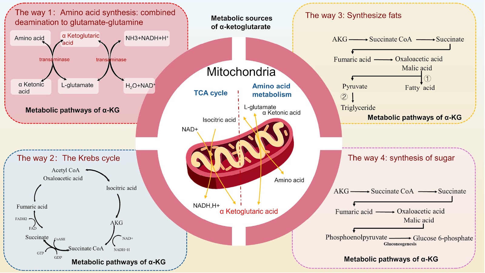
Bone metabolism in bone tissue is constantly maintained in a state of dynamic equilibrium. The mass of bone and joint tissues is determined by both bone formation and bone resorption. It is hypothesized that disrupted metabolic balance leads to osteoporosis, osteoarthritis, rheumatoid arthritis, and bone tumors. Such disruptions often manifest as either a reduction or abnormality in bone mass and are frequently accompanied by pathological changes such as inflammation, fractures, and pain. α-Ketoglutarate (α-KG) serves as a pivotal intermediate in various metabolic pathways in mammals, significantly contributing to cellular energy metabolism, amino acid metabolism, and other physiological processes. α-KG may be a therapeutic target for a variety of bone-related diseases, such as osteoporosis, osteoarthritis, and rheumatoid arthritis, because of its role in maintaining the metabolic balance of bone. After the application of α-KG, bone loss and inflammation in bone tissue are alleviated. This review focuses on the regulatory effects of α-KG on various cells in bone and joint tissues. Owing to the regulatory effect of α-KG on the balance of bone metabolism, the application of α-KG in the treatment of osteoporosis, osteoarthritis, rheumatoid arthritis, bone tumors, and other bone tissue diseases has been clarified.

Essential role of the metabolite α-ketoglutarate in bone tissue and bone-related diseases
Graphical Abstract
Original ResearchVol. 57, Issue 8 • pp. 1304-1311DOI: 10.3724/abbs.2025039

Puerarin prevents cadmium-induced endoplasmic reticulum stress via SIRT1-dependent PERK-CHOP pathway in HepG2 cells

Authors: Di Huang, Mengqi Qiu, Kuanhong Luo, Yanzhe Zhu, Siyu Zhang, Zhen He, Xiaobo Hu, Zhaohui Cao

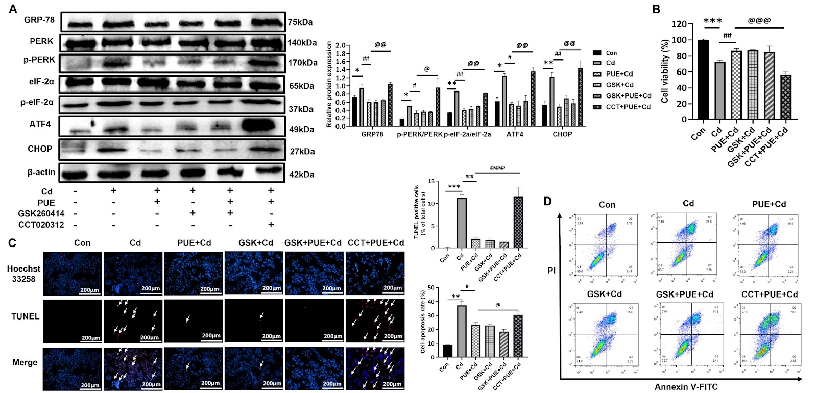
Cadmium (Cd) is a high-risk heavy metal that induces oxidative stress, endoplasmic reticulum (ER) stress and inflammation, damaging organs such as the liver. Puerarin (PUE) has been shown to treat liver injury and especially prevent Cd-induced hepatic damage via its antioxidant activity. Sirtuin 1 (SIRT1), a histone deacetylase, is a key protector against various stress insults. However, its role in the protection of PUE against Cd-induced liver damage has not been clarified. Thus, this study is designed to elucidate the molecular mechanism in the human hepatoma cell line HepG2. The results first reveal that Cd-induced apoptosis is significantly restored by PUE pretreatment, as confirmed by the CCK-8, flow cytometric, Hoechst 33258 and TUNEL assays. Mechanistically, PUE significantly decreases ROS production and increases SOD levels in Cd-treated HepG2 cells. Moreover, PUE pretreatment alleviates ER stress by inhibiting the PERK-eIF2α-ATF4-CHOP axis and subsequently partially restores ER function as revealed by decreased Ca2+ release from the ER. In addition, further study demonstrates that PUE upregulates SIRT1 expression, which suppresses the PERK signaling cascade and reduces CHOP levels. Collectively, our results first demonstrate that PUE protects HepG2 cells from Cd-induced apoptosis at least partially by inhibiting the PERK-eIF2α-ATF4-CHOP pathway in a SIRT1 expression-dependent manner. Puerarin appears to have great potential as a hepatoprotective agent.

Puerarin prevents cadmium-induced endoplasmic reticulum stress via SIRT1-dependent PERK-CHOP pathway in HepG2 cells
Graphical Abstract
Original ResearchVol. 57, Issue 8 • pp. 1234-1243DOI: 10.3724/abbs.2024217

Ageing-associated gut dysbiosis deteriorates mouse cognition

Authors: Huihui Ju, Yile Zhou, Wanting Wei, Yan Hu, Hongwei Fang, Zhouyi Chen, Xia Sun, Yi Shi, Hao Fang

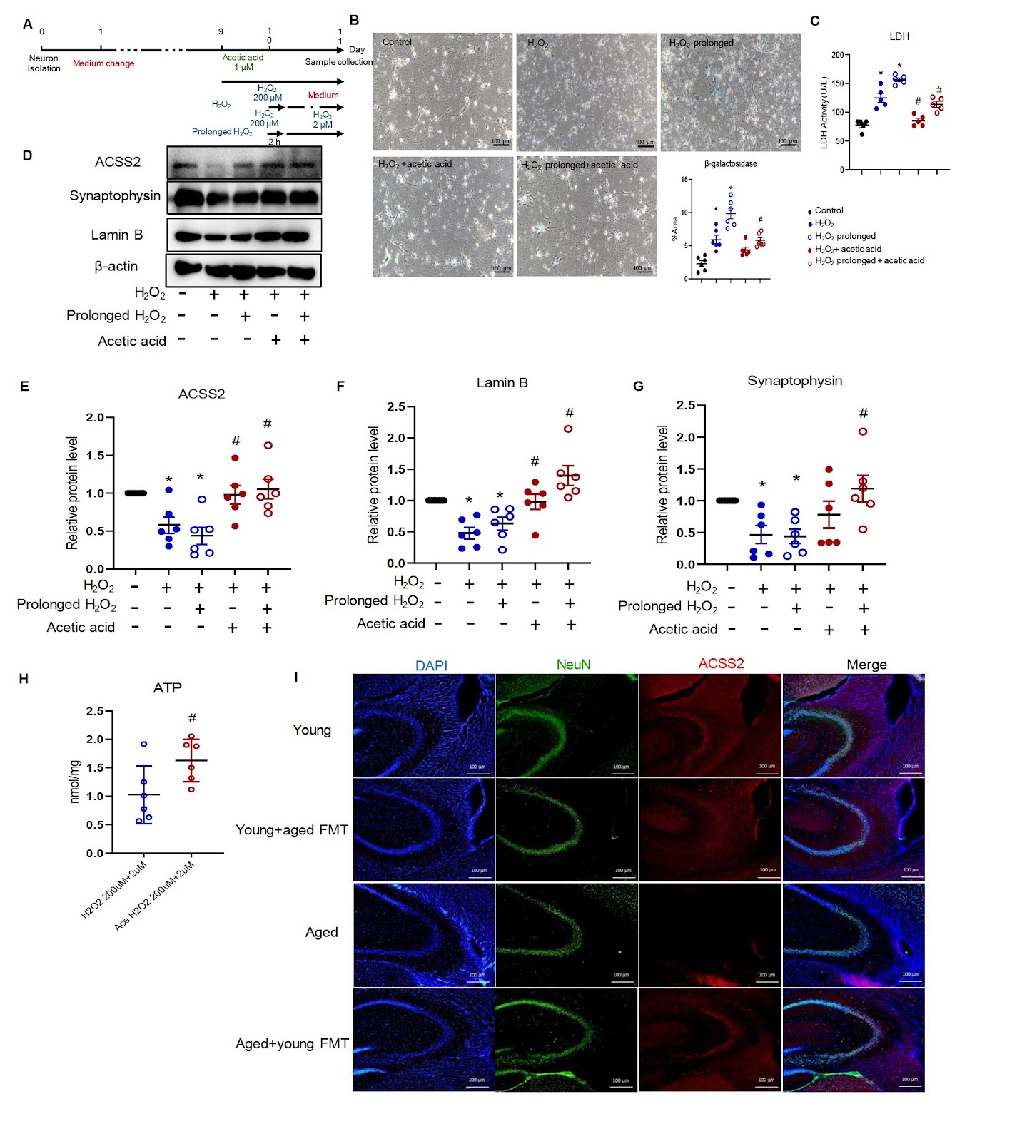
Ageing is an independent factor for cognitive dysfunction. Ageing-associated alterations in the gut microbiota also affect cognition. The present study is designed to investigate changes in the gut microbiota and their participation in ageing-associated cognitive impairment. Both 10-week-old and 18-month-old mice are used. Mouse cognition is examined by novel object recognition and T-maze tests. Mouse feces are collected for sequencing and transplantation. Protein expression in the mouse intestine and hippocampus is studied using immunohistochemistry and immunofluorescence staining. Senescent neurons are induced by hydrogen peroxide in vitro. The cell lysates are used for western blot analysis and adenosine triphosphate (ATP) measurement. Our results show that 18-month-old mice exhibit cognitive dysfunction compared with young mice. In aged mice, transplanting the microbiota of young mice increases the protein presence of synaptophysin in the hippocampus and partially restores cognition. The protein expressions of mucin-2 and E-cadherin in the intestine are reduced in aged mice but are increased by transplantation. Gut microbiota analyses reveal that the reduced abundance of the microbe Bacilli-Lactobacillales-Lactobacillaceae-Lactobacillus in aged mice is restored by transplantation. Fecal microbiota transplantation in young mice increases the serum level of acetic acid in aged mice. Hydrogen peroxide stimulation induces senescence and reduces the protein expression levels of synaptophysin and acetyl-coenzyme A synthetase member 2 (ACSS2) in primary neurons. Incubation with acetic acid upregulates the protein expressions of ACSS2 and synaptophysin and further increases ATP production in senescent neurons. In summary, gut microbiota transplantation increases the abundance of Lactobacillales, elevates serum acetic acid level, and improves cognitive function in aged mice. Gut microbiota transplantation has therapeutic importance for ageing-associated cognitive decline.

Ageing-associated gut dysbiosis deteriorates mouse cognition
Graphical Abstract
Original ResearchVol. 57, Issue 8 • pp. 1244-1251DOI: 10.3724/abbs.2024208

D-CAPS: an efficient CRISPR-Cas9-based phage defense system for E. coli

Authors: Mingjun Sun, Jie Gao, Hongjie Tang, Hengyi Wang, Liyan Zhou, Chuan Song, Yongqiang Tian, Qi Li

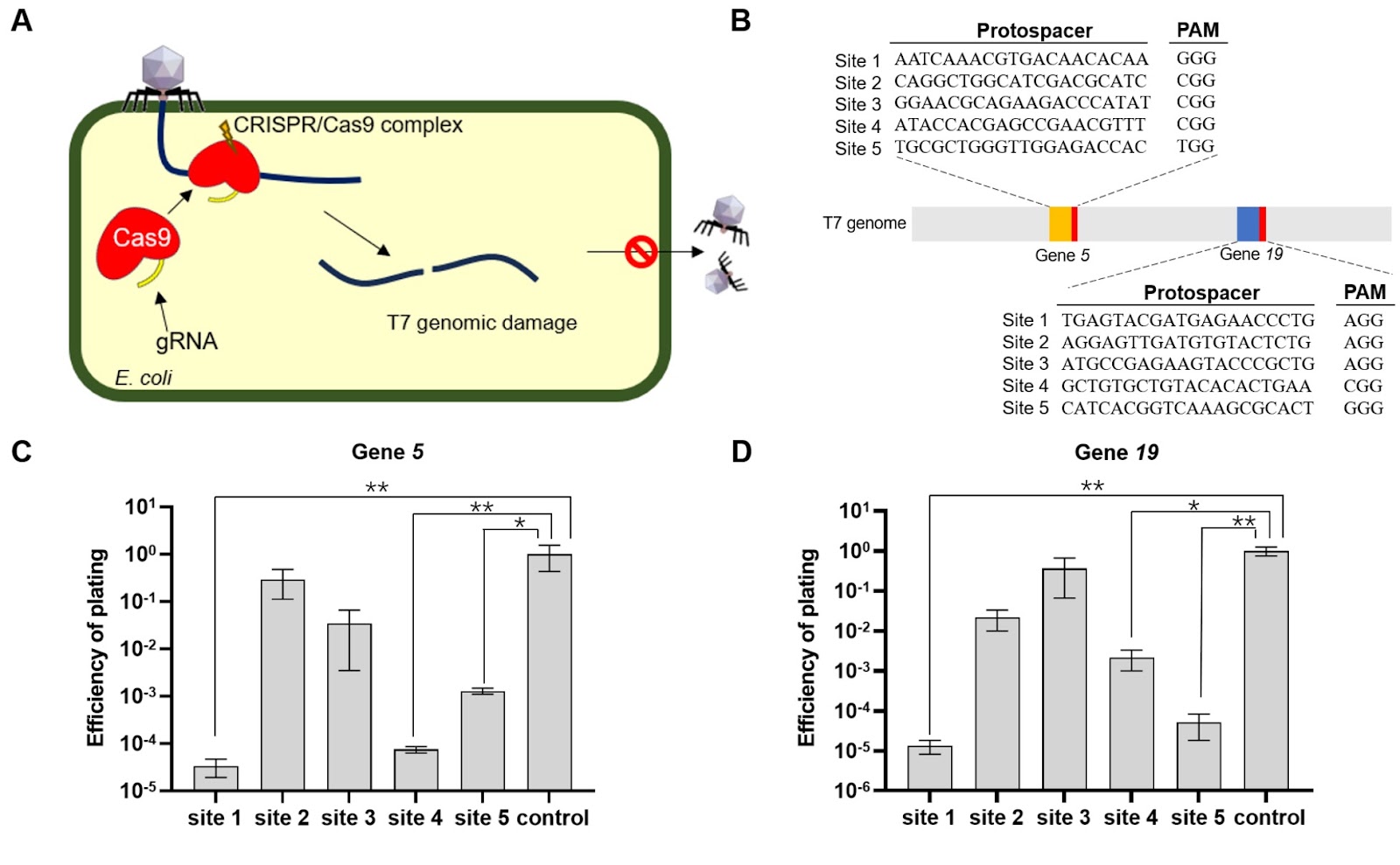
Escherichia coli is widely used in industrial chemical synthesis but faces significant challenges due to bacteriophage contamination, which reduces product quality and yield. Therefore, developing an efficient antiphage system is essential. In this study, we develop a CRISPR-Cas9-based antiphage system (CAPS) targeting essential genes of the T7 phage (gene 5 and gene 19) with single gRNAs transformed into MG1655 strains expressing Cas9. While CAPS provides limited resistance, with plating efficiencies ranging from 10–5 to 10–1, further optimization is needed. To enhance efficacy, we design a double-site-targeting CRISPR-Cas9-based antiphage system (D-CAPS). D-CAPS demonstrates complete resistance, with no plaques observed even at a high multiplicity of infection (MOI of 2), and growth curve analysis reveals that antiphage E. coli strains grow normally, similar to the wild-type strain, even at a high multiplicity of infection. Furthermore, D-CAPS is effective against BL21(DE3) strains, showing strong resistance and demonstrating its versatility across different E. coli strains. Protein expression analysis via green fluorescent protein confirms that E. coli carrying D-CAPS could maintain normal protein expression levels even in the presence of phages, comparable to wild-type strains. Overall, D-CAPS offers a robust and versatile approach to enhancing E. coli resistance to phages, providing a practical solution for protecting industrial E. coli strains and improving fermentation processes.

D-CAPS: an efficient CRISPR-Cas9-based phage defense system for E. coli
Graphical Abstract
Original ResearchVol. 57, Issue 8 • pp. 1252-1259DOI: 10.3724/abbs.2024229

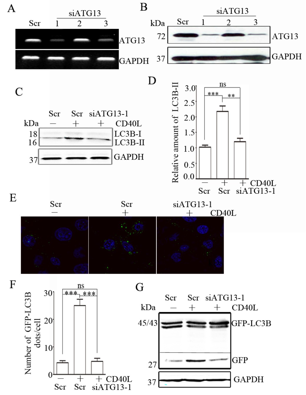
CD40 ligation-induced ERK activation leads to enhanced radiosensitivity in cervical carcinoma cells via promoting autophagy

Authors: Baocai Liu, Yadong Zhang, Quan Wang, Qian Wang, Zhixin Wang, Li Feng

CD40, a member of the tumor necrosis factor (TNF) receptor superfamily, plays an important role not only in the immune system but also in tumor progression. CD40 ligation reportedly promotes autophagy in immune cells. However, the effects of CD40 ligation on autophagy and its mechanism in solid tumor cells are still unclear. In this study, we find that CD40 ligation promotes autophagosome formation and consequently promotes autophagic flux in cervical cancer cells. Mechanistically, this effect relies on ERK contributing to CD40 ligation-induced ATG13 upregulation by p53. Furthermore, we demonstrate that CD40 ligation-induced autophagy increases the radiosensitivity of cervical cancer cells. Taken together, our results provide new evidence for the involvement of the CD40 pathway in autophagy and radiotherapy in cervical cancer cells.

CD40 ligation-induced ERK activation leads to enhanced radiosensitivity in cervical carcinoma cells via promoting autophagy
Graphical Abstract