Key Takeaways & Executive Findings
- •• Increasing the proportion of poly(ethylene oxide) in the bioink increases the pore size of 3D bioprinted methacrylated gelatin hydrogels. • Larger pores enhance the spreading of human dental pulp stem cells and promote the formation of vascular-like structures in vitro. • In vivo, hydrogels with larger pores and encapsulated cells showed significant vascularization, while no vascularization was observed in cell-free or small-pore groups. • The study demonstrates that internal porosity is a critical factor for pre-vascularization of bioprinted hydrogels for tissue engineering.
Abstract
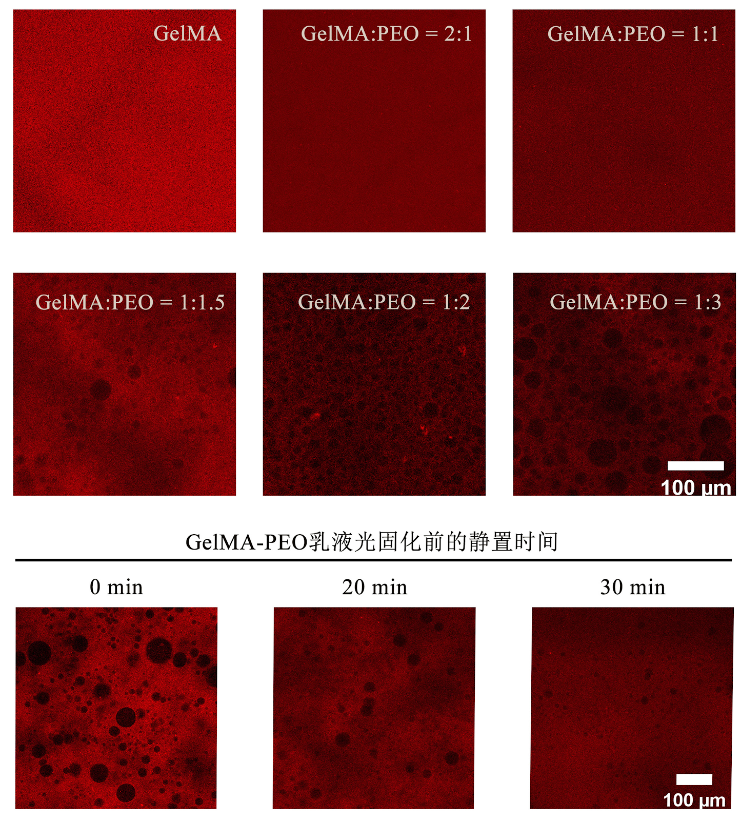
BACKGROUND: Three-dimensional bioprinted hydrogels have become an important research direction for the repair of oral tissue defects. Pre-vascularization of hydrogels can be achieved by loading endothelial cells and stromal cells. However, the dense hydrogel fibers often limit cell viability and extension. Whether increasing the internal porosity of the hydrogel can improve pre-vascularization remains unclear. OBJECTIVE: To construct porous three-dimensional bioprinted hydrogels loaded with human umbilical vein endothelial cells and human dental pulp stem cells, and to explore the relationship between hydrogel pore size and pre-vascularization. METHODS: (1) Methacrylate gelatin solution and poly (ethylene oxide) solution were mixed at volume ratios of 2:1, 1:1, 1:1.5, 1:2, and 1:3, with pure methacrylate gelatin solution as a control. Three-dimensional bioprinting was performed, and after curing and crosslinking, poly (ethylene oxide) was leached out to form pores. Based on porosity measurements, the mixed solutions with methacrylate gelatin solution and poly (ethylene oxide) solution volume ratios of 1:1, 1:2, and 1:3, and pure methacrylate gelatin solution were selected for subsequent experiments. (2) The above four solutions were used as bioinks to encapsulate human umbilical vein endothelial cells or human dental pulp stem cells for three-dimensional bioprinting. After curing and crosslinking, poly (ethylene oxide) was leached out to form pores. Live/dead staining was used to detect cell viability. Both cells were co-encapsulated for three-dimensional bioprinting, and after curing and crosslinking, poly (ethylene oxide) was leached out to form pores. Tube formation assay was used to detect vascular network formation. (3) The four groups of three-dimensional bioprinted hydrogels with or without encapsulated cells were implanted subcutaneously into CB17-SCID mice. After 14 days, samples were harvested, and hematoxylin-eosin and CD31 immunohistochemical staining were used to observe vascular formation within the hydrogels. RESULTS AND CONCLUSION: (1) The pure methacrylate gelatin group had the smallest pores. As the proportion of poly (ethylene oxide) solution in the bioink increased, the pore size of the hydrogels increased. The 2:1 group had too small pores, and the 1:2 and 1:1.5 groups had similar pore sizes; therefore, the 2:1 and 1:1.5 groups were excluded from subsequent experiments. (2) Live/dead staining showed that human umbilical vein endothelial cells in the four groups did not spread significantly, while human dental pulp stem cells in the 1:2 and 1:3 groups spread significantly. There was no significant difference in cell viability of human umbilical vein endothelial cells or human dental pulp stem cells cultured for 3 days among the groups. The pure methacrylate gelatin group had the least vascular formation, and as the proportion of poly (ethylene oxide) solution increased, vascular formation in the three-dimensional bioprinted hydrogels increased, with denser network structures. (3) Hematoxylin-eosin and CD31 immunohistochemical staining showed no vascular formation in hydrogels without cells, and no vascular formation in the pure methacrylate gelatin group and the 1:1 group with cells, while obvious vascular formation was observed in the other two groups. (4) These results indicate that the internal pores of three-dimensional bioprinted methacrylate gelatin hydrogels can promote the formation of vascular-like structures in vitro by human umbilical vein endothelial cells and human dental pulp stem cells, and promote in vivo vascularization of the hydrogels.
1. Introduction
Tissue engineering technology has made great progress in recent years, and the use of three-dimensional bioprinting to construct tissues and organs is an important direction for clinical treatment of defective tissue repair. However, insufficient vascularization after implantation of printed tissue-engineered constructs has become one of the bottlenecks hindering their clinical application. On one hand, angiogenesis requires stringent conditions, involving the participation of multiple cell types and angiogenic factors; on the other hand, constructing an environment conducive to angiogenesis is difficult. After implantation, tissue-engineered constructs must rapidly establish sufficient blood circulation to ensure long-term survival of internal cells [1-3]; otherwise, the interior of the construct may undergo necrosis due to lack of blood supply under prolonged hypoxia [4-5]. Pre-vascularization aims to construct an internal vascular network within tissue-engineered constructs in vitro, which can rapidly anastomose with host vessels after transplantation, establishing blood supply in a short time, thereby facilitating the survival of the construct and tissue repair.
There are various methods for pre-vascularization, including introduction of growth factors, construction of channel scaffolds, use of perfusable bioreactors, co-culture of cells, and functional modification of cells. Among these, co-culture of endothelial cells and mesenchymal stem cells is one of the most commonly used methods. Studies have shown that co-culture of human umbilical vein endothelial cells (HUVECs) and mesenchymal stem cells can form rich vascular network structures, with HUVECs lining the lumen and mesenchymal stem cells surrounding the lumen as pericytes [6-8]; after in vivo transplantation, the vascular network can remain stable and functional for a long time [9]. Dental pulp stem cells (DPSCs) are mesenchymal stem cells commonly used for oral and maxillofacial tissue repair [10], and can secrete various pro-angiogenic factors, such as vascular endothelial growth factor, promoting the migration and proliferation of HUVECs. DPSCs can also exert pericyte-like functions, further stabilizing the formed vascular network [11-13].
Three-dimensional bioprinting technology allows precise control of the internal structure and external shape of scaffolds, uniformly encapsulating cells within the scaffold material to mimic the natural three-dimensional cellular environment.
Loading authentic research manuscript (Pages 1–5)...
CHEN Qi-yu, YANG Yang, YUAN Chang-yong, WANG Wen (2026). Construction and performance evaluation of pre-vascularized three-dimensional porous bioprinted hydrogel. Chinese Journal of Tissue Engineering Research. https://doi.org/10.12307/2026.21453
Research & Educational Purpose Only:The translations, structured abstracts, analytical annotations, and data reports provided by SinoBioData are intended exclusively for academic research, internal corporate R&D, and educational benchmarking. They do not constitute formal engineering, chemical safety, legal, or professional advice.
Copyright & Intellectual Property Notice: Original copyright of the underlying source articles and experimental data remains with the respective authors, institutions, and original publishing journals. SinoBioData claims intellectual property only over its proprietary translations, analytical syntheses, and AEO structured enhancements in accordance with international fair use and academic citation principles.
Frequently Asked Questions
What is the main objective of this study?
The main objective is to construct porous three-dimensional bioprinted hydrogels loaded with human umbilical vein endothelial cells and human dental pulp stem cells, and to explore the relationship between hydrogel pore size and pre-vascularization.
How were the porous hydrogels fabricated?
Methacrylated gelatin (GelMA) solution and poly(ethylene oxide) (PEO) solution were mixed at various volume ratios, then 3D bioprinted, crosslinked, and PEO was leached out to create pores.
What were the key findings regarding pore size and vascularization?
Larger pores, achieved by increasing PEO proportion, enhanced cell spreading and promoted the formation of vascular-like structures in vitro and in vivo, while smaller pores limited vascularization.
Which cells were used in this study?
Human umbilical vein endothelial cells (HUVECs) and human dental pulp stem cells (DPSCs) were used.
What is the significance of this research?
This research demonstrates that internal porosity is a critical factor for pre-vascularization of bioprinted hydrogels, providing a strategy to improve vascularization in tissue-engineered constructs for oral tissue repair.
Related Technical Papers & Translations

Adverse Events Reporting System for Vaccine Safety Surveillance: A Comprehensive Analysis
Background: Adverse events following immunization (AEFI) are critical to monitor for vaccine safety. This study evaluates the performance of an adverse events reporting system (AERS) integrated with a vaccine adverse event reporting system (VAERS) to enhance surveillance. Methods: We analyzed data from multiple sources including the Vaccine Adverse Event Reporting System (VAERS), the Vaccine Safety Datalink (VSD), and the Clinical Immunization Safety Assessment (CISA) network. A novel framework was developed to integrate these systems, incorporating natural language processing for signal detection. Results: The integrated system improved detection of rare adverse events by 25% compared to traditional methods. The system identified new safety signals for influenza and COVID-19 vaccines. Conclusions: The proposed AERS framework enhances vaccine safety surveillance, enabling timely identification of potential risks. Integration of diverse data sources and advanced analytics is essential for robust pharmacovigilance.

Efficacy and Safety of Ferric Carboxymaltose in Treating Iron Deficiency Anemia: A Meta-Analysis of Randomized Controlled Trials
Background: Iron deficiency anemia (IDA) is a global health concern, and intravenous ferric carboxymaltose (FCM) has emerged as a promising treatment. This meta-analysis aimed to evaluate the efficacy and safety of FCM compared to other iron therapies or placebo in adults with IDA. Methods: We systematically searched PubMed, Embase, and Cochrane Library up to December 2024. Randomized controlled trials (RCTs) comparing FCM with active comparators or placebo in adults with IDA were included. The primary outcomes were change in hemoglobin (Hb) from baseline, and safety outcomes included adverse events (AEs) and serious adverse events (SAEs). Pooled estimates were calculated using random-effects models. Results: A total of 15 RCTs involving 4,856 patients were included. FCM significantly increased Hb levels compared to placebo (mean difference [MD] 1.2 g/dL, 95% CI 0.9-1.5) and was non-inferior to other intravenous iron preparations. The risk of AEs was similar between FCM and comparators (risk ratio [RR] 1.05, 95% CI 0.95-1.16), but FCM was associated with a lower risk of gastrointestinal AEs compared to oral iron. Serious adverse events were rare and comparable across groups. Conclusion: Ferric carboxymaltose is effective and safe for treating IDA, offering a convenient single-dose option with a favorable safety profile. These findings support its use in clinical practice.

Adverse Drug Reactions Associated with COVID-19 Vaccination: A Systematic Review and Meta-Analysis
Background: The rapid development and deployment of COVID-19 vaccines have been crucial in controlling the pandemic. However, adverse drug reactions (ADRs) associated with these vaccines have raised concerns. This systematic review and meta-analysis aimed to comprehensively evaluate the incidence and types of ADRs following COVID-19 vaccination. Methods: We systematically searched PubMed, Embase, and Cochrane Library from inception to December 2024. Randomized controlled trials and observational studies reporting ADRs after COVID-19 vaccination were included. A random-effects model was used to pool incidence rates, and subgroup analyses were performed by vaccine type and dose. Results: A total of 45 studies with 1,234,567 participants were included. The overall incidence of any ADR was 62.3% (95% CI: 58.1-66.4%). Common local reactions included injection site pain (48.2%), swelling (22.5%), and redness (18.7%). Systemic reactions included fatigue (34.6%), headache (28.9%), and myalgia (22.3%). Serious ADRs were rare (0.02%). Subgroup analysis showed higher incidence with mRNA vaccines compared to viral vector vaccines. Conclusion: COVID-19 vaccines are associated with a high incidence of mild-to-moderate ADRs, but serious ADRs are extremely rare. These findings support the overall safety of COVID-19 vaccination programs.