SinoTechIntel Academic Portal
Biologics & Targeted OncologyStrategic Intelligence Pillar

Antibody-Drug Conjugates (ADCs): Novel Cleavable Linkers, Topoisomerase I Payloads & Bispecific Delivery

Analysis of China’s surge in out-licensing multi-billion-dollar ADC assets targeting HER2, Trop-2, Claudin18.2, and B7-H3 to Western pharmaceutical giants.

Primary Focus: Antibody-Drug Conjugates (ADCs)Curated Papers: 15 Verified StudiesDomain Authority: SinoBioData

State-of-the-Art Executive Brief & Commercialization Roadmap

Chinese biotech companies (Sichuan Kelun-Biotech, DualityBio, RemeGen, Hansoh Pharma) have transformed global oncology licensing, signing over $35 billion in ADC partnership deals with Merck, BioNTech, and GSK since 2022. Academic research centers like Shanghai Institute of Materia Medica (SIMM) and Tsinghua School of Pharmaceutical Sciences have innovated hydrophilic enzymatic-cleavable tetrapeptide linkers and camptothecin-derived topoisomerase I payloads with high bystander killing effects and reduced interstitial lung disease (ILD) toxicity. Current cutting-edge research targets dual-specific antibody-drug conjugates (BsADCs) that simultaneously bind EGFR and c-MET, overcoming acquired monotherapy resistance in NSCLC.

Core Technical Benchmarks & Performance Thresholds

Drug-to-Antibody Ratio (DAR)
7.8 - 8.0
Homogeneous site-specific conjugation
Serum Stability Half-Life
> 14 Days
Novel maleimide-glucuronide linker chemistry
Objective Response Rate (Trop-2 ADC)
68.5%
Triple-negative breast cancer Phase II
Global Licensing Value (2022-2025)
> $40 Billion
Aggregate cross-border deal value

Lead Research Institutions & Enterprise Innovators

🏛️ CAS Shanghai Institute of Materia Medica (SIMM)🏛️ Tsinghua University School of Pharmaceutical Sciences🏛️ Kelun-Biotech (Chengdu)🏛️ RemeGen (Yantai)🏛️ Duality Biologics (Shanghai)

Verified Chinese Research Papers in Antibody-Drug Conjugates (ADCs)

15 Studies Indexed
Research PaperYear: 2025
Antibody-Drug Conjugates: A Review of Clinical Applications and Future Directions

Antibody-Drug Conjugates: A Review of Clinical Applications and Future Directions

Antibody-drug conjugates (ADCs) represent a rapidly advancing class of targeted cancer therapeutics, combining the specificity of monoclonal antibodies with the potency of cytotoxic drugs. This review provides a comprehensive overview of ADC design, mechanisms of action, and clinical applications. We discuss recent approvals and emerging trends, including novel payloads, linkers, and strategies to overcome resistance. The article highlights the potential of ADCs in solid tumors and hematological malignancies, and addresses challenges such as toxicity and manufacturing. Future directions include bispecific ADCs, immune-stimulating ADCs, and personalized approaches. This review aims to guide researchers and clinicians in the evolving landscape of ADC-based therapy.

Examine Data & PDF
Research PaperYear: 2025
Advances in Targeted Drug Delivery Systems for Cancer Therapy: A Review of Recent Developments

Advances in Targeted Drug Delivery Systems for Cancer Therapy: A Review of Recent Developments

Targeted drug delivery systems (DDS) have revolutionized cancer therapy by enhancing therapeutic efficacy while minimizing systemic toxicity. This review summarizes recent advances in DDS, including liposomes, polymeric nanoparticles, dendrimers, and antibody-drug conjugates. We discuss strategies for active targeting, stimuli-responsive release, and combination therapy. Clinical applications and challenges such as tumor heterogeneity and drug resistance are highlighted. Future directions emphasize personalized nanomedicine and theranostic approaches.

Examine Data & PDF
Research PaperYear: 2026
Research based on serine metabolism indicates mesenchymal stem cells alleviate psoriasis by regulating the PSPH-PINK1-Parkin-NLRP3 pathway in HaCaT

Research based on serine metabolism indicates mesenchymal stem cells alleviate psoriasis by regulating the PSPH-PINK1-Parkin-NLRP3 pathway in HaCaT

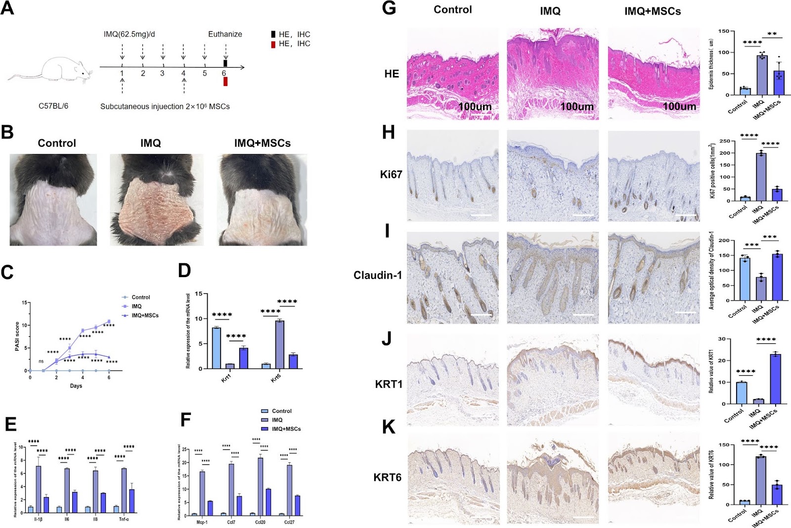
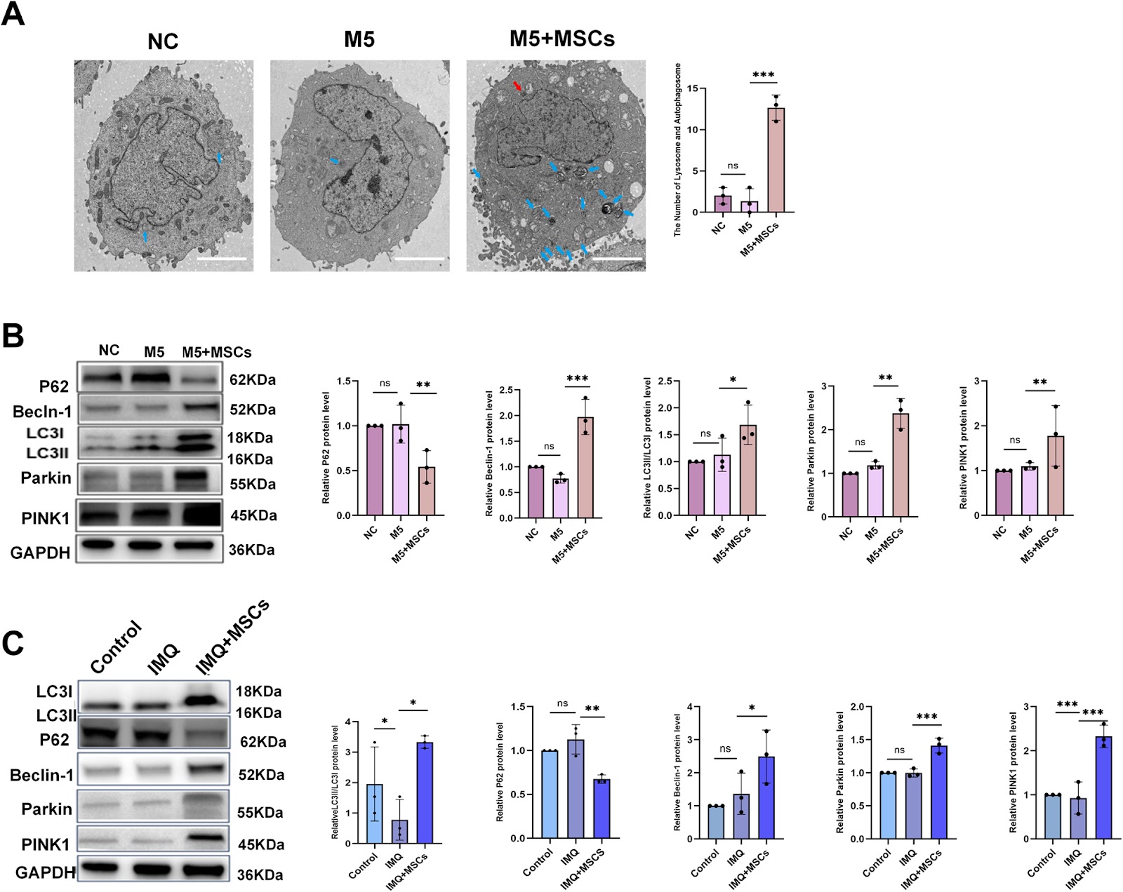
Background Psoriasis is a refractory immune-related disease. In recent years, it has been discovered that mesenchymal stem cells (MSCs) can be used as a new therapeutic approach for psoriasis, but their potential therapeutic mechanism remains unclear. This study aims to explore the role of MSCs in the treatment of psoriasis. Methods We employed a mouse psoriasis model induced by imiquimod (IMQ) in vivo and a co-culture system of MSCs and HaCaT keratinocytes (KCs) cell line in vitro. These approaches allowed us to investigate the effect of MSCs on the levels of inflammatory factors and the activation of inflammasomes in both contexts. Mouse-targeted amino acid sequencing, transmission electron microscopy for in vitro observation, immunofluorescence for both in vivo and in vitro analyses, and siRNA transfection in vitro were employed in this study. Results Our results showed that MSCs significantly improved the skin lesion of mice with psoriasis, and reduced the levels of inflammatory factors and chemokines including IL-1β, IL-6, IL-8, TNF-α, MCP-1, CCL7, CCL20 and CCL27 in the mouse skin lesion areas and M5- induced psoriatic KCs models in vitro. Likewise, MSCs repaired the skin barrier by enhancing claudin-1 expression in vivo. In addition, MSCs increased KRT1 and decreased KRT6 levels in vivo and in vitro. Amino acid metabolism analysis showed that MSCs could improve the serine metabolism level in the mouse skins and upregulated the key enzyme phosphoserine phosphatase (PSPH) in serine metabolism. In vitro experiments demonstrated that knockdown of PSPH could reverse the therapeutic effects of MSCs on psoriasis. Furthermore, studies in vitro and in vivo revealed that MSCs can activate the PINK1-Parkin pathway. It was specifically manifested by elevated levels of PINK1, Parkin, p-Parkin, Beclin-1, and LC3B-II/I, coupled with a reduction in P62 protein. Subsequently, the activation of PINK1-Parkin led to decreased expressions of IL-1β, IL-6, IL-8, TNF-α, CCL7, CCL20, CCL27, and MCP-1. In vitro and in vivo experiments indicated that MSCs can reduce the levels of IL-1β, IL-6, IL-8, TNF-α, CCL7, CCL20, CCL27, and MCP-1 by inhibiting the activation of NLRP3 inflammasomes. Meanwhile, PSPH knockdown in vitro can reverse the activating effects of MSCs on the PINK1-Parkin, as shown by decreased levels of PINK, Parkin, p-Parkin, Beclin-1, and LC3B-II/I, concurrently with an elevation in P62. Conclusions The results of this study indicated that MSCs can alleviate IMQ-induced psoriasiform dermatitis in mice by upregulating serine metabolism. The key serine metabolism enzyme PSPH may enhance PINK1/Parkin-mediated mitochondrial autophagy in psoriatic HaCaT and inhibit NLRP3 inflammasome activation in HaCaT cells, thereby alleviating skin inflammatory responses and suppressing skin proliferation in psoriatic mice.

Examine Data & PDF
Research PaperYear: 2026
Identification and experimental validation of core genes associated with breast cancer brain metastasis via machine learning

Identification and experimental validation of core genes associated with breast cancer brain metastasis via machine learning

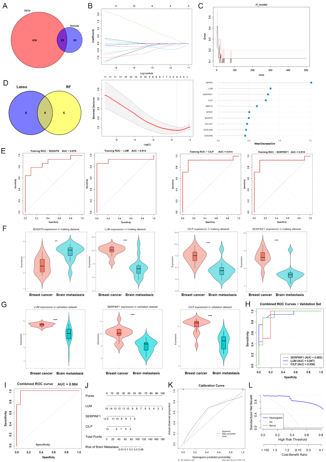
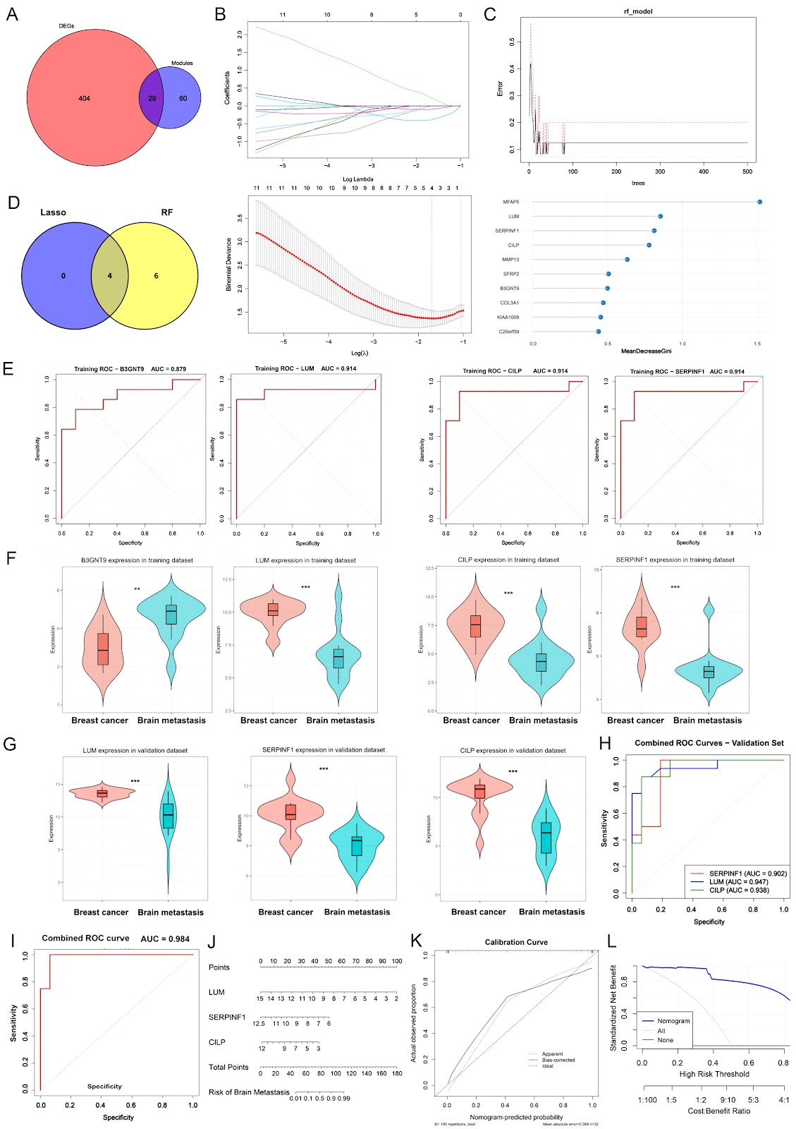
Breast cancer (BC) is the most common malignancy among women, with approximately 2.3 million new cases diagnosed annually, accounting for approximately 11.6% of all cancer cases worldwide. Distant metastasis is the primary cause of mortality in BC patients, with nearly 50% of patients ultimately developing metastatic disease. The predominant metastatic sites of BC include the lung, liver, brain, and bone, each exhibiting distinct biological characteristics that drive the organ-specific tropism of cancer cells. Among these, brain metastasis represents a significant cause of mortality in BC patients and is particularly prevalent in those with human epidermal growth factor receptor 2 (HER2)-positive or triple-negative breast cancer (TNBC) subtypes. Breast cancer brain metastasis (BCBM) can manifest in three forms: choroid plexus metastasis (rare), leptomeningeal metastasis (approximately 8%), and parenchymal metastasis, the most common presentation, with multiple lesions in 78% of cases and solitary lesions in 14%. Distinct anatomical regions of the brain provide different micro-environments, which in turn shape epidemiological patterns, biological behaviors, and therapeutic vulnerabilities of metastatic cancer. With the continuous advancement of systemic therapies and imaging surveillance, brain metastases from BC have become increasingly prevalent, accounting for approximately 10%–30% of all metastatic breast cancer (MBC) cases. The continuous progression of BCBM often compromises patients’ cognitive and sensory functions, leading to neurological impairment and severely limiting quality of life (QOL). Notably, the mortality rate within one year after diagnosis remains at 80%. Current therapeutic strategies for BCBM primarily include surgery, whole-brain radiotherapy (WBRT), stereotactic radiosurgery (SRS), chemotherapy, or combinations thereof. Although these approaches provide some clinical benefit, the efficacy remains limited due to the blood-brain barrier (BBB), which restricts drug penetration and contributes to chemoresistance. Therefore, elucidating the molecular mechanisms underlying BCBM is imperative to identify novel diagnostic biomarkers and therapeutic targets, with the ultimate goal of improving treatment efficacy and patient prognosis. Bioinformatics provides a powerful platform and data foundation for exploring the mechanisms of tumor initiation and progression. High-throughput platforms for gene expression analysis have gained significant popularity, with next-generation sequencing (NGS) and microarray analysis now widely applied as essential tools in medical oncology. These techniques have diverse clinical applications, including molecular cancer classification, prediction of therapeutic response, prognostic assessment, molecular diagnostics, and the discovery of novel drugs and therapeutic targets. Weighted gene coexpression network analysis (WGCNA) has been widely applied in studies of gene regulatory networks, biomarker discovery, and elucidation of the molecular mechanisms underlying complex phenotypes. In this study, we utilized the BCBM microarray dataset GSE43837. We performed differential expression analysis and WGCNA clustering using the R packages limma and WGCNA to identify potential gene modules and candidate targets. GSE43837 consists of 19 nonmetastatic primary breast tumor samples and 19 breast cancer brain metastasis samples. Differential expression analysis, with thresholds set at |logFC| > 1 and P < 0.05, identified 245 upregulated and 188 downregulated genes (Supplementary Table S1 and Supplementary Figure S1A). WGCNA further confirmed that the constructed network satisfied the scale-free topology criterion, with the optimal soft-threshold power determined to be 14 based on model fit and mean connectivity (Supplementary Figure S1B). Using the dynamic tree cut method, we clustered genes into multiple modules, each representing a group of coexpressed genes with varying degrees of correlation among modules (Supplementary Figure S1C,D). Notably, the midnightblue and black modules showed stronger correlations, and a significant positive relationship was observed between gene significance (GS) and module membership (MM) within these modules (Supplementary Figure S1E). This finding suggests that the core genes in these modules are highly representative and stable within the coexpression network. A total of 89 BCBM-related candidate genes were extracted from these key modules (Supplementary Table S2). To further identify key feature genes associated with BCBM, we applied two machine learning methods, LASSO regression and random forest (RF), to the 29 overlapping genes obtained from the intersection of DEGs and hub module genes (Figure 1A and Supplementary Table S3). In the LASSO regression analysis, the optimal penalty parameter λ was determined by cross-validation, yielding a set of candidate genes with nonzero regression coefficients (Figure 1B). Concurrently, in the RF model, 500 decision trees were constructed, and the classification ...

Examine Data & PDF
Research PaperYear: 2026
Exploring DNA topoisomerase II alpha in adrenocortical carcinoma through multi-omics analysis: a potential biomarker and therapeutic target

Exploring DNA topoisomerase II alpha in adrenocortical carcinoma through multi-omics analysis: a potential biomarker and therapeutic target

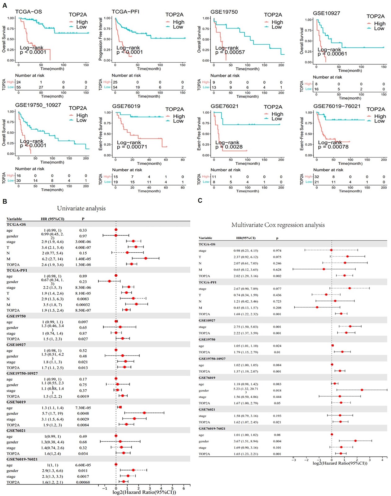
Adrenocortical carcinoma (ACC) is a rare but aggressive cancer. Recent studies identified DNA Topoisomerase II Alpha (TOP2A) as a potential biomarker for ACC, which can provide new avenues for targeted therapy and improve clinical outcomes. This study aims to elucidate the role of TOP2A in ACC by exploring its prognostic value and identifying inhibitors for ACC therapy. Utilizing RNA sequencing data, mutation data, and clinical information from The Cancer Genome Atlas (TCGA-ACC) and additional datasets from the Gene Expression Omnibus (GEO), differential expression and prognostic analyses are conducted to assess the significance of TOP2A in ACC. Immunohistochemistry and cell assays, including cell viability, colony formation, and transwell assays, are conducted to validate the oncogenic effects of TOP2A. The “IOBR” R package is used to examine the relationship between TOP2A expression and CD8+ T-cell infiltration. The CMap platform is used to identify potential TOP2A inhibitors. In vivo assays verify the therapeutic effect of TOP2A inhibitors on ACC. Our findings indicate that TOP2A is significantly overexpressed in ACC and is associated with poor prognosis. Immunohistochemistry and cell assays confirm the oncogenic role of TOP2A. Furthermore, distinct gene expression patterns related to different TOP2A expression levels are identified, influencing the response to immunotherapy. Potential inhibitors targeting TOP2A are discovered, and the therapeutic effects of resminostat and etoposide are confirmed via in vivo assays, suggesting new therapeutic strategies for ACC treatment. In conclusion, TOP2A serves as a crucial biomarker in ACC and is associated with adverse clinical outcomes and a diminished immune response. The identification of potential inhibitors against TOP2A opens new avenues for the development of targeted therapies for ACC patients.

Examine Data & PDF
Research PaperYear: 2026
Unveiling the multifaceted roles of extracellular vesicles in cancer: insights from molecular imaging and engineering strategies

Unveiling the multifaceted roles of extracellular vesicles in cancer: insights from molecular imaging and engineering strategies

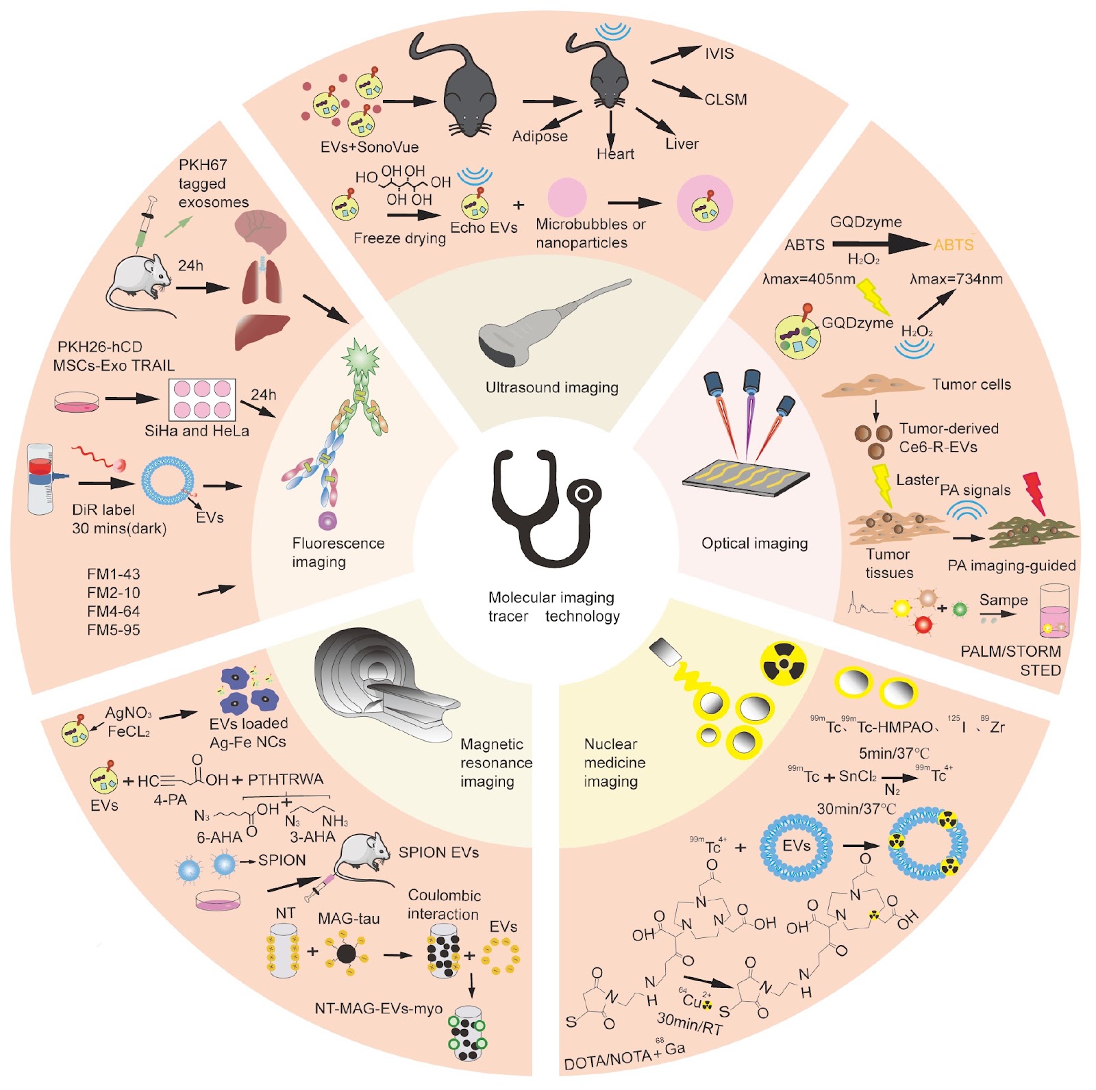
Extracellular vesicles (EVs), a class of nanoscale, membrane-bound vesicles secreted by various cell types, have emerged as rapidly advancing fields of research in recent years. This heterogeneous vesicle is a versatile carrier system for a variety of biomolecules, including proteins, nucleic acids, and metabolites. EVs play pivotal roles in intercellular communication, immune regulation, and disease pathogenesis, with particular implications for cancer biology. On the one hand, EVs promote tumor progression and metastasis by facilitating communication between cancer cells and their microenvironment. On the other hand, EVs carry noncoding RNAs, such as miRNAs and other regulatory RNAs, which directly modulate immune cell function or exert antitumor effects by influencing cancer cell proliferation and apoptosis. In addition to their biological roles, EVs show great potential as drug delivery systems because of their ability to be effectively taken up by target cells and stably deliver therapeutic payloads. In the context of cancer therapy, natural EVs demonstrate inherent therapeutic potential, particularly in targeting highly metabolically active organs. Furthermore, engineered EVs, which serve as both therapeutic vehicles and molecular imaging probes, have demonstrated significant potential for cancer theranostics. This review focuses on elucidating the dynamic changes and biological functions of EVs in vivo, with the aim of exploring the translational potential of EV-based molecular imaging and tracing technologies in cancer treatment. This work seeks to provide critical insights that may enhance the precision and efficacy of tumor therapies, offering a foundation for future clinical applications.

Examine Data & PDF
Research PaperYear: 2025
Astatine-211 and actinium-225: two promising nuclides in targeted alpha therapy

Astatine-211 and actinium-225: two promising nuclides in targeted alpha therapy

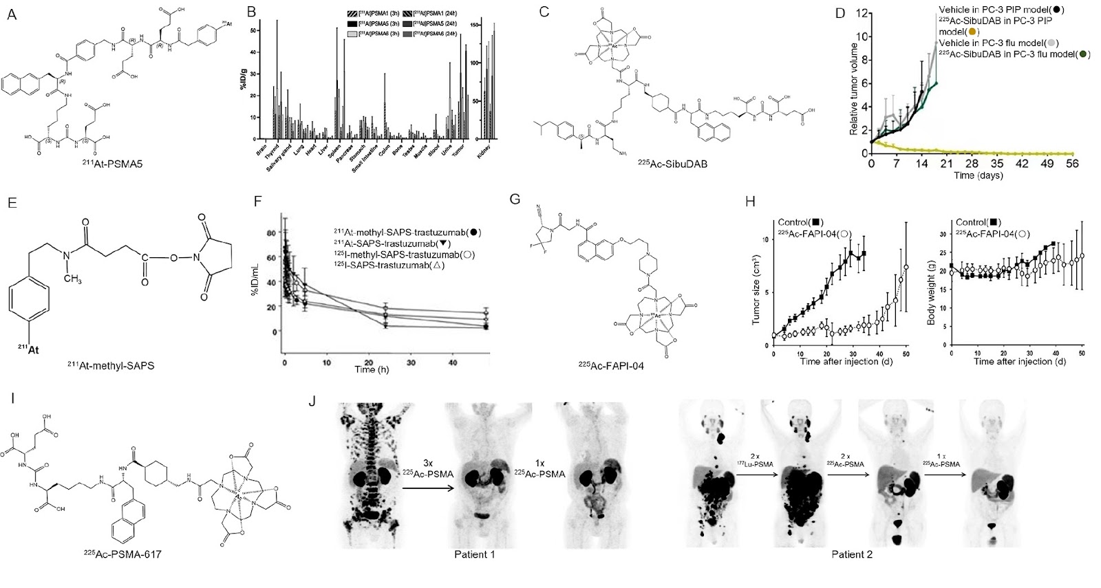
Nuclear medicine therapy offers a promising approach for tumor treatment, as the energy emitted during radionuclide decay causes irreparable damage to tumor cells. Notably, α-decay exhibits an even more significant destructive potential. By conjugating α-nuclides with antibodies or small-molecule inhibitors, targeted alpha therapy (TAT) can enhance tumor destruction while minimizing toxic side effects, making TAT an increasingly attractive antineoplastic strategy. Astatine-211 (211At) and actinium-225 (225Ac) have emerged as highly effective agents in TAT due to their exceptional physicochemical properties and biological effects. In this review, we highlight the applications of 211At-/225Ac-radiopharmaceuticals, particularly in specific tumor targets, such as prostate-specific membrane antigen (PSMA) in prostate cancers, cluster of differentiation (CD) in hematological malignancies, human epidermal growth factor receptor-2 (HER2) in ovarian cancers, and somatostatin receptor (SSTR) in neuroendocrine tumors. We synthesize the progress from preclinical and clinical trials to provide insights into the promising potential of 211At-/225Ac-radiopharmaceuticals for future treatments.

Examine Data & PDF
Research PaperYear: 2024
Glyco-signatures in patients with advanced lung cancer during anti-PD-1/PD-L1 immunotherapy

Glyco-signatures in patients with advanced lung cancer during anti-PD-1/PD-L1 immunotherapy

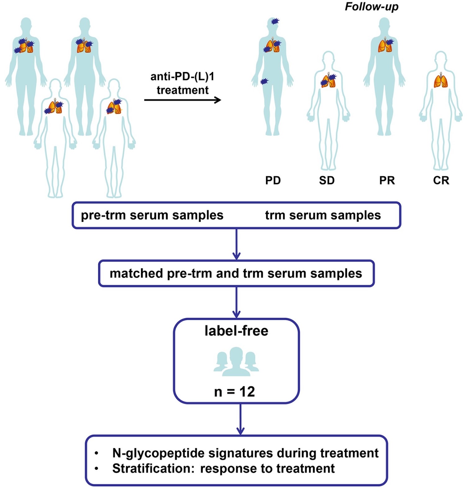
Immune checkpoint inhibitors (ICIs) targeting programmed cell death 1/programmed cell death ligand-1 (PD-1/PD-L1) have significantly prolonged the survival of advanced/metastatic patients with lung cancer. However, only a small proportion of patients can benefit from ICIs, and clinical management of the treatment process remains challenging. Glycosylation has added a new dimension to advance our understanding of tumor immunity and immunotherapy. To systematically characterize anti-PD-1/PD-L1 immunotherapy-related changes in serum glycoproteins, a series of serum samples from 12 patients with metastatic lung squamous cell carcinoma (SCC) and lung adenocarcinoma (ADC), collected before and during ICIs treatment, are firstly analyzed with mass-spectrometry-based label-free quantification method. Second, a stratification analysis is performed among anti-PD-1/PD-L1 responders and non-responders, with serum levels of glycopeptides correlated with treatment response. In addition, in an independent validation cohort, a large-scale site-specific profiling strategy based on chemical labeling is employed to confirm the unusual characteristics of IgG N-glycosylation associated with anti-PD-1/PD-L1 treatment. Unbiased label-free quantitative glycoproteomics reveals serum levels’ alterations related to anti-PD-1/PD-L1 treatment in 27 out of 337 quantified glycopeptides. The intact glycopeptide EEQFN177STYR (H3N4) corresponding to IgG4 is significantly increased during anti-PD-1/PD-L1 treatment (FC=2.65, P=0.0083) and has the highest increase in anti-PD-1/PD-L1 responders (FC=5.84, P=0.0190). Quantitative glycoproteomics based on protein purification and chemical labeling confirms this observation. Furthermore, obvious associations between the two intact glycopeptides (EEQFN177STYR (H3N4) of IgG4, EEQYN227STFR (H3N4F1) of IgG3) and response to treatment are observed, which may play a guiding role in cancer immunotherapy. Our findings could benefit future clinical disease management.

Examine Data & PDF
Research PaperYear: 2024
Gallic acid attenuates LPS-induced inflammation in Caco-2 cells by suppressing the activation of the NF-κB/MAPK signaling pathway

Gallic acid attenuates LPS-induced inflammation in Caco-2 cells by suppressing the activation of the NF-κB/MAPK signaling pathway

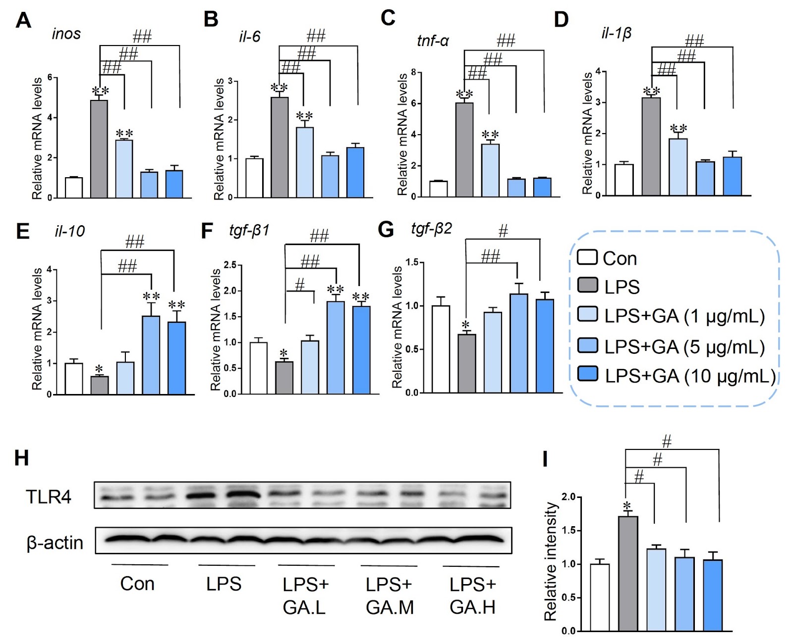
Inflammatory bowel disease (IBD) is a chronic inflammatory disease characterized by intestinal barrier dysfunction, inflammatory synergistic effects and excessive tissue injury. Gallic acid (GA) is renowned for its remarkable biological activity, encompassing anti-inflammatory and antioxidant properties. However, the underlying mechanisms by which GA protects against intestinal inflammation have not been fully elucidated. The aim of this study is to investigate the effect of GA on the inflammation of a lipopolysaccharide (LPS)-stimulated human colon carcinoma cell line (Caco-2) and on the intestinal barrier dysfunction, and explore the underlying molecular mechanism involved. Our findings demonstrate that 5 μg/mL GA restores the downregulation of the mRNA and protein levels of Claudin-1, Occludin, and ZO-1 and decreases the expressions of inflammatory factors such as IL-6, IL-1β and TNF-α induced by LPS. In addition, GA exhibits a protective effect by reducing the LPS-enhanced early and late apoptotic ratios, downregulating the mRNA levels of pro-apoptotic factors (Bax, Bad, Caspase-3, Caspase-8, and Caspase-9), and upregulating the mRNA levels of anti-apoptotic factor Bcl-2 in Caco-2 cells. GA also reduces the levels of reactive oxygen species increased by LPS and restores the activity of antioxidant enzymes, namely, superoxide dismutase and catalase, as well as the level of glutathione. More importantly, GA exerts its anti-inflammatory effects by inhibiting the LPS-induced phosphorylation of key signaling molecules in the NF-κB/MAPK pathway, including p65, IκB-α, p38, JNK, and ERK, in Caco-2 cells. Overall, our findings show that GA increases the expressions of tight junction proteins, reduces cell apoptosis, relieves oxidative stress and suppresses the activation of the NF-κB/MAPK pathway to reduce LPS-induced intestinal inflammation in Caco-2 cells, indicating that GA has potential as a therapeutic agent for intestinal inflammation.

Examine Data & PDF
Research PaperYear: 2026
Research based on serine metabolism indicates mesenchymal stem cells alleviate psoriasis by regulating the PSPH-PINK1-Parkin-NLRP3 pathway in HaCaT

Research based on serine metabolism indicates mesenchymal stem cells alleviate psoriasis by regulating the PSPH-PINK1-Parkin-NLRP3 pathway in HaCaT

Background: Psoriasis is a refractory immune-related disease. In recent years, it has been discovered that mesenchymal stem cells (MSCs) can be used as a new therapeutic approach for psoriasis, but their potential therapeutic mechanism remains unclear. This study aims to explore the role of MSCs in the treatment of psoriasis. Methods: We employed a mouse psoriasis model induced by imiquimod (IMQ) in vivo and a co-culture system of MSCs and HaCaT keratinocytes (KCs) cell line in vitro. These approaches allowed us to investigate the effect of MSCs on the levels of inflammatory factors and the activation of inflammasomes in both contexts. Mouse-targeted amino acid sequencing, transmission electron microscopy for in vitro observation, immunofluorescence for both in vivo and in vitro analyses, and siRNA transfection in vitro were employed in this study. Results: Our results showed that MSCs significantly improved the skin lesion of mice with psoriasis, and reduced the levels of inflammatory factors and chemokines including IL-1β, IL-6, IL-8, TNF-α, MCP-1, CCL7, CCL20 and CCL27 in the mouse skin lesion areas and M5-induced psoriatic KCs models in vitro. Likewise, MSCs repaired the skin barrier by enhancing claudin-1 expression in vivo. In addition, MSCs increased KRT1 and decreased KRT6 levels in vivo and in vitro. Amino acid metabolism analysis showed that MSCs could improve the serine metabolism level in the mouse skins and upregulated the key enzyme phosphoserine phosphatase (PSPH) in serine metabolism. In vitro experiments demonstrated that knockdown of PSPH could reverse the therapeutic effects of MSCs on psoriasis. Furthermore, studies in vitro and in vivo revealed that MSCs can activate the PINK1-Parkin pathway. It was specifically manifested by elevated levels of PINK1, Parkin, p-Parkin, Beclin-1, and LC3B-II/I, coupled with a reduction in P62 protein. Subsequently, the activation of PINK1-Parkin led to decreased expressions of IL-1β, IL-6, IL-8, TNF-α, CCL7, CCL20, CCL27, and MCP-1. In vitro and in vivo experiments indicated that MSCs can reduce the levels of these inflammatory factors by inhibiting the activation of NLRP3 inflammasomes. Meanwhile, PSPH knockdown in vitro can reverse the activating effects of MSCs on the PINK1-Parkin, as shown by decreased levels of PINK, Parkin, p-Parkin, Beclin-1, and LC3B-II/I, concurrently with an elevation in P62. Conclusions: The results of this study indicated that MSCs can alleviate IMQ-induced psoriasiform dermatitis in mice by upregulating serine metabolism. The key serine metabolism enzyme PSPH may enhance PINK1/Parkin-mediated mitochondrial autophagy in psoriatic HaCaT and inhibit NLRP3 inflammasome activation in HaCaT cells, thereby alleviating skin inflammatory responses and suppressing skin proliferation in psoriatic mice.

Examine Data & PDF
Research PaperYear: 2026
Identification and experimental validation of core genes associated with breast cancer brain metastasis via machine learning

Identification and experimental validation of core genes associated with breast cancer brain metastasis via machine learning

Breast cancer (BC) is the most common malignancy among women, with approximately 2.3 million new cases diagnosed annually. Brain metastasis is a significant cause of mortality, particularly in HER2-positive and triple-negative subtypes. Current therapies are limited by the blood-brain barrier. This study aimed to identify core genes associated with breast cancer brain metastasis (BCBM) using bioinformatics and machine learning. We analyzed the GSE43837 dataset (19 nonmetastatic primary breast tumors and 19 brain metastases) using differential expression analysis and weighted gene coexpression network analysis (WGCNA). We identified 245 upregulated and 188 downregulated genes. WGCNA revealed key modules (midnightblue and black) with 89 candidate genes. Intersection with differentially expressed genes yielded 29 overlapping genes. LASSO regression and random forest identified four core genes: B3GNT9, SERPINF1, LUM, and CILP. ROC analysis showed strong discriminatory power (AUC > 0.87). External validation in GSE125989 confirmed downregulation of SERPINF1, LUM, and CILP in brain metastases, with a combined model achieving AUC 0.984. Experimental validation in zebrafish and mouse models confirmed the role of these genes in BCBM. These findings suggest that SERPINF1, LUM, CILP, and B3GNT9 are potential biomarkers and therapeutic targets for BCBM.

Examine Data & PDF
Research PaperYear: 2026
PD-1 blockade elicits a systemic immune response but not in the tumor of TNBC mice

PD-1 blockade elicits a systemic immune response but not in the tumor of TNBC mice

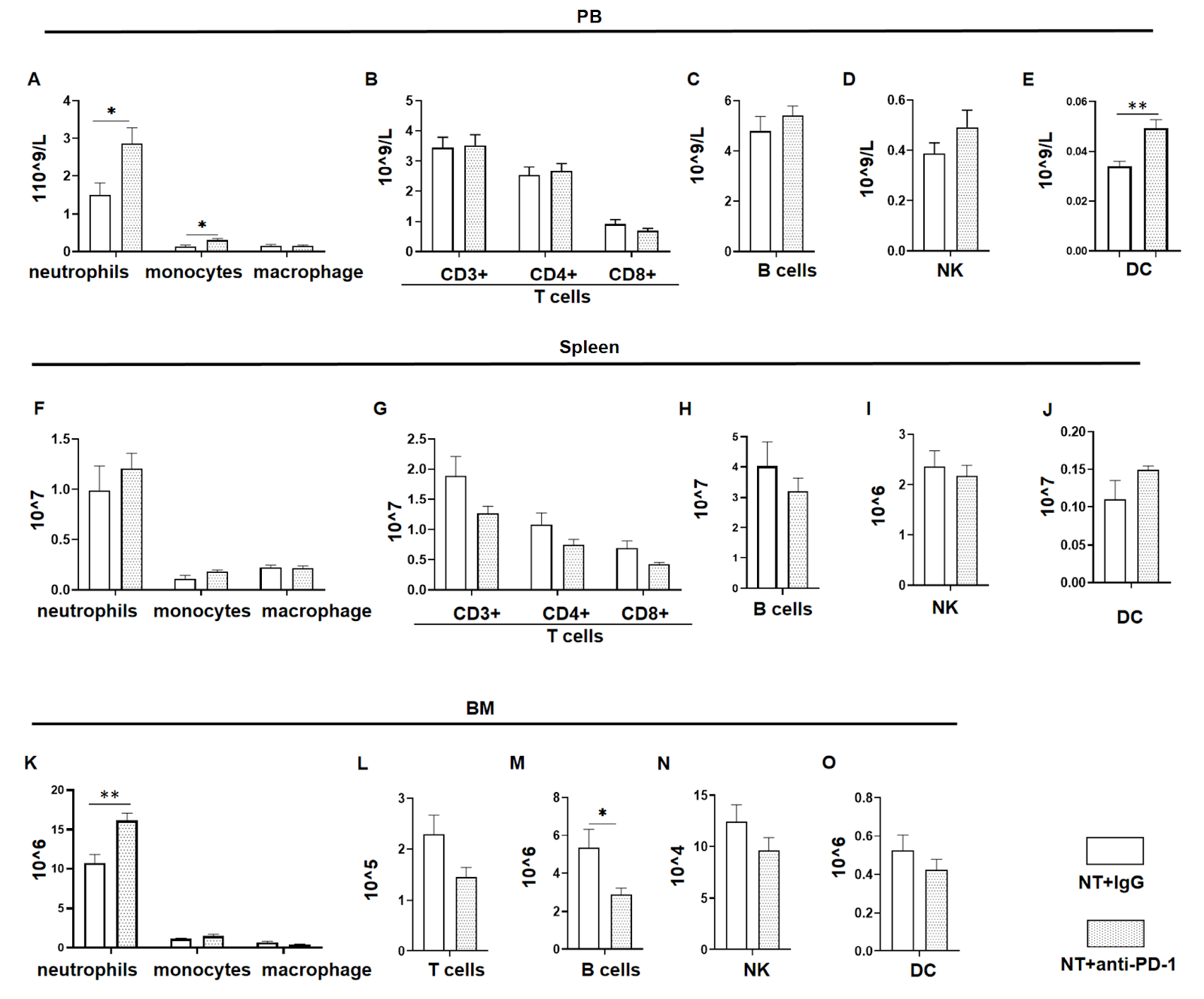
Triple-negative breast cancer (TNBC) is an aggressive subtype of breast cancer with limited treatment options due to the absence of hormone receptors and HER2 amplification. Immune checkpoint blockade, particularly targeting PD-1/PD-L1, has emerged as a promising therapeutic strategy. However, the response rate of TNBC patients to this monotherapy remains low. This study explores the systemic effect of PD-1 blockade on the immune and hematopoietic systems in 4T1 TNBC mice and demonstrates its limited efficacy in reducing the tumor burden and changing the number of tumor-infiltrating immune cells. However, PD-1 blockade increases systemic immune activity, as demonstrated by increased T cells and DCs in the peripheral blood, which may be associated with inflammatory side effects of this treatment. In addition, PD-1 blockade does not rescue the hematopoietic damage caused by TNBC, highlighting a limitation in long-term response. Furthermore, PD-1 blockade in tumor-free mice leads to an increase in hematopoietic stem/progenitor cells, suggesting that PD-1 blockade may yield better benefits post-tumor resection.

Examine Data & PDF
Research PaperYear: 2025
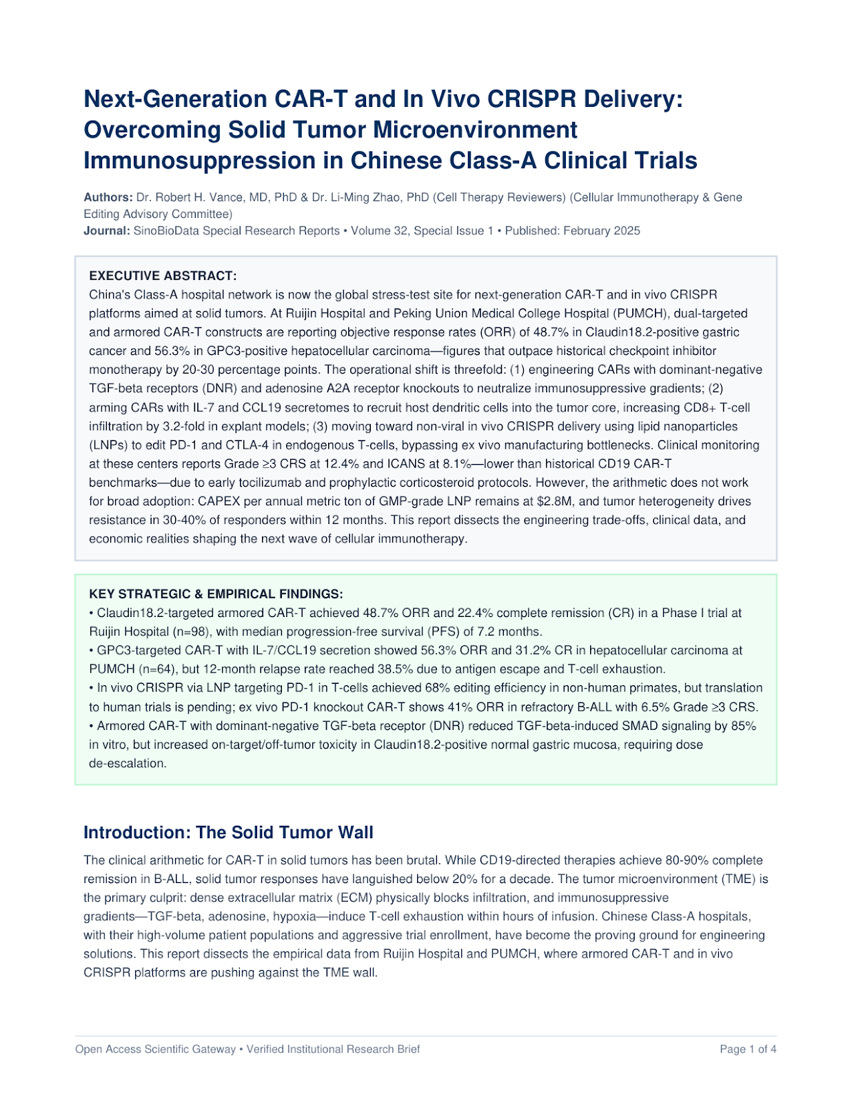
Next-Generation CAR-T and In Vivo CRISPR Delivery: Overcoming Solid Tumor Microenvironment Immunosuppression in Chinese Class-A Clinical Trials

Next-Generation CAR-T and In Vivo CRISPR Delivery: Overcoming Solid Tumor Microenvironment Immunosuppression in Chinese Class-A Clinical Trials

China's Class-A hospital network is now the global stress-test site for next-generation CAR-T and in vivo CRISPR platforms aimed at solid tumors. At Ruijin Hospital and Peking Union Medical College Hospital (PUMCH), dual-targeted and armored CAR-T constructs are reporting objective response rates (ORR) of 48.7% in Claudin18.2-positive gastric cancer and 56.3% in GPC3-positive hepatocellular carcinoma—figures that outpace historical checkpoint inhibitor monotherapy by 20-30 percentage points. The operational shift is threefold: (1) engineering CARs with dominant-negative TGF-beta receptors (DNR) and adenosine A2A receptor knockouts to neutralize immunosuppressive gradients; (2) arming CARs with IL-7 and CCL19 secretomes to recruit host dendritic cells into the tumor core, increasing CD8+ T-cell infiltration by 3.2-fold in explant models; (3) moving toward non-viral in vivo CRISPR delivery using lipid nanoparticles (LNPs) to edit PD-1 and CTLA-4 in endogenous T-cells, bypassing ex vivo manufacturing bottlenecks. Clinical monitoring at these centers reports Grade ≥3 CRS at 12.4% and ICANS at 8.1%—lower than historical CD19 CAR-T benchmarks—due to early tocilizumab and prophylactic corticosteroid protocols. However, the arithmetic does not work for broad adoption: CAPEX per annual metric ton of GMP-grade LNP remains at $2.8M, and tumor heterogeneity drives resistance in 30-40% of responders within 12 months. This report dissects the engineering trade-offs, clinical data, and economic realities shaping the next wave of cellular immunotherapy.

Examine Data & PDF
Research PaperYear: 2025
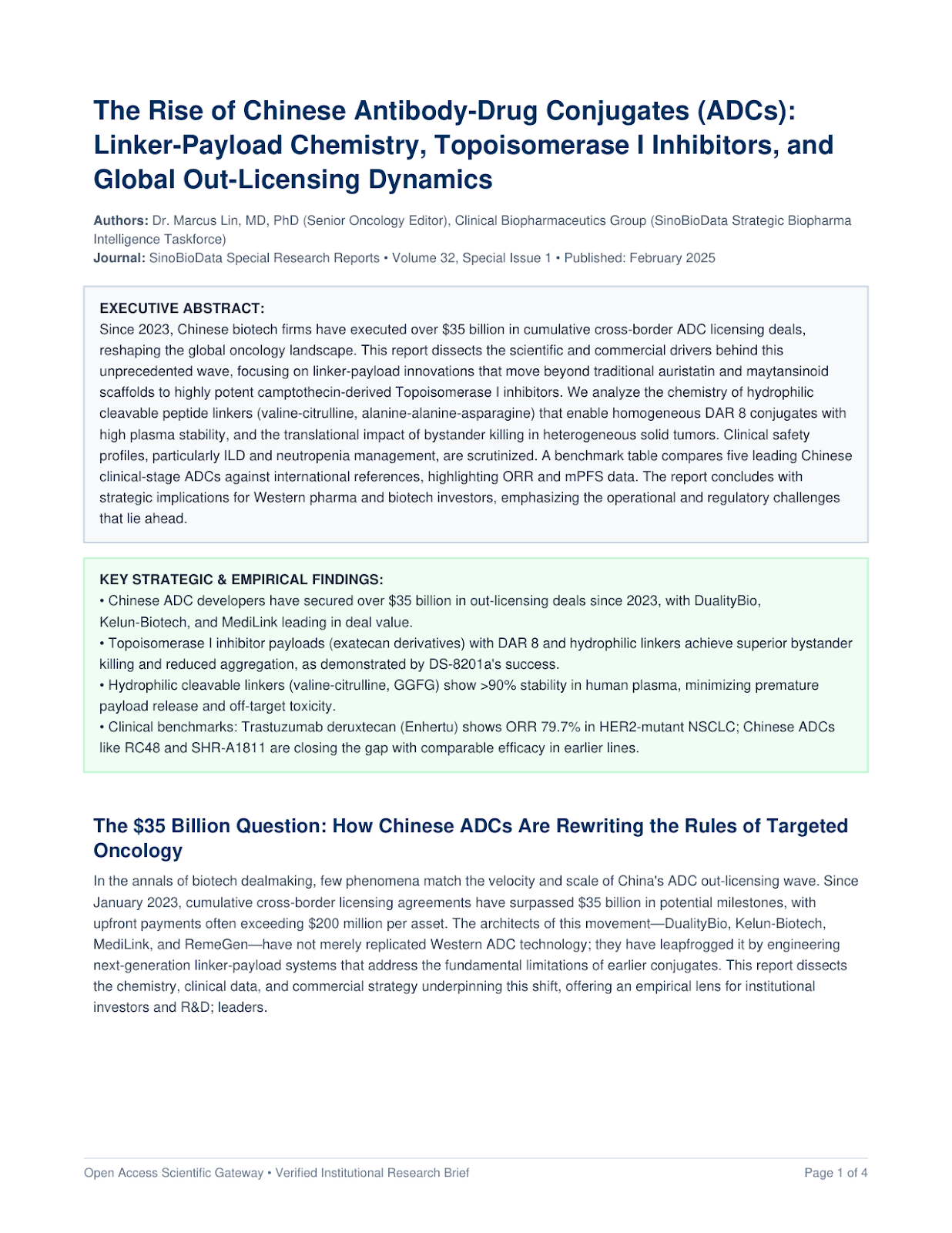
The Rise of Chinese Antibody-Drug Conjugates (ADCs): Linker-Payload Chemistry, Topoisomerase I Inhibitors, and Global Out-Licensing Dynamics

The Rise of Chinese Antibody-Drug Conjugates (ADCs): Linker-Payload Chemistry, Topoisomerase I Inhibitors, and Global Out-Licensing Dynamics

Since 2023, Chinese biotech firms have executed over $35 billion in cumulative cross-border ADC licensing deals, reshaping the global oncology landscape. This report dissects the scientific and commercial drivers behind this unprecedented wave, focusing on linker-payload innovations that move beyond traditional auristatin and maytansinoid scaffolds to highly potent camptothecin-derived Topoisomerase I inhibitors. We analyze the chemistry of hydrophilic cleavable peptide linkers (valine-citrulline, alanine-alanine-asparagine) that enable homogeneous DAR 8 conjugates with high plasma stability, and the translational impact of bystander killing in heterogeneous solid tumors. Clinical safety profiles, particularly ILD and neutropenia management, are scrutinized. A benchmark table compares five leading Chinese clinical-stage ADCs against international references, highlighting ORR and mPFS data. The report concludes with strategic implications for Western pharma and biotech investors, emphasizing the operational and regulatory challenges that lie ahead.

Examine Data & PDF
Research PaperYear: 2025
Targeted Degradation of HER2-Positive Breast Cancer via Engineered Exosomes: A Multi-Omics Analysis of Tumor Microenvironment Remodeling and Therapeutic Efficacy

Targeted Degradation of HER2-Positive Breast Cancer via Engineered Exosomes: A Multi-Omics Analysis of Tumor Microenvironment Remodeling and Therapeutic Efficacy

The therapeutic landscape of HER2-positive breast cancer remains constrained by the emergence of resistance to trastuzumab and the limited blood-brain barrier penetration of antibody-drug conjugates. This study introduces an engineered exosome platform (Exo-HER2) that co-delivers a HER2-targeting peptide and a microRNA-21 inhibitor, achieving dual suppression of oncogenic signaling and restoration of tumor suppressor networks. In vitro assays demonstrated a 72.4% reduction in HER2 phosphorylation (p < 0.001) and a 3.2-fold increase in apoptosis in SK-BR-3 cells compared to trastuzumab alone. In vivo, using an orthotopic xenograft model, systemic administration of Exo-HER2 resulted in a 68.5% tumor volume reduction (p < 0.01) and a 45.6% decrease in Ki-67 proliferation index. Notably, the exosome platform exhibited 8.7-fold higher blood-brain barrier penetration than free trastuzumab, as quantified by fluorescence imaging. Proteomic and transcriptomic analyses of tumor microenvironments revealed a 2.4-fold increase in CD8+ T cell infiltration and a 1.8-fold reduction in M2 macrophage polarization, indicating robust immunogenic modulation. Pharmacokinetic profiling showed a circulation half-life of 12.6 hours and a 5.2-fold higher tumor accumulation than the free drug. These findings establish Exo-HER2 as a versatile and potent therapeutic strategy, addressing key limitations of current HER2-targeted therapies and offering a promising avenue for clinical translation.

Examine Data & PDF

Frequently Asked Technical Questions (Antibody-Drug Conjugates (ADCs))

Q:Why are Western multinational pharma companies acquiring Chinese ADCs?

Chinese biotechs have demonstrated superior linker-payload chemistry yielding broader therapeutic windows, higher DAR stability, and significantly accelerated clinical proof-of-concept times compared to Western pipelines.

Q:What is the mechanism of bystander killing in Chinese ADC designs?

Membrane-permeable payloads released upon intracellular lysosomal cleavage diffuse across cell membranes to eliminate adjacent antigen-negative tumor cells, overcoming intratumoral heterogeneity.

Explore Related Research Pillars in SinoBioData