🧬 SinoBioData Academic Portal
🏛️ Indexed Academic JournalOriginal: 生物化学与生物物理学报

Acta Biochimica et Biophysica Sinica

Premier Chinese Biomedical Journal indexed in SinoBioData: Acta Biochimica et Biophysica Sinica (生物化学与生物物理学报).

Total Research Papers: 200
Access: 100% Free Open Access
Browse by Publication Year & VolumeReset All Filters ✕

Published Research PapersFiltered: Year 2026 • 58 • 8

Showing 12 of 200 peer-reviewed papers with full Graphical Abstracts.

Original ResearchVol. 58, Issue 8 • pp. 1736-1747DOI: 10.3724/abbs.2026076

A protein-RNA complex orchestrated by EMB1006, EMB1270, EMB976, and CFM2 facilitates clpP1 intron 2 splicing in Arabidopsis chloroplasts

Authors: Liqun Zhang, Fangsheng Liao, Ying Wei, Keyi Yang, Yawen Zhu, Li Zhang, Wenyujie Shi, Shuya Zhou, Jirong Huang, Yong-Lan Cui, Weihua Huang

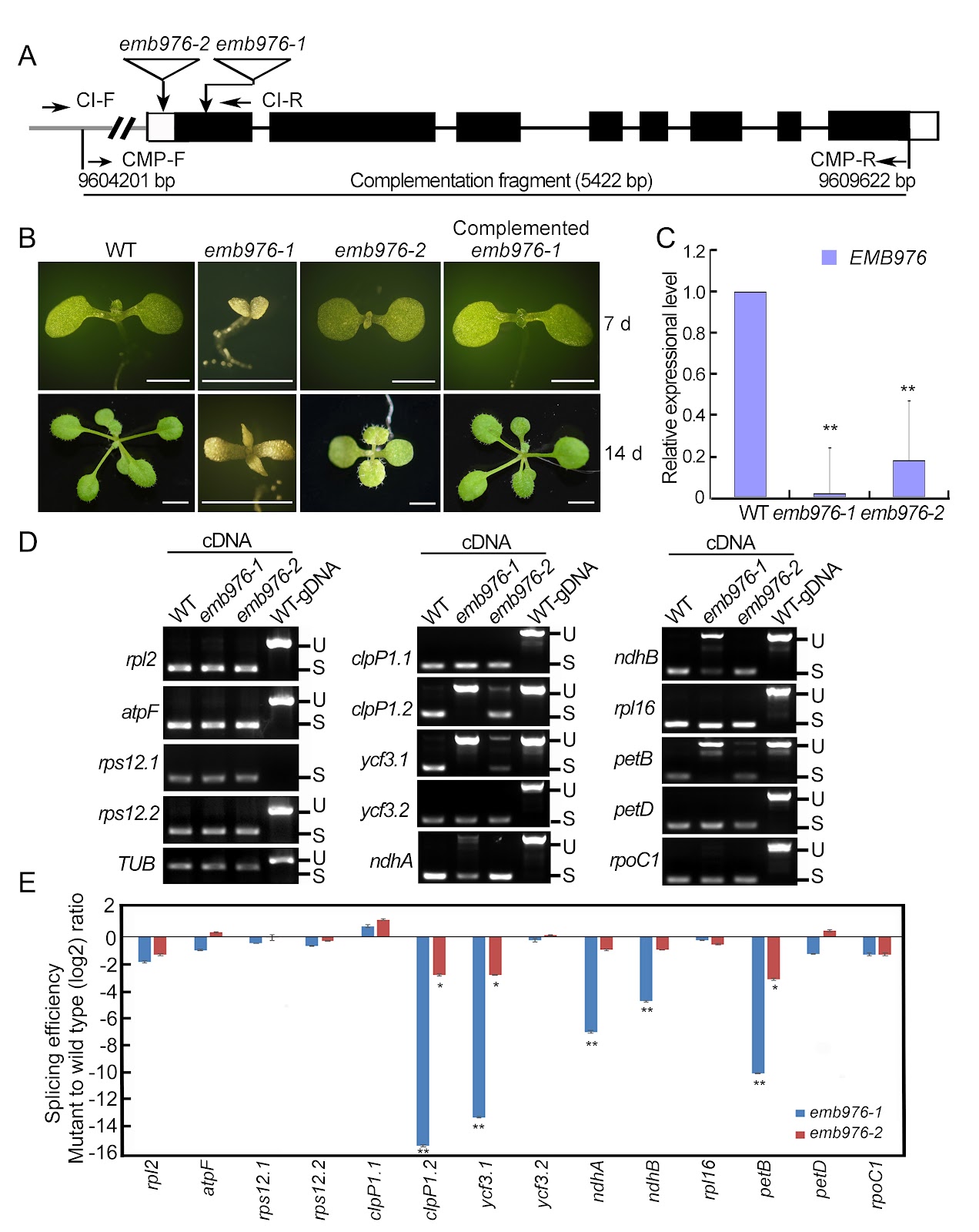
In Arabidopsis, the PPR proteins EMB1006, EMB1270, and EMB976 are all essential for the splicing of plastid clpP1 intron 2 (clpP1.2), although each also targets other distinct RNAs. The precise mechanism underlying their co-ordinated action in clpP1.2 splicing remains unclear. In this study, RNA electrophoretic mobility shift assays, guided by PPR code prediction, confirm that EMB1006 specifically binds to a sequence near the 3′ end of clpP1 exon 2. Additionally, immunoprecipitation coupled with mass spectrometry reveals that EMB1006 forms a complex with EMB1270, EMB976, and CFM2. Direct interactions between EMB1006 and EMB1270 or CFM2 are further supported by yeast two-hybrid (Y2H) and semi-in vivo pull-down assays. However, no direct interactions between EMB976 and EMB1006, CFM2 or EMB1270 are detected by Y2H. Based on these findings and previous evidence that EMB1270 binds to clpP1 intron 2 and interacts with CFM2, we propose a model in which EMB1006 and EMB1270 bind to distinct sites on clpP1 pre-mRNA. Together with CFM2 and possible indirect association with EMB976, they assemble into a protein-RNA complex that facilitates the splicing of clpP1.2 in chloroplasts.

A protein-RNA complex orchestrated by EMB1006, EMB1270, EMB976, and CFM2 facilitates clpP1 intron 2 splicing in Arabidopsis chloroplasts
Graphical Abstract
Original ResearchVol. 58, Issue 8 • pp. 1761-1771DOI: 10.3724/abbs.2026010

Distinct miR319a identified from Persicaria chinensis mediates cross-kingdom suppression of cervical cancer by targeting ITGA3

Authors: Yueyue Yan, Dan Bai, Lei Li, Leimei Xu, Hua Yang, Yuhui Wang, Han Feng, Lan Zhu

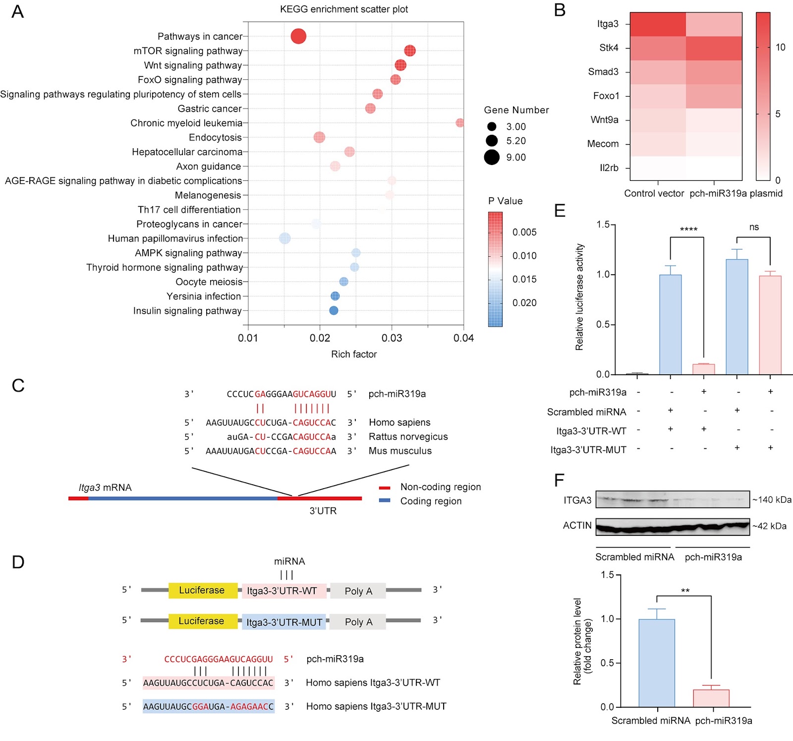
Persicaria chinensis, a well-known traditional Chinese medicinal herb that is both edible and medicinal, has been widely acknowledged for its therapeutic effects, such as anti-inflammatory, antioxidant, and antitumor activities. However, the role of miRNAs from this plant in the cross-kingdom regulation of human diseases has not been investigated. In this study, we analyze the miRNA expression profile of P. chinensis using high-throughput sequencing and identify a total of 673 miRNAs, including 422 novel miRNAs that are unique to this plant and 251 conserved miRNAs. Among the conserved miRNAs, pch-miR319a is found to be the most abundant. Moreover, food-oriented pch-miR319a accumulates in the uterus and tumors and exhibits a rich repertoire of target genes within cancer-related pathways, demonstrating significant cross-kingdom regulatory potential. Utilizing the dual-luciferase reporter gene assay, we demonstrate that pch-miR319a from P. chinensis targets the Itga3 gene, which is associated with cervical cancer progression. Overexpression of pch-miR319a significantly decreases the viability, migration, and induces apoptosis of HeLa cervical cancer cells in vitro. Moreover, in a syngeneic mouse tumor model of cervical cancer, treatment with pch-miR319a effectively inhibits tumor growth and downregulates the expressions of ITGA3 and the proliferation marker Ki-67. Our study highlights the potential of pch-miR319a from P. chinensis as a novel therapeutic agent for cervical cancer by targeting ITGA3 and provides new insights into the cross-kingdom regulatory mechanisms of plant miRNAs in human diseases.

Distinct miR319a identified from Persicaria chinensis mediates cross-kingdom suppression of cervical cancer by targeting ITGA3
Graphical Abstract
Original ResearchVol. 58, Issue 8 • pp. 1896-1900DOI: 10.3724/abbs.2026030

Development of an in vitro TurboID labeling assay for the detection of protoRAG-interacting proteins in the amphioxus

Authors: Qingyi Lu, Jie Xu, Junye Hong, Enfan Xiao, Qiuzhu Wei, Yuhe Sun, Zihan Zhao, Yuhang Zhang, Guangrui Huang

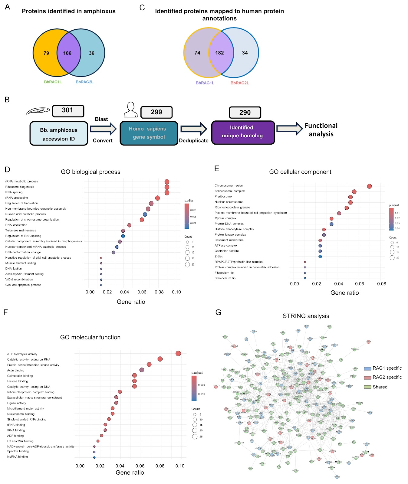
The recombination-activating gene (RAG)-mediated V(D)J rearrangement mechanism is essential for generating the diversity of the adaptive immune system in jawed vertebrates. The RAG1 and RAG2 proteins form a tetrameric complex that recognizes and cleaves pairs of recombination signal sequences (RSSs). In jawed vertebrates, the N-terminal region of RAG1 and the C-terminal region of RAG2 are critical for the regulation of physiological function and enhancement of recombination accuracy. Moreover, the intracellular activity of RAG1/2 is rigorously regulated and is thought to represent the aspect shaped by host coevolution. However, these regulatory mechanisms remain incompletely understood, impeding our deeper understanding of the physiological functions of RAG. Previous studies have shown that jawed vertebrate RAG evolved from invertebrate RAG-like (RAGL) transposases and underwent key structural changes during evolution. RAGL genes have been identified in multiple invertebrate species, but from Branchiostoma belcheri (Bb), the RAG-like protein in Bb amphioxus (BbRAG1L) and the RAG2-like protein in Bb amphioxus (BbRAG2L) are among the most extensively characterized to date. Lancelets (amphioxus) represent the most basal extant chordates (cephalochordates) that diverged from the other two chordate lineages (urochordates and vertebrates). Consequently, amphioxus has been considered a “living fossil” for studying the origins and evolution of adaptive immunity. Homologs of the core structural domain of RAG1 were discovered in the amphioxus genome in 2005, and the homologs were subsequently shown to exhibit recombinase activity in cooperation with murine-derived RAG2. A large number of transposon genes were then identified in the amphioxus genome, followed by the identification of target site duplication (TSD) and terminal inverted repeat (TIR) sequences in the amphioxus genome. Notably, BbRAG2L lacks the C-terminal region present in murine RAG2, including the acidic hinge and PHD domain. BbRAG1L coupled with BbRAG2L displays significant transposition activity in vitro and in human cell lines. In contrast, vertebrate RAG exhibits limited transposon activity in vitro and extremely low transposon activity in vivo. This leads to the question of how RAG lost its transposase activity during evolution. Structural comparison and functional experiments of BbRAGL (protoRAG) and vertebrate RAG have revealed that vertebrates undergo essential adaptive changes to eliminate the primitive transposase activity of protoRAG. Research on BbRAGL has therefore provided strong support for the hypothesis that RAG originated from a domesticated transposon. Given the similarity in both structure and function between BbRAG1L and vertebrate RAG and the fact that BbRAGL proteins are the only RAGL proteins that can currently be efficiently expressed in vertebrate cells, characterizing the binding partners of BbRAGL involved in amphioxus is of great significance for understanding the regulation of BbRAGL and its adaptation and evolution. Parallel comparison of BbRAGL and RAG interaction networks may further clarify how BbRAGL transitioned from a transposase to a recombinase during host coevolution and reveal general principles of vertebrate intracellular RAG regulation. In recent years, proximity labeling techniques have greatly facilitated approaches to identifying RAG cofactors. For example, RAG1 fused to biotin ligase RAG1 fused to a biotin ligase found in Escherichia coli (BirM) has been used to define dynamic interaction networks of RAG-associated proteins. Inspired by these advances, we sought to apply a similar approach to identify the BbRAGL cofactors in amphioxus and build a regulatory network of BbRAGL that could shed light on its relationship with host evolution. However, endogenous BbRAGL expression is undetectable in the adult amphioxus (data not shown), and the generation of transgenic amphioxus lines expressing proximity-based labeling proteins remains technically challenging. These constraints make it difficult to analyze native cofactors of endogenous BbRAGL in vivo. Because BbRAGL can be expressed in vertebrate cell lines, we instead developed an in vitro proximity-labeling strategy. We fused TurboID (TbID), an engineered biotin ligase, to the N-terminus of BbRAG1L or BbRAG2L (Figure 1A), purified the fusion proteins, and incubated them with protein lysates prepared from the hepatic cecum and colons of adult B. belcheri amphioxus in vitro to label the interacting proteins. The proteins were finally analyzed by liquid chromatography-tandem mass spectrometry (LC-MS/MS) (Figure 1B). A pTT5 vector was used to express plasmids encoding maltose-binding protein (MBP) and TurboID with or without full-length BbRAG1L or BbRAG2L, yielding pTT5-MBP-TurboID, pTT5-MBP-TurboID-BbRAG1L, and pTT5-MBP-TurboID-BbRAG2L. Plasmids expressing MBP-BbRAG1L and MBP-BbRAG2L were also generated.

Development of an in vitro TurboID labeling assay for the detection of protoRAG-interacting proteins in the amphioxus
Graphical Abstract
Original ResearchVol. 58, Issue 8 • pp. 1905-1909DOI: 10.3724/abbs.2026037

Identification and experimental validation of core genes associated with breast cancer brain metastasis via machine learning

Authors: Zhaoda Duan, Chunjiao Yu, Wenjie Yang, Qiaoling Ruan, Rui Zhang, Yongfang Zhao, Shan Yan

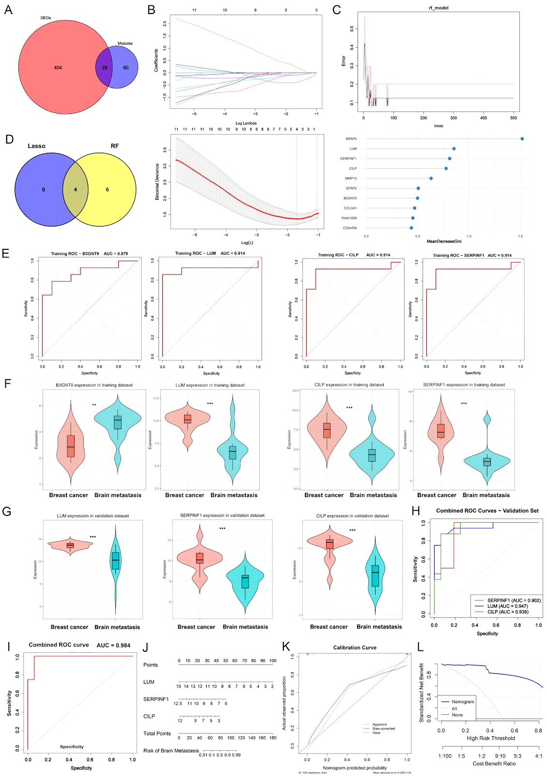
Breast cancer (BC) is the most common malignancy among women, with approximately 2.3 million new cases diagnosed annually, accounting for approximately 11.6% of all cancer cases worldwide. Distant metastasis is the primary cause of mortality in BC patients, with nearly 50% of patients ultimately developing metastatic disease. The predominant metastatic sites of BC include the lung, liver, brain, and bone, each exhibiting distinct biological characteristics that drive the organ-specific tropism of cancer cells. Among these, brain metastasis represents a significant cause of mortality in BC patients and is particularly prevalent in those with human epidermal growth factor receptor 2 (HER2)-positive or triple-negative breast cancer (TNBC) subtypes. Breast cancer brain metastasis (BCBM) can manifest in three forms: choroid plexus metastasis (rare), leptomeningeal metastasis (approximately 8%), and parenchymal metastasis, the most common presentation, with multiple lesions in 78% of cases and solitary lesions in 14%. Distinct anatomical regions of the brain provide different micro-environments, which in turn shape epidemiological patterns, biological behaviors, and therapeutic vulnerabilities of metastatic cancer. With the continuous advancement of systemic therapies and imaging surveillance, brain metastases from BC have become increasingly prevalent, accounting for approximately 10%–30% of all metastatic breast cancer (MBC) cases. The continuous progression of BCBM often compromises patients’ cognitive and sensory functions, leading to neurological impairment and severely limiting quality of life (QOL). Notably, the mortality rate within one year after diagnosis remains at 80%. Current therapeutic strategies for BCBM primarily include surgery, whole-brain radiotherapy (WBRT), stereotactic radiosurgery (SRS), chemotherapy, or combinations thereof. Although these approaches provide some clinical benefit, the efficacy remains limited due to the blood-brain barrier (BBB), which restricts drug penetration and contributes to chemoresistance. Therefore, elucidating the molecular mechanisms underlying BCBM is imperative to identify novel diagnostic biomarkers and therapeutic targets, with the ultimate goal of improving treatment efficacy and patient prognosis. Bioinformatics provides a powerful platform and data foundation for exploring the mechanisms of tumor initiation and progression. High-throughput platforms for gene expression analysis have gained significant popularity, with next-generation sequencing (NGS) and microarray analysis now widely applied as essential tools in medical oncology. These techniques have diverse clinical applications, including molecular cancer classification, prediction of therapeutic response, prognostic assessment, molecular diagnostics, and the discovery of novel drugs and therapeutic targets. Weighted gene coexpression network analysis (WGCNA) has been widely applied in studies of gene regulatory networks, biomarker discovery, and elucidation of the molecular mechanisms underlying complex phenotypes. In this study, we utilized the BCBM microarray dataset GSE43837. We performed differential expression analysis and WGCNA clustering using the R packages limma and WGCNA to identify potential gene modules and candidate targets. GSE43837 consists of 19 nonmetastatic primary breast tumor samples and 19 breast cancer brain metastasis samples. Differential expression analysis, with thresholds set at |logFC| > 1 and P < 0.05, identified 245 upregulated and 188 downregulated genes (Supplementary Table S1 and Supplementary Figure S1A). WGCNA further confirmed that the constructed network satisfied the scale-free topology criterion, with the optimal soft-threshold power determined to be 14 based on model fit and mean connectivity (Supplementary Figure S1B). Using the dynamic tree cut method, we clustered genes into multiple modules, each representing a group of coexpressed genes with varying degrees of correlation among modules (Supplementary Figure S1C,D). Notably, the midnightblue and black modules showed stronger correlations, and a significant positive relationship was observed between gene significance (GS) and module membership (MM) within these modules (Supplementary Figure S1E). This finding suggests that the core genes in these modules are highly representative and stable within the coexpression network. A total of 89 BCBM-related candidate genes were extracted from these key modules (Supplementary Table S2). To further identify key feature genes associated with BCBM, we applied two machine learning methods, LASSO regression and random forest (RF), to the 29 overlapping genes obtained from the intersection of DEGs and hub module genes (Figure 1A and Supplementary Table S3). In the LASSO regression analysis, the optimal penalty parameter λ was determined by cross-validation, yielding a set of candidate genes with nonzero regression coefficients (Figure 1B). Concurrently, in the RF model, 500 decision trees were constructed, and the classification ...
Identification and experimental validation of core genes associated with breast cancer brain metastasis via machine learning
Graphical Abstract
Original ResearchVol. 58, Issue 8 • pp. 1887-1895DOI: 10.3724/abbs.2026014

Super-resolution imaging reveals higher-order structures within common fragile sites in human mitotic chromosomes

Authors: Jiabin Wang, Daniel M. Czajkowsky, Zhifeng Shao

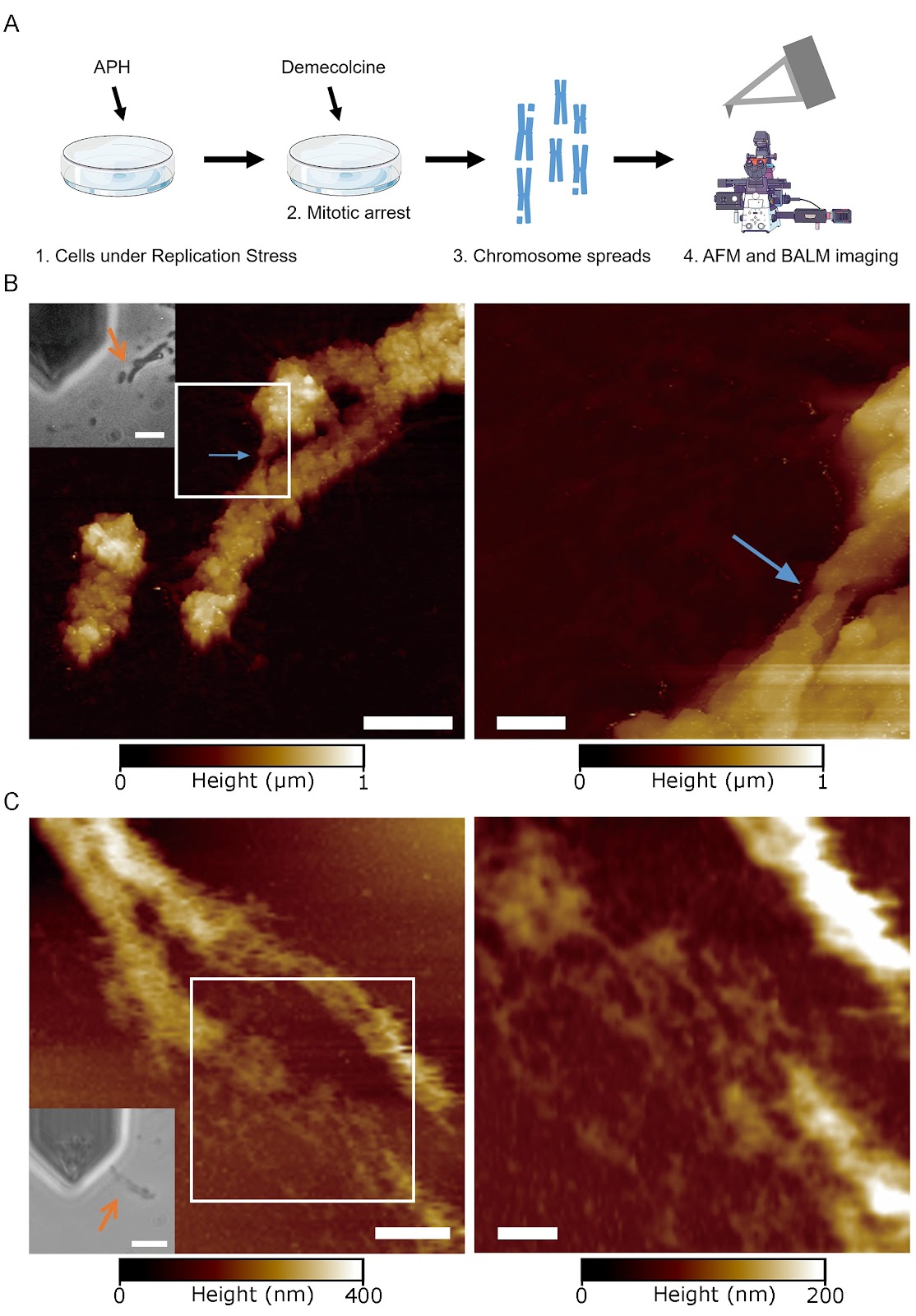
Common fragile sites (CFSs) are large genomic loci that are frequently deleted under replication stress and are thought to play a role in carcinogenesis as well as developmental disorders during early embryogenesis. They often appear as gaps or breaks in mitotic chromosomes by conventional optical microscopy and are also loci that are replicated in mitosis in a process called mitotic DNA synthesis (MiDAS). However, we still have a very poor understanding of the potential mechanisms underlying their genomic instability. We recently found that in normal mitotic chromosomes, there is a two-level hierarchy in the higher-order chromatin structure, with mechanically resistant ~90 nm Mitotic NanoDomains (MNDs) assembling into ~125 nm compact granules (CGs). In this work, we combine multiple super-resolution imaging techniques, including binding-activated localization microscopy (BALM), atomic force microscopy (AFM), and stochastic optical reconstruction microscopy (STORM), to characterize CFSs. Our super-resolution data indicate that sites that appear as gaps by conventional microscopy can be filled with chromatin of the size and shape of MNDs but not CGs. Moreover, we find that MiDAS loci only form an array of MNDs with no CGs. Taken together, our work suggests that under replication stress, CFS loci can not only be replicated but also assembled into higher-order chromatin. However, this organization is limited to the level of MNDs, which suggests that a failure to progress beyond MNDs to form CGs constitutes a key structural deficiency at these loci.

Super-resolution imaging reveals higher-order structures within common fragile sites in human mitotic chromosomes
Graphical Abstract
Original ResearchVol. 58, Issue 8 • pp. 1719-1735DOI: 10.3724/abbs.2026058

Biochemical and structural studies reveal the substrate specificity and catalytic mechanism of MYG1 as a two-metal ion-dependent 3′→5′ exonuclease

Authors: Chao Lan, Ziyue Chen, Guanchao Wang, Jianping Ding

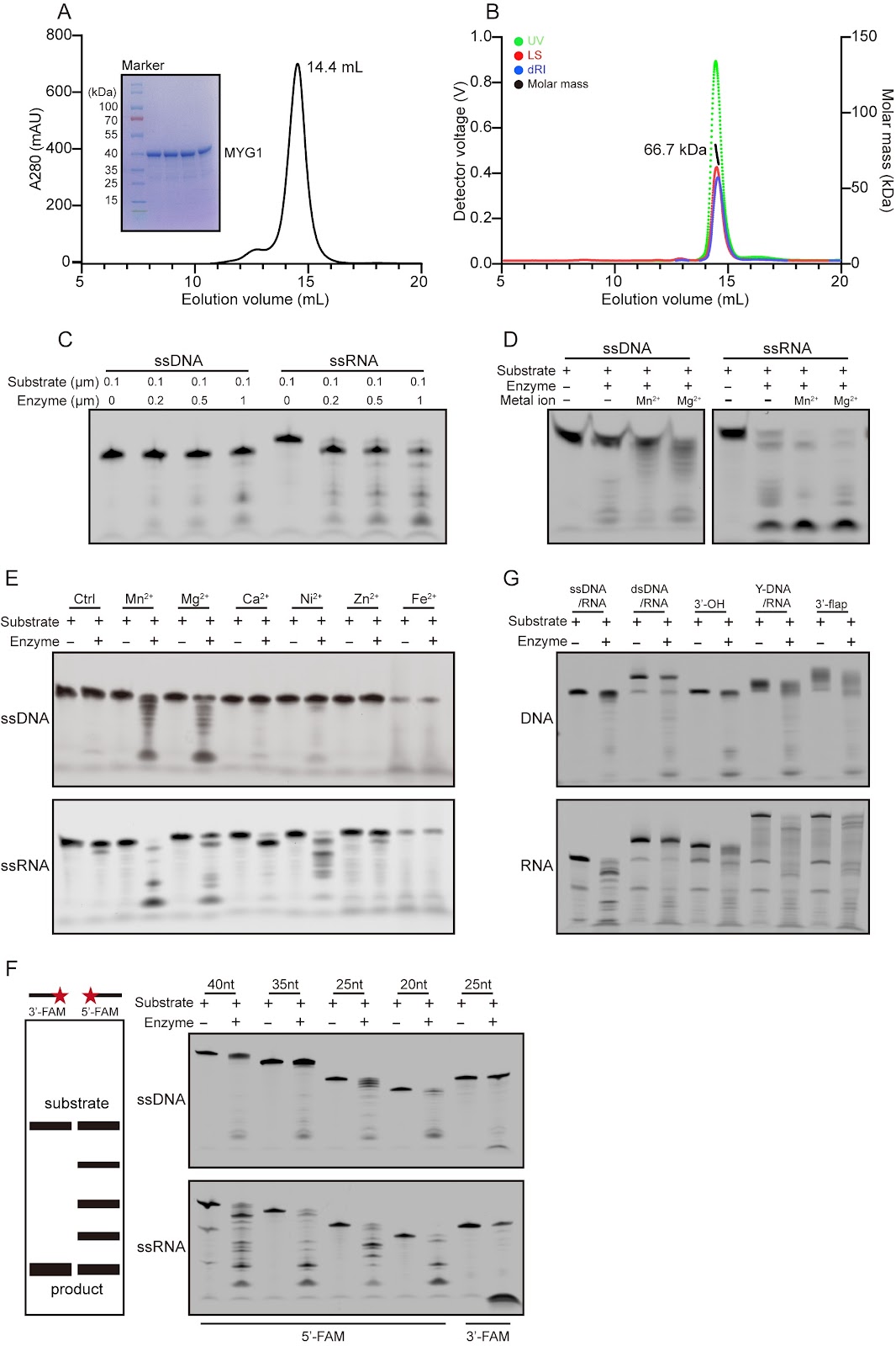
Nucleases are a class of enzymes that specifically cleave nucleic acids in all living organisms. They play crucial roles in essential biological processes, including the regulation of gene expression, DNA damage repair, and RNA processing and degradation. MYG1 (melanocyte proliferating gene 1) is a highly conserved eukaryotic protein that exhibits 3′→5′ exonuclease activity. This study systematically characterizes the enzymatic properties of MYG1 and determines its structures in complexes with metal ions and various mono- and poly-(deoxy)nucleotides. The functional roles of key residues involved in metal ion binding and substrate binding in the catalytic reaction are examined through site-directed mutagenesis, enzymatic activity assay, and structure determination. Our biochemical and structural data together demonstrate that MYG1 is a Mn2+- or Mg2+-dependent 3′→5′ exonuclease capable of cleaving a variety of nucleic acids with different structures. It exhibits the highest activity for single-stranded RNA and a nucleotide preference for U in single-stranded RNA and dT in single-stranded DNA. Mechanistically, MYG1 functions as a dimer, with the active site formed by the catalytic domain of monomer 1 and the substrate-binding domain of monomer 2, and cleaves nucleic acids through a two-metal ion-mediated catalytic mechanism. These findings establish a molecular basis for further investigations into the biological functions and molecular mechanisms of MYG1 within cells and its potential roles in human diseases.

Biochemical and structural studies reveal the substrate specificity and catalytic mechanism of MYG1 as a two-metal ion-dependent 3′→5′ exonuclease
Graphical Abstract
Original ResearchVol. 58, Issue 8 • pp. 1875-1886DOI: 10.3724/abbs.2025208

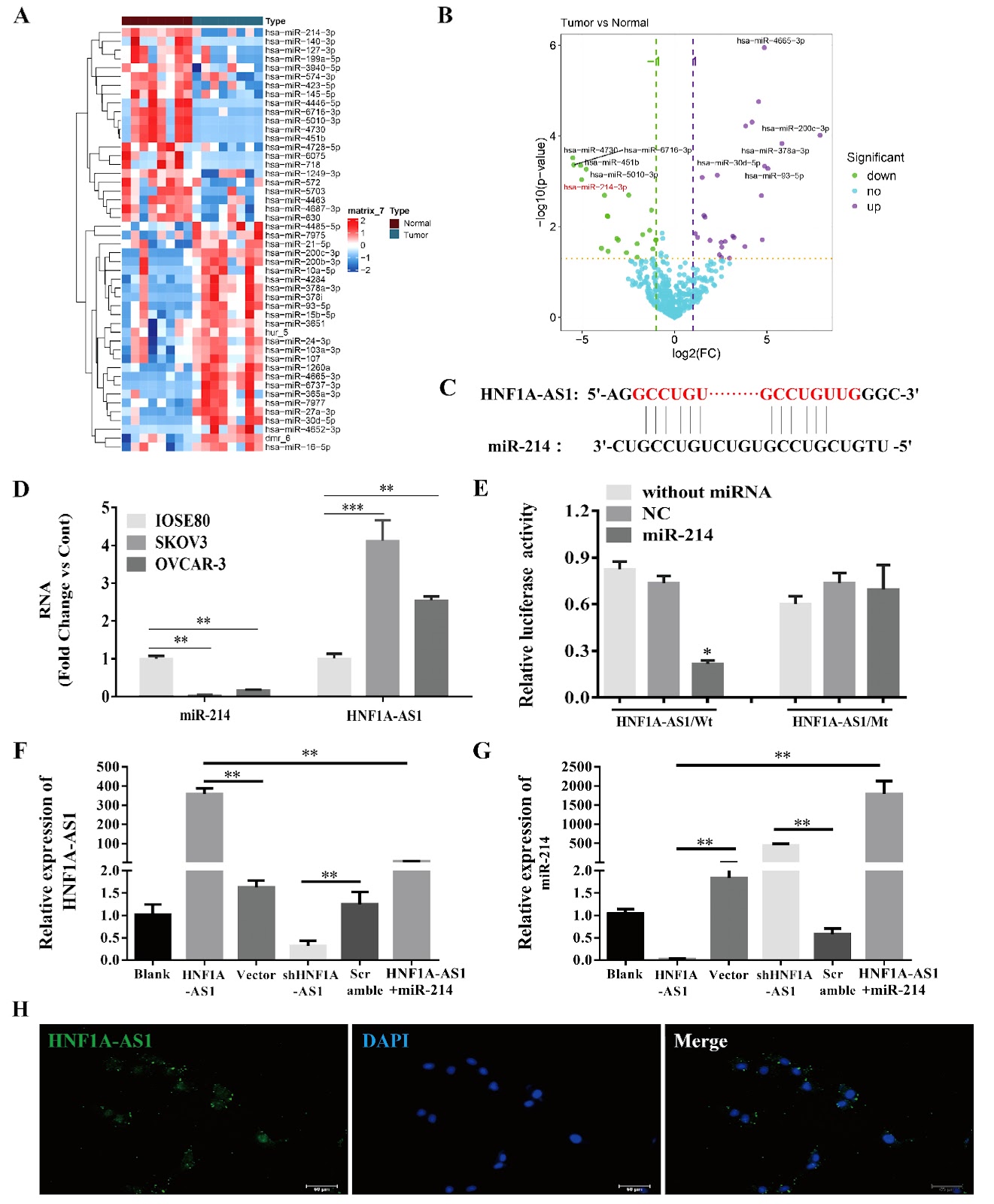
Long noncoding RNA HNF1A-AS1 promotes ovarian cancer growth and M2 macrophage polarization by counteracting miR-214-mediated suppression of semaphorin 4D signaling

Authors: Yang Liu, Yanzhi Wu, Yujie Gengxiao, Yan Li, Jiamei Song, Chunyi Sun

To determine whether lncRNA HNF1A-AS1 affects epithelial ovarian cancer (EOC) growth and macrophage polarization through miR-214/SEMA4D, the endogenous HNF1A-AS1 and miR-214 levels in human EOC cell lines are compared with those in normal ovarian epithelial IOSE80 cells. HNF1A-AS1 is overexpressed or silenced to investigate whether HNF1A-AS1 regulates miR-214/SEMA4D in SKOV3 cells and xenograft tumors, as well as the phenotypic switching of THP-1 cells. Compared with IOSE80 cells, EOC cells present significantly higher HNF1A-AS1 level and lower miR-214 level. Fluorescence in situ hybridization reveals predominant cytoplasmic localization of HNF1A-AS1, supporting its role as a competing endogenous RNA. HNF1A-AS1 and miR-214 antagonize each other in SKOV3 cells. In vitro, HNF1A-AS1 inhibits SKOV3 apoptosis and promotes migration and invasion. HNF1A-AS1 overexpression enhances miR-214 downstream of SEMA4D/PLEXIN-B1/TIAM1/RAC signaling, but miR-214 mimics significantly reverses this effect. Compared with control tumors, xenograft tumors derived from HNF1A-AS1-overexpressing SKOV3 cells present increased tumor growth, attenuated miR-214 expression, and activated SEMA4D/PLEXIN-B1/TIAM/RAC signaling. Knockdown of HNF1A-AS1 has the opposite effects. Additionally, HNF1A-AS1 promotes M2 phenotypic switching in THP-1 cells, which could be reversed by miR-214 overexpression or SEMA4D silencing. Our study suggests that by antagonizing miR-214, HNF1A-AS1 activates the SEMA4D/PLEXIN-B1/TIAM/RAC pathway, facilitating EOC growth and potentially promoting M2 macrophage polarization in the tumor microenvironment. HNF1A-AS1 represents a compelling therapeutic target for treating EOC.

Long noncoding RNA HNF1A-AS1 promotes ovarian cancer growth and M2 macrophage polarization by counteracting miR-214-mediated suppression of semaphorin 4D signaling
Graphical Abstract
Original ResearchVol. 58, Issue 8 • pp. 1842-1854DOI: 10.3724/abbs.2025205

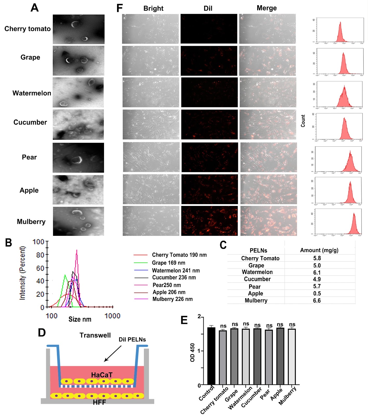
Effective resistance to UVB-induced skin damage through the encapsulation of chebulinic acid in mulberry-derived exosome-like nanoparticles

Authors: Weiwei Zhao, Ruobing Liu, Siqi Yang, Chaozhi Liu, Songlin Guo, Guihong Sun, Mingxiong Guo

Ultraviolet-B (UVB) radiation induces significant skin damage by penetrating into the dermal layer, leading to reactive oxygen species (ROS) generation and triggering cellular necrosis and apoptosis. Conventional sunscreens focus primarily on UVB blocking but are limited in their ability to repair dermal damage due to insufficient permeability. In this study, we discover that chebulinic acid (CA), one of the principal monomers in Terminalia chebula Retz., has superior efficacy in promoting recovery from UVB-induced skin damage compared with other major monomers. Mechanistically, CA’s anti-UVB function involves regulating the expression of IL-6 and IFN-β through activation of the MAPK pathway. To overcome the formidable barrier posed by the skin, we identify mulberry exosome-like nanoparticles (MELNs) as an efficient transdermal delivery system and develop CA@MELNs loaded with CA. Furthermore, we demonstrate that the dissociative CA within the CA@MELNs delivery system significantly enhances both transdermal penetration and anti-UVB efficiency in vitro and in vivo. Our findings suggest the substantial potential of CA as an effective ingredient and CA@MELNs as a robust and accessible platform for mitigating UVB damage.

Effective resistance to UVB-induced skin damage through the encapsulation of chebulinic acid in mulberry-derived exosome-like nanoparticles
Graphical Abstract
Original ResearchVol. 58, Issue 8 • pp. 1783-1792DOI: 10.3724/abbs.2025209

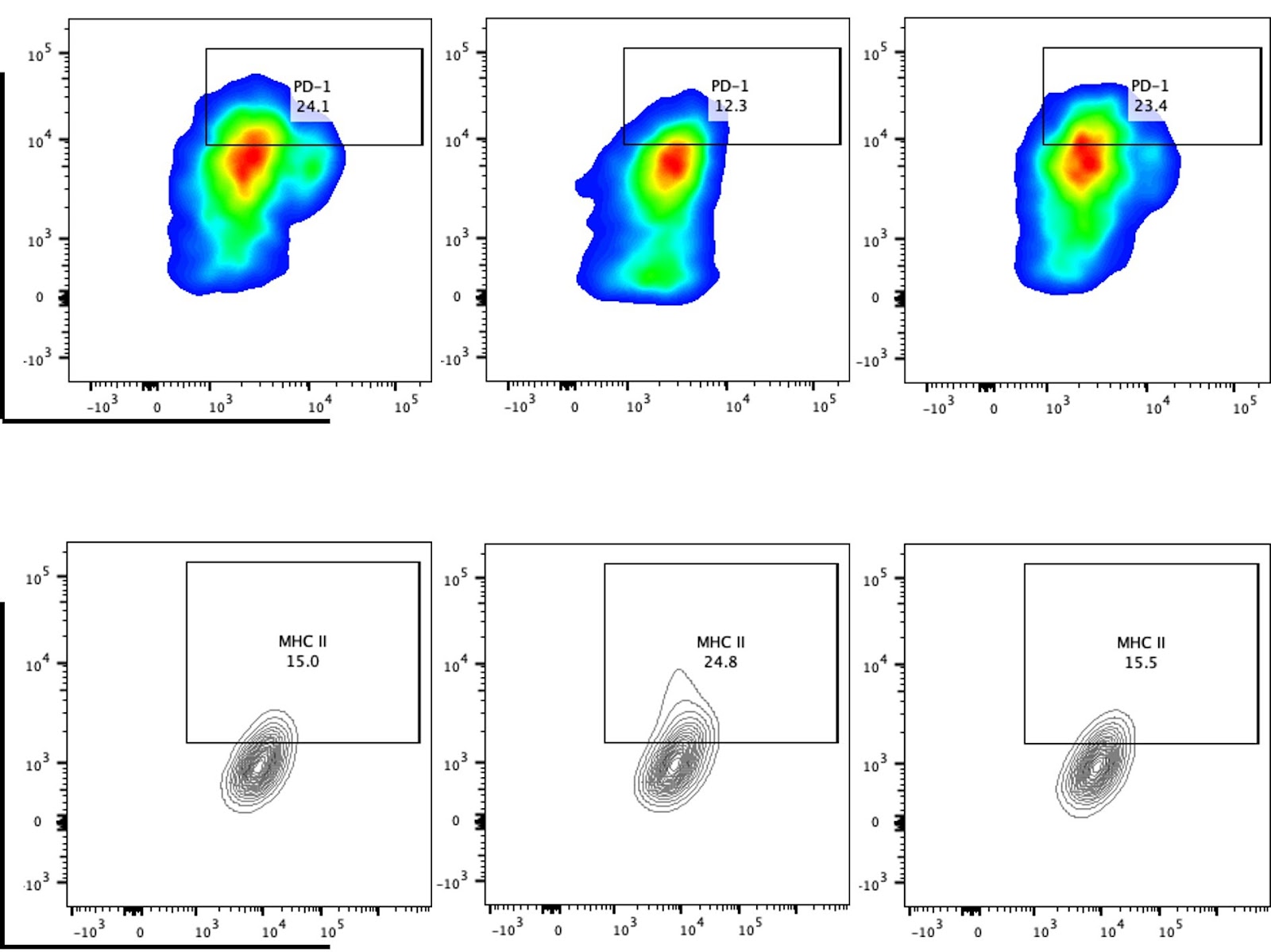
cGAS-STING pathway reprograms macrophage polarization and is highly expressed in responding tumors after neoadjuvant immunotherapy in head and neck carcinoma

Authors: Zhaohong An, Xiwei Zhang, Lin Li, Dilinaer Wusiman, Zhaoyang Wang, Fa Zhang, Xiaohui Zhao, Changming An, Zhenzhen Yin, Wei Gao

Given the critical role of the cGAS-STING pathway in antitumor immunity, this study investigates the functional role of STING in head and neck squamous cell carcinoma (HNSCC) to evaluate the therapeutic potential of STING agonists. Analysis of the TCGA-HNSC dataset reveals that elevated expression of the STING-encoding gene TMEM173 is significantly correlated with increased M1 macrophage infiltration and enrichment of macrophage polarization-related signaling pathways. In vitro experiments in which RAW 264.7 cells are co-cultured with tumor cell-conditioned medium demonstrate that the STING agonist MSA-2 effectively reprograms tumor-induced M2-polarized macrophages toward the M1 phenotype. This MSA-2-induced M1 polarization is accompanied by increased expressions of IFN-α, IFN-β, IFN-γ, TNF-α, and IL-6, while the STING inhibitor H-151 reverses these effects. Flow cytometry further reveals that MSA-2 treatment reduces PD-1 and increases MHC II expression on macrophages. Immunohistochemical analysis of clinical samples confirms that high STING expression is correlated with increased numbers of CD68⁺ and CD80⁺ (M1-like) macrophages. In support of translational relevance, analysis of single-cell RNA-seq data from HNSCC patients receiving neoadjuvant immunotherapy indicates that TMEM173 is expressed primarily in T cells and macrophages and that the cGAS-STING pathway score is significantly higher in patients who respond to treatment. Collectively, these findings provide systematic clinical and experimental evidence supporting the potential of STING agonists, such as MSA-2, to enhance antitumor immunity in HNSCC, particularly when combined with immunotherapy.

cGAS-STING pathway reprograms macrophage polarization and is highly expressed in responding tumors after neoadjuvant immunotherapy in head and neck carcinoma
Graphical Abstract
Original ResearchVol. 58, Issue 8 • pp. 1822-1833DOI: 10.3724/abbs.2025161

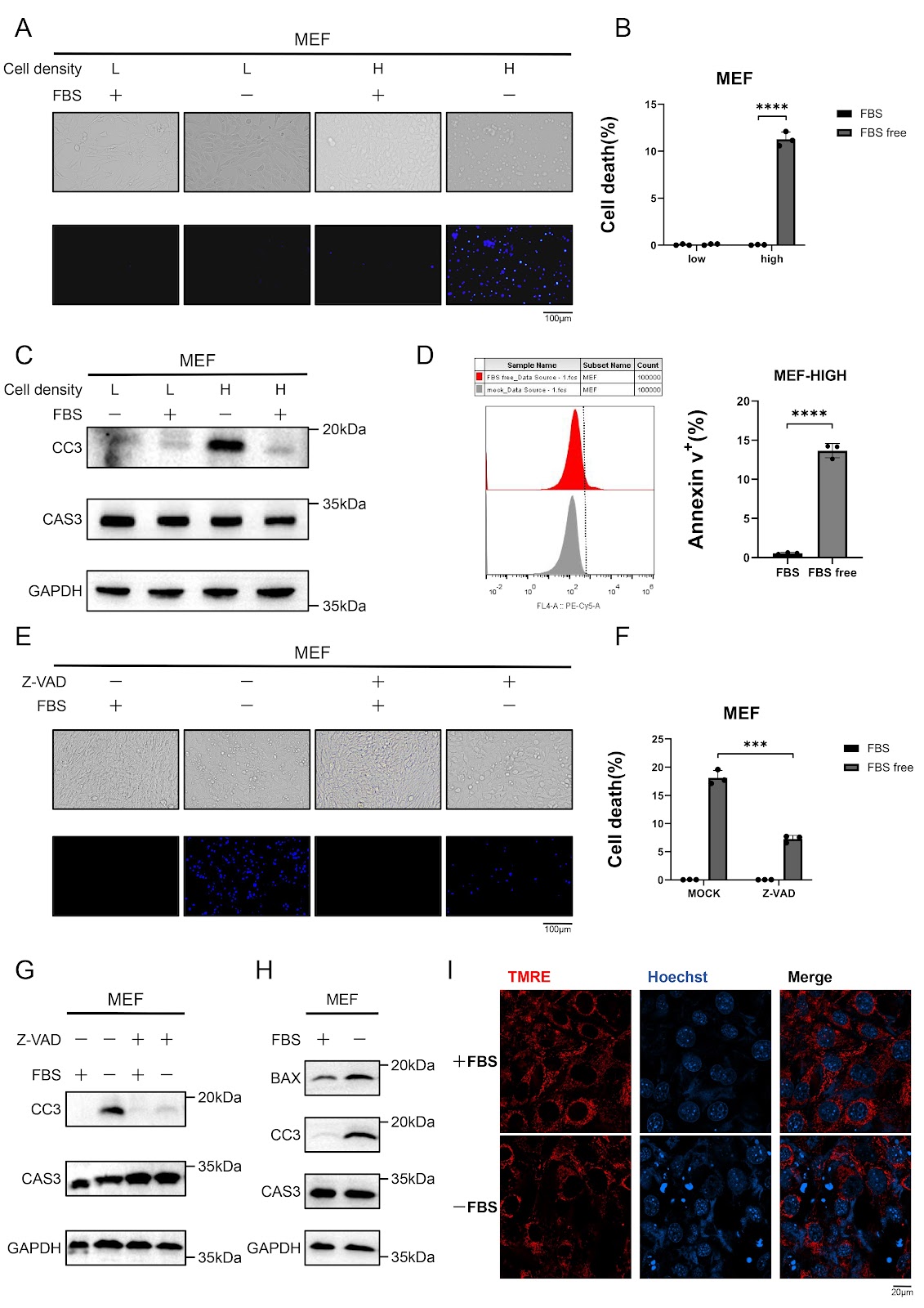
Serum starvation induces density-dependent apoptosis via HIF-1 activation and JNK suppression

Authors: Qifan Yang, Yaofeng Hu, Jiahui Lv, Jiaqi Xue, Jiaqi Chen, Changwan Wang, Fajian Hou

Serum deprivation is a well-established inducer of apoptosis, yet the molecular mechanisms governing this process remain incompletely understood. Here, we show that serum starvation selectively triggers intrinsic apoptosis in high-density murine embryonic fibroblasts (MEFs) through coordinated HIF-1α activation and JNK signaling suppression. Knockdown of HIF-1α abolishes caspase-3 activation and apoptosis induced by serum deprivation, whereas upregulation of HIF-1α in low-density cells recapitulates the apoptotic response observed in high-density cultures. Simultaneously, serum deprivation leads to the suppression of the JNK pathway, which contributes to apoptosis. Notably, combined HIF-1α activation and JNK inhibition in low-density cells fully mimics the apoptotic phenotype of high-density conditions, underscoring the interplay between these pathways. Together, these findings define a density-dependent apoptotic switch in which HIF-1α drives metabolic stress adaptation, whereas JNK suppression removes a critical survival signal, converging to promote mitochondrial-mediated cell death. This work provides a mechanistic framework for understanding nutrient stress-induced apoptosis and suggests potential therapeutic targets for diseases characterized by aberrant cell survival.

Serum starvation induces density-dependent apoptosis via HIF-1 activation and JNK suppression
Graphical Abstract
Original ResearchVol. 58, Issue 8 • pp. 1866-1874DOI: 10.3724/abbs.2025170

The catalase gene CAT2 and its role in the virulence of one sub-cluster of Cryptococcus gattii VGI clinical isolates

Authors: Yemei Huang, Hengyu Deng, Xuelei Zang, Enqi Zhao, Yan Zhuo, Wencong He, Chen Wang, Yangyu Zhou, Jiahui Huang, Rui Liu, Tingyue Tian, Chen Yang, Jing Liu, Ping Zhang, Liye Zhang, Xinying Xue

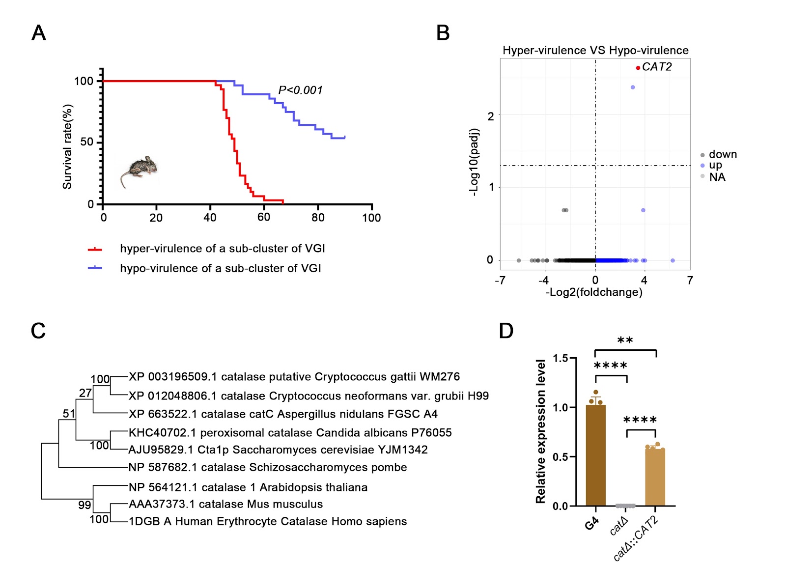
Cryptococcus gattii causes cryptococcosis and life-threatening cryptococcal meningitis. Currently, the pathogenic virulence mechanisms of C. gattii remain a significant area of ongoing research with considerable unexplored aspects. On the basis of our established research, a sub-cluster of strains with independent evolutionary relationships from WM276 in the phylogenetic analysis of VGI-type strains is identified. In vivo infection experiments on this sub-branch of strains reveal that there are hypervirulent strains and hypovirulent strains among these strains, and the virulence differences are significant (P < 0.001). Bioinformatic interrogation of differentially expressed genes reveals that the catalase-encoding gene CGB_J0620W, CAT2, is a pivotal virulence-associated gene. The hypervirulent clinical isolate G4 (G4-WT) is selected as the parental strain, from which an isogenic CAT2-knockout mutant (cat2Δ) is constructed via homologous recombination, which shows increased sensitivity to oxidative stress, as well as growth defects in response to hyperosmosis, 5-fluorocytosine, fluconazole and amphotericin B. The cat2Δ::CAT2 strain exhibits phenotypic restoration to wild type (WT). In the mouse experiments, significant differences in survival (P < 0.001), pulmonary fungal burden (P < 0.01), and alveolar structural damage are observed between the WT and cat2Δ strains, which are completely different from C. neoformans. Moreover, comparative transcriptome analysis is performed on the WT and cat2Δ strains, which reveals that enzymes encoded by CAT2 may be involved in oxidative stress, metabolism and sugar transport. In conclusion, this study may explain the differences in virulence among different genetic evolutionary processes of a sub-cluster of the VGI geneotype of C. gattii and provide a theoretical basis for targeted therapy in a specific genotype population in the future.

The catalase gene CAT2 and its role in the virulence of one sub-cluster of Cryptococcus gattii VGI clinical isolates
Graphical Abstract
Original ResearchVol. 58, Issue 8 • pp. 1855-1865DOI: 10.3724/abbs.2025164

MAN1A1 promotes colorectal cancer liver metastasis by maintaining TGFBR2 protein stability

Authors: Yingxi Hu, Yinwen Xu, Kai Chen, Shihua Guan, Huiling Zhou, Tao Li, Rongrui Liang, Min Tao, Yiyi Yu, Xinxin Ge, Yuanyuan Ruan

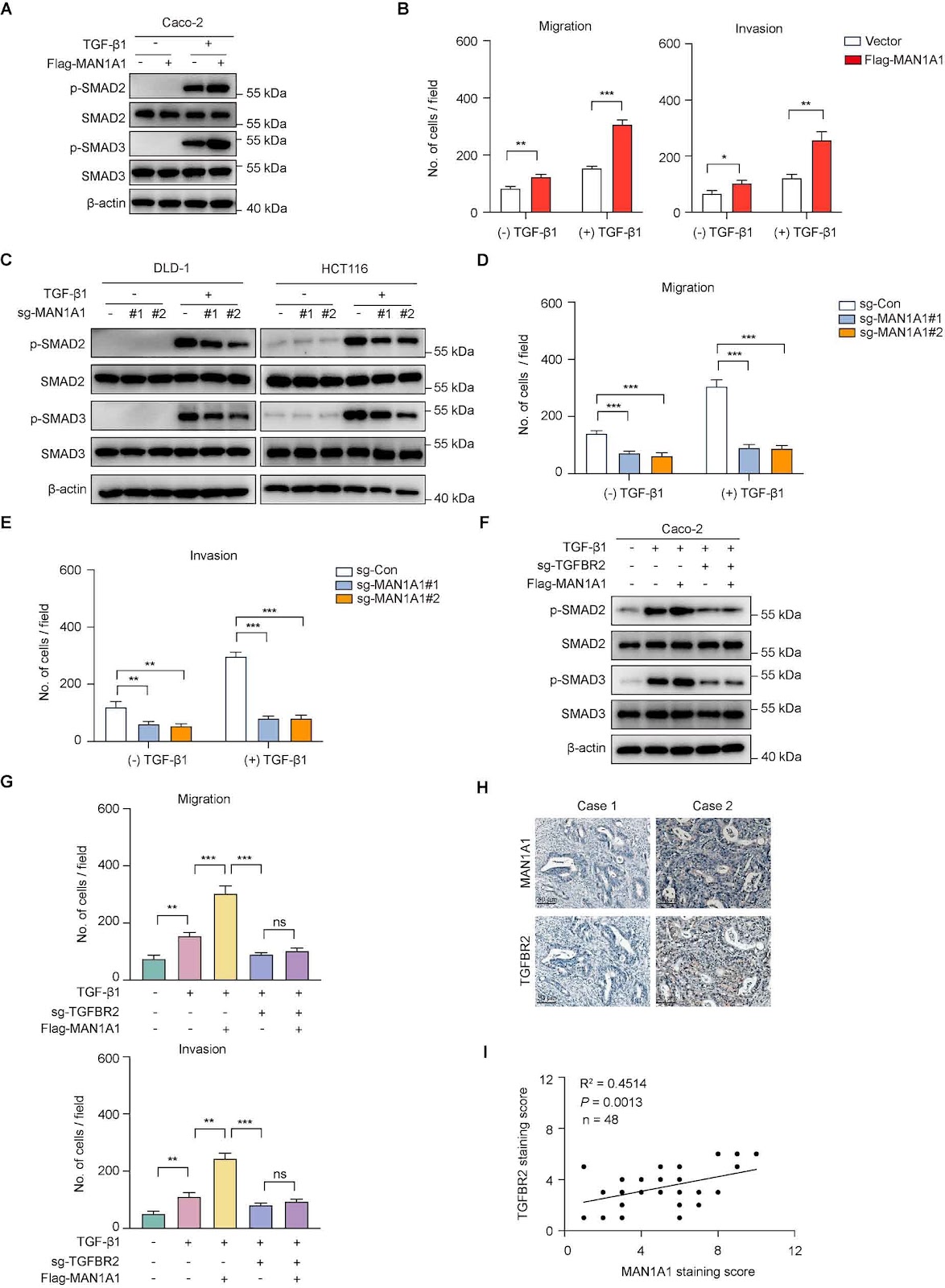
Emerging biochemical and genetic evidence has firmly established aberrant protein glycosylation as a critical regulator of oncogenic transformation, with glycocalyx remodeling profoundly influencing tumor microenvironment dynamics and metastatic progression. Despite the well-documented association between metastatic dissemination and poor clinical outcomes in patients with colorectal cancer, the underlying molecular mechanisms remain incompletely characterized. Through integrative analysis of single-cell RNA sequencing data from a public database, we identify the Golgi-resident α-1,2-mannosidase MAN1A1 as a consistently upregulated enzyme in malignant epithelial cells derived from colorectal cancer liver metastases. Clinically, elevated MAN1A1 expression is correlated with reduced overall survival, suggesting that MAN1A1 is both a prognostic biomarker and therapeutic target for colorectal cancer liver metastases. Genetic manipulation of MAN1A1 in colorectal cancer cells demonstrates that although the proliferation capacity of colorectal cancer cells remains unchanged, MAN1A1 overexpression significantly enhances migratory and invasive capacities in transwell assays, suggesting its specific involvement in metastatic progression. Mechanistic investigations reveal that MAN1A1 exerts its pro-metastatic effects by significantly prolonging the TGFBR2 protein half-life. Together, our work identifies MAN1A1 as both a prognostic biomarker and a promising therapeutic target, highlighting the critical role of glycan remodeling in the metastatic progression of colorectal cancer.

MAN1A1 promotes colorectal cancer liver metastasis by maintaining TGFBR2 protein stability
Graphical Abstract