🧬 SinoBioData Academic Portal
🏛️ Indexed Academic JournalOriginal: 生物化学与生物物理学报

Acta Biochimica et Biophysica Sinica

Premier Chinese Biomedical Journal indexed in SinoBioData: Acta Biochimica et Biophysica Sinica (生物化学与生物物理学报).

Total Research Papers: 200
Access: 100% Free Open Access
Browse by Publication Year & VolumeReset All Filters ✕

Published Research PapersFiltered: Year 2025 • 57 • 7

Showing 8 of 200 peer-reviewed papers with full Graphical Abstracts.

Original ResearchVol. 57, Issue 7 • pp. 1164-1174DOI: 10.3724/abbs.2024240

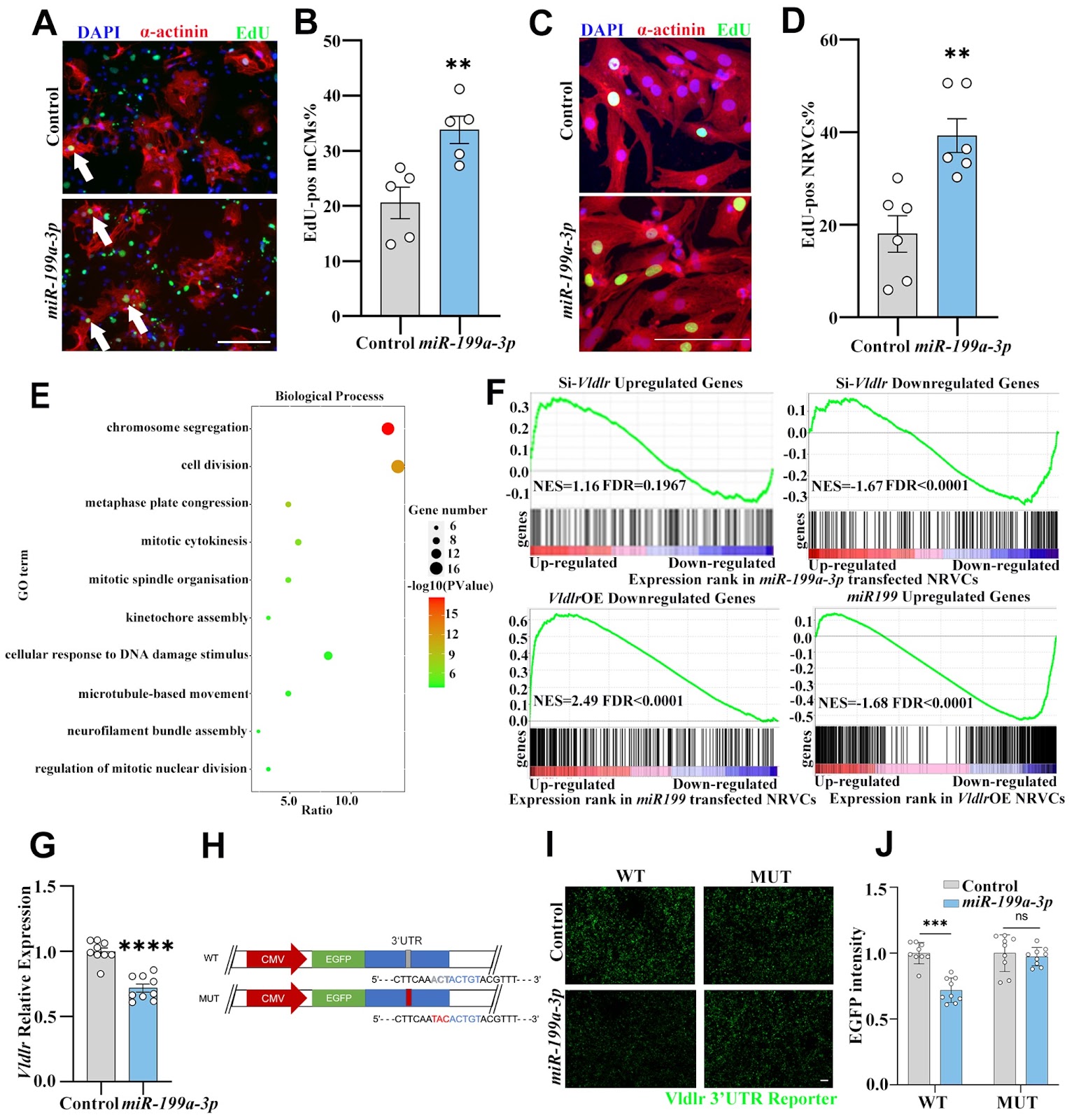
miR-199a-3p suppresses Vldlr expression to promote cardiomyocyte proliferation

Authors: Rui Jiang, Lijuan Pei, Hongjie Zhang, Fenglian He, Yuhan Min, Xinhang Li, Ke Wei

The proliferative capacity of cardiomyocytes is limited in adult mammals, and replacing lost tissue following acute ischemic injury is challenging. Previous studies have demonstrated that miR-199a-3p can promote cardiomyocyte proliferation, but the exact mechanism by which this occurs remains unclear, although multiple targets of miR-199a-3p have been identified. We recently showed that very-low-density-lipoprotein receptor (Vldlr) inhibits cardiomyocyte proliferation, and in this study we aim to test whether Vldlr is a functional target gene of miR-199a-3p. 3′UTR reporter assays demonstrate that miR-199a-3p directly binds to the 3′UTR of Vldlr and inhibits its translation. Overexpressing Vldlr blunts the pro-proliferative effect of miR-199a-3p on cardiomyocytes, suggesting that Vldlr is indeed a functional target of miR-199a-3p. Mechanistically, Vldlr reduces S807/811 phosphorylation of RB1, and inhibiting CDK4/6 to prevent RB1 phosphorylation can block the pro-proliferative effect of both Vldlr knockdown and miR-199a-3p, suggesting that RB1 phosphorylation is required for the cardiomyocyte proliferation induced by miR-199a-3p and Vldlr knockdown. The findings of this study reveal Vldlr as a novel functional target of miR-199a-3p in cardiomyocytes and identify RB1 as a downstream effector of cardiomyocyte proliferation. The identification of the role of the miR-199a-3p-Vldlr-RB1 axis in cardiomyocyte proliferation may provide potential therapeutic targets for cardiac regenerative medicine.
miR-199a-3p suppresses Vldlr expression to promote cardiomyocyte proliferation
Graphical Abstract
Original ResearchVol. 57, Issue 7 • pp. 1139-1150DOI: 10.3724/abbs.2024215

High-resolution imaging atlas reveals the context-dependent role of pancreatic sympathetic innervation in diabetic mice

Authors: Qingqing Xu, Yuxin Chen, Xinyan Ni, Hanying Zhuang, Shenxi Cao, Liwei Zhao, Leying Wang, Jianhui Chen, Wen Z Yang, Wenwen Zeng, Xi Li, Hongbin Sun, Wei L Shen

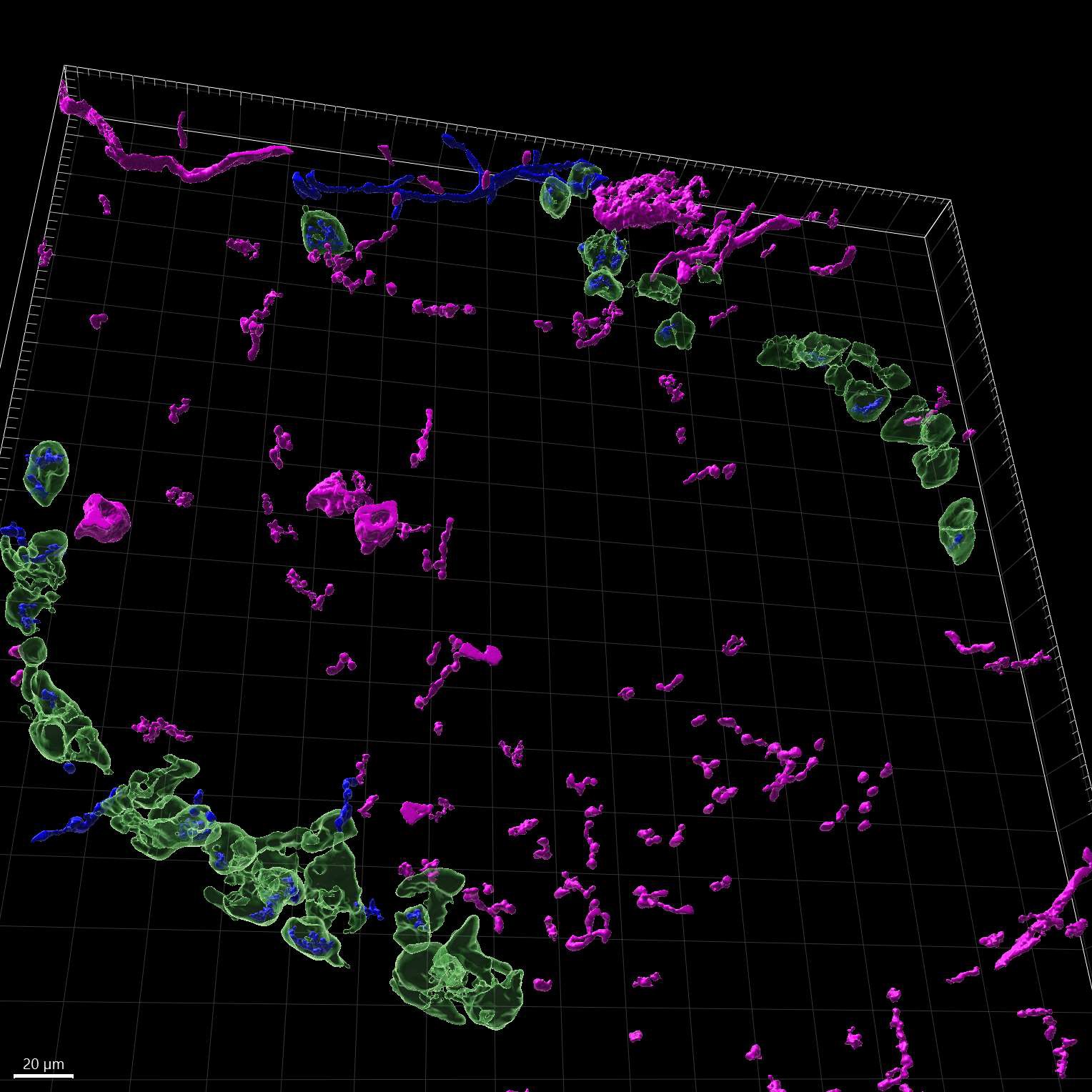
A better understanding of how sympathetic nerves impact pancreatic function is helpful for understanding diabetes. However, there is still uncertainty and controversy surrounding the roles of sympathetic nerves within the pancreas. To address this, we utilize high-resolution imaging and advanced three-dimensional (3D) reconstruction techniques to study the patterns of sympathetic innervation and morphology in the islets of adult wild-type (WT) and diabetic mice. Our data show that more than ~30% of α/β-cells are innervated by sympathetic nerves in both WT and diabetic mice. Additionally, sympathetic innervated α/β-cells are reduced in diet-induced obese (DIO) mice, whereas sympathetic innervated β-cells are increased in db/db mice. In addition, in situ chemical pancreatic sympathetic denervation (cPSD) improves glucose tolerance in WT and db/db mice but decreases glucose tolerance in DIO mice. In situ cPSD also enhances insulin sensitivity in diabetic mice without affecting WT mice. Overall, our findings advance our understanding of diabetes by highlighting the distinctive impact of pancreatic sympathetic innervation on glucose regulation.

High-resolution imaging atlas reveals the context-dependent role of pancreatic sympathetic innervation in diabetic mice
Graphical Abstract
Original ResearchVol. 57, Issue 7 • pp. 1037-1046DOI: 10.3724/abbs.2025025

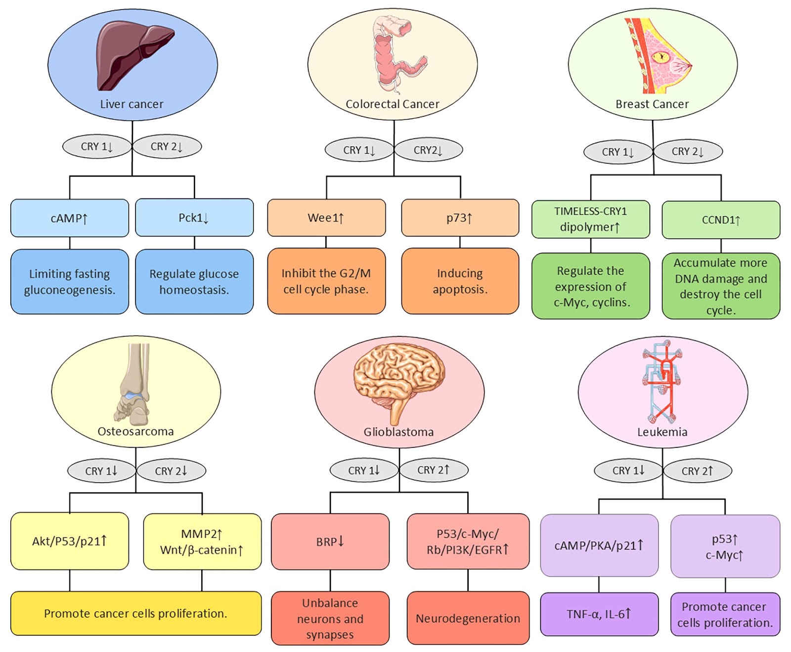
The role of cryptochrome (CRY) in cancer: molecular mechanisms and clock-based therapeutic strategies

Authors: Shuzhao Zhang, Xue Chen, Jiayi Li, An’an Xu, Ann M. Bode, Xiangjian Luo

The circadian rhythm is a phenomenon in which physiological, behavioral, and biochemical processes within an organism naturally fluctuate over a period of approximately 24 hours. This phenomenon is ubiquitous in living organisms. Disruption of circadian rhythms in mammals leads to different diseases, such as cancer, and neurodegenerative and metabolic disorders. In specific tissues, numerous genes have been found to have circadian oscillations, suggesting a broad role for rhythm genes in the regulation of gene expression. This review systematically summarizes the role of cryptochromes (CRYs) in the initiation and progression of different types of cancer and discusses the relationships between clock genes and the tumor microenvironment (TME), as well as clock-based therapeutic strategies.

The role of cryptochrome (CRY) in cancer: molecular mechanisms and clock-based therapeutic strategies
Graphical Abstract
Original ResearchVol. 57, Issue 7 • pp. 1068-1080DOI: 10.3724/abbs.2025026

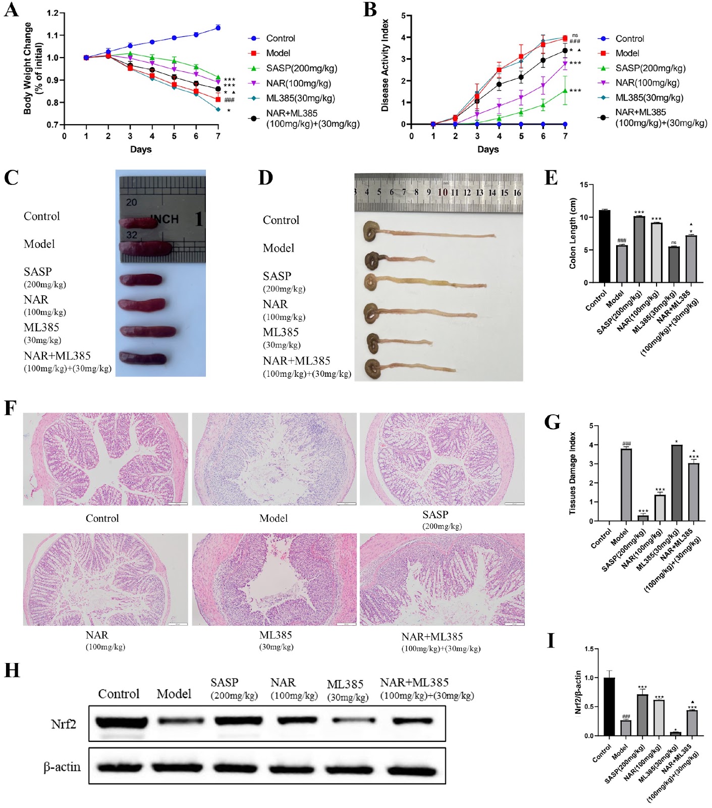
The protective effect of naringenin on ulcerative colitis in mice through increasing Nrf2 pathway activity

Authors: Jiaxiang Li, Li Hua, Meichun Hu, Ni Zhu, Sijin Dong, Xiaoli Jing, Zihuan Zhu, Yifei Liu, Yanhong Zhou

Ulcerative colitis (UC) is a chronic inflammatory disease with an increasing prevalence worldwide. Naringenin (NAR) has been proven effective in preventing UC, but its mechanism has not been fully elucidated. In this study, network pharmacology and bioinformatics methods are used to screen the genes associated with NAR and UC. A mouse model of dextran sulfate sodium (DSS)-induced UC is established. After treatment with NAR, the disease activity index (DAI) is scored, and colonic histopathology is observed via hematoxylin-eosin (HE) staining. The expressions of the nuclear factor erythroid 2-related factor 2 (Nrf2) signaling pathway and inflammation-related factors in the colons of UC mice are examined via western blot analysis and immunohistochemistry (IHC). The results of the animal experiments reveal that the model group of UC mice present the most severe weight loss and the highest DAI scores. After the administration of NAR, weight loss is alleviated, and DAI scores are reduced (P < 0.05). NAR improves pathological manifestations in the mouse colon, such as reducing inflammatory cell infiltration and restoring goblet cell loss (P < 0.05). NAR significantly increases the protein expression levels of Nrf2, heme oxygenase 1 (HO-1), and NAD(P)H dehydrogenase [quinone] 1 (NQO1) in the colon (P < 0.05) but decreases the protein expression levels of nuclear factor kappa-B (NF-κB), tumor necrosis factor-α (TNF-α), and interleukin-1β (IL-1β) (P < 0.05), thus alleviating the inflammatory response in UC model mice.

The protective effect of naringenin on ulcerative colitis in mice through increasing Nrf2 pathway activity
Graphical Abstract
Original ResearchVol. 57, Issue 7 • pp. 1081-1092DOI: 10.3724/abbs.2025003

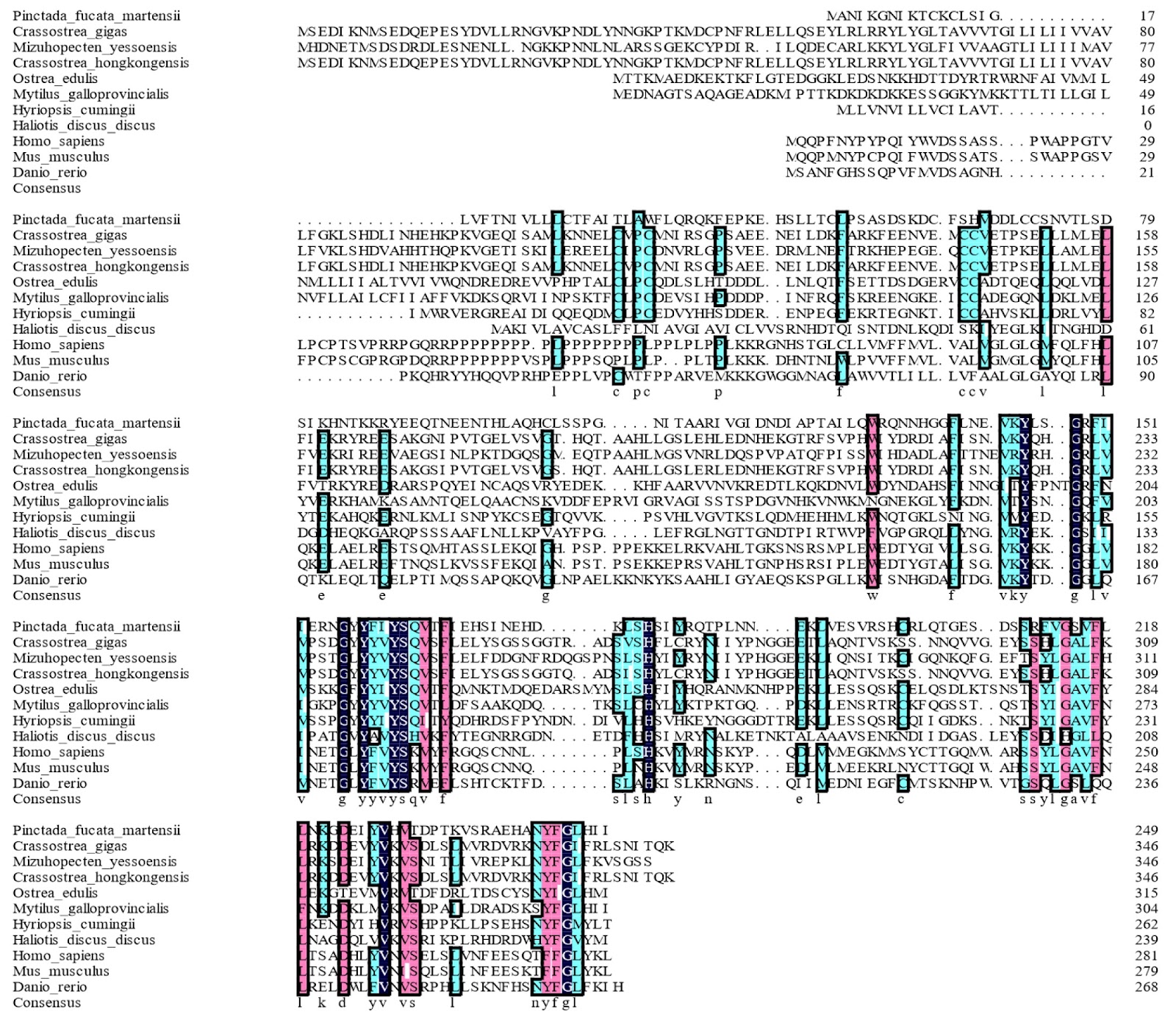
Regulation of immune responses by a tumor necrosis factor in pearl oysters: insights from PmTNF gene expression and function

Authors: Yifan Wu, Bidan Liang, Haiying Liang

Tumor necrosis factor (TNF) is a multifunctional cytokine that regulates cellular processes such as inflammation, apoptosis, differentiation, and proliferation and activates various functions of the immune system. This article reports the discovery and characterization of a novel tumor necrosis factor gene in the pearl oyster Pinctada fucata martensii, which is named PmTNF. The deduced PmTNF protein sequence displays the typical structural characteristics of a TNF domain, and phylogenetic analysis of the sequences of PmTNF and its putative orthologs shows that they conform to the current taxonomy. Analysis of PmTNF mRNA expression via real-time PCR reveals its constitutive expression in all the examined tissues, with the highest expression in the gills. Furthermore, PmTNF expression in the gills varies upon exposure to pathogen-derived stimuli, with modest upregulation in response to lipopolysaccharides, but with significant downregulation in response to polyinosinic:polycytidylic acid. Nucleus insertion surgery induces an increase in PmTNF mRNA level in the gills at 12 h postoperation. Knocking down PmTNF through RNA interference significantly inhibits the expressions of immune-related genes in the NF-κB signaling pathway in the gills by 24 h (P < 0.05). The function of PmTNF is further characterized by studying the activity of an engineered recombinant PmTNF protein (rPmTNF) in vivo. Upon nuclear insertion, treatment with rPmTNF for 6 h upregulates several genes in the NF-κB pathway. Similarly, rPmTNF increases the activities of the antioxidant enzymes, including superoxide dismutase, glutathione and peroxidase, which reflect the total antioxidant capacity. Collectively, these results indicate that PmTNF participates in pearl oyster immunity by modulating the NF-κB pathway and activating the antioxidant defense system.

Regulation of immune responses by a tumor necrosis factor in pearl oysters: insights from PmTNF gene expression and function
Graphical Abstract
Original ResearchVol. 57, Issue 7 • pp. 1115-1124DOI: 10.3724/abbs.2025018

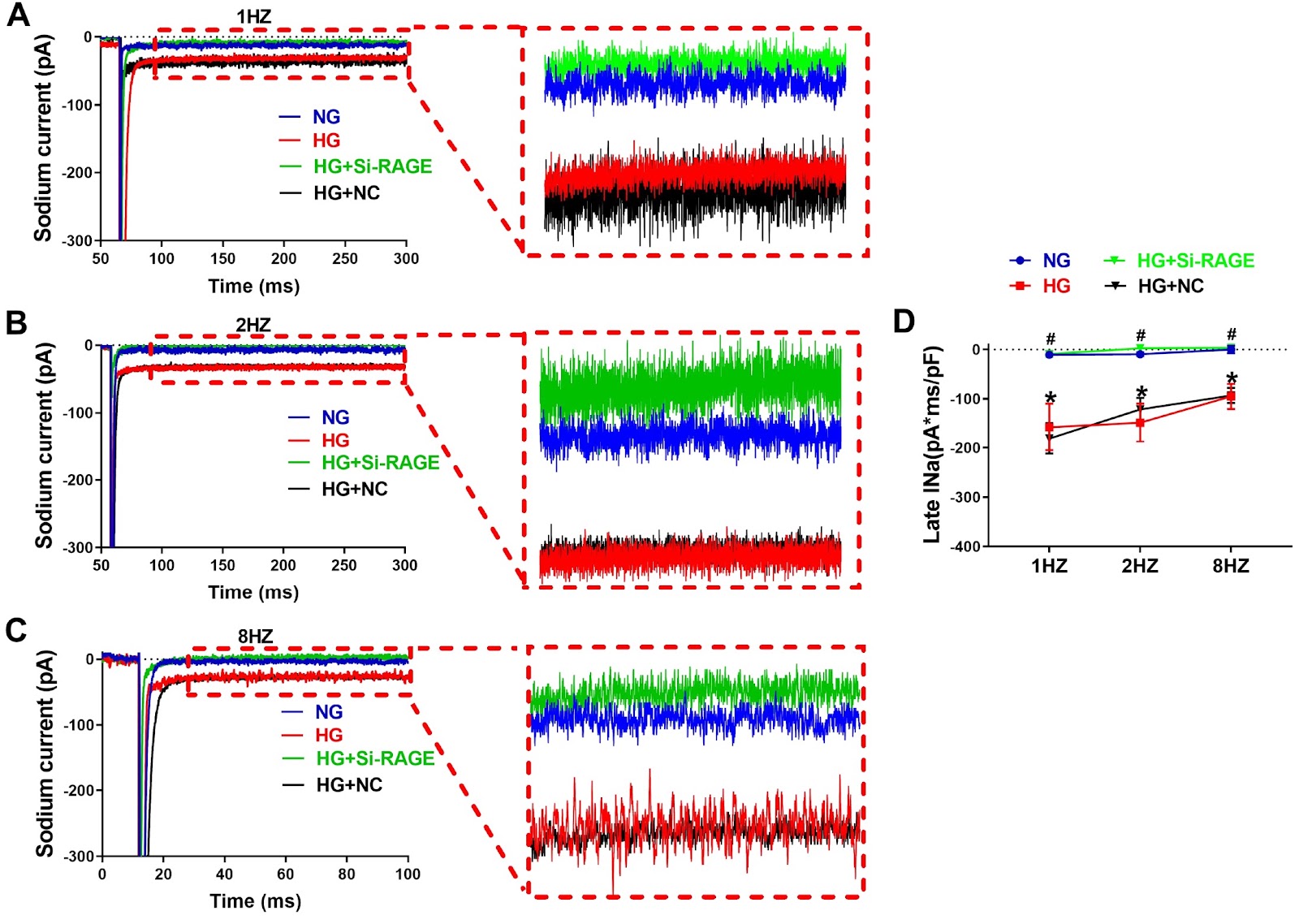
Atrial APD prolongation caused by the upregulation of RAGE and subsequent INaL increase in diabetic patients

Authors: Yingchun Luo, Wenbo Ma, Qi Kang, Han Pan, Ling Shi, Jiudong Ma, Jiahui Song, Dongmei Gong, Kai Kang, Xuexin Jin

Diabetes mellitus (DM) is a risk factor for the development of atrial fibrillation (AF). The action potential duration (APD) has been demonstrated to be prolonged in the atrium of diabetic mice. In contrast, the APD is generally shortened in AF patients. It is unclear what change occurs in the atrial APD of diabetic patients. In this study, we explore the APD change of atrial myocytes from diabetic patients and the underlying molecular mechanisms. The whole-cell patch-clamp technique is used to detect single-cell electrical activity in diabetic and nondiabetic human samples. The results show that both APD50 and APD90, the APD at 50% and 90% repolarization, are increased in diabetic patients compared with those in nondiabetic controls. The density of late sodium current (INaL) in the atrial myocytes of diabetic patients is greater than that in the myocytes of nondiabetic patients. The expression of receptor for advanced glycation end products (RAGE) is increased in the atria of diabetic patients. In cultured HL-1 cells, high glucose (HG) treatment increases INaL, and the expression of RAGE prolongs APD. The siRNA-mediated knockdown of RAGE reduces the INaL and shortens the APD. The APD is prolonged in the atria of diabetic patients because of the upregulation of RAGE and the subsequent increase in INaL. Our findings provide novel insights into atrial electrical remodeling in diabetic patients.

Atrial APD prolongation caused by the upregulation of RAGE and subsequent INaL increase in diabetic patients
Graphical Abstract
Original ResearchVol. 57, Issue 7 • pp. 1151-1163DOI: 10.3724/abbs.2024230

AKR1C3 protects cardiomyocytes against hypoxia-induced cell apoptosis through the Nrf-2/NF-κB pathway

Authors: Wenlu Zhang, Wei Tian, Xin Xia, Hua Tian, Ting Sun

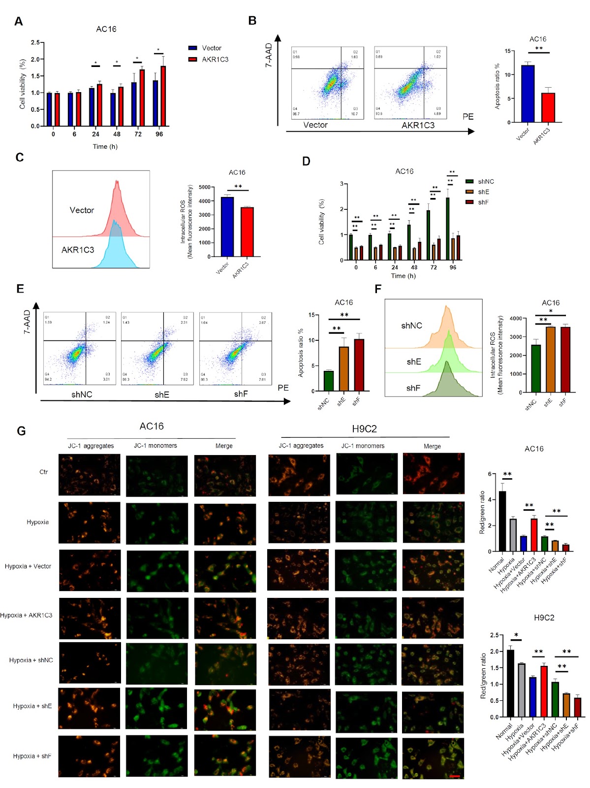
Hypoxia-induced apoptosis plays a critical role in the progression of various cardiac diseases, such as heart failure and acute myocardial infarction (AMI). Aldosterone reductase 1C3 (AKR1C3), a member of the aldo-keto reductase superfamily, participates in the metabolism of steroid hormones and redox reactions in vivo. Imbalances in prostaglandin levels have been linked to coronary events. However, the function and molecular mechanism by which AKR1C3 influences AMI are not yet fully understood. This study aims to investigate the role of AKR1C3 in hypoxia-induced myocardial cell damage and elucidate its mechanism. Our findings reveal that a hypoxic microenvironment triggers cardiomyocyte apoptosis and elevates AKR1C3 expression in H9C2 and AC16 cells, as well as in cardiac tissue from rats and mice with AMI. The overexpression of AKR1C3 promotes cardiomyocyte proliferation and cell vitality, whereas the silencing of AKR1C3 exerts the opposite effects in vitro. AKR1C3 protects cardiomyocytes against hypoxia-induced cell apoptosis by reducing ROS levels, preventing mitochondrial damage, and maintaining the oxygen consumption rate (OCR) and ATP production; conversely, AKR1C3 knockdown leads to adverse outcomes. Moreover, the application of a ROS inhibitor (MitoQ10) mitigates the increase in mitochondrial ROS in cardiomyocytes induced by AKR1C3 knockdown under hypoxic conditions. Mechanically, AKR1C3 increases Nrf-2 expression through the ubiquitin-proteasome pathway in cardiomyocytes and subsequently inhibits the NF-κB signaling pathway, thereby inhibiting Bax/caspase-3 signaling. Collectively, these results suggest that AKR1C3 prevents hypoxia-induced cardiomyocyte injury by modulating the Nrf-2/NF-κB axis, suggesting new insights into the mechanisms underlying myocardial protection.

AKR1C3 protects cardiomyocytes against hypoxia-induced cell apoptosis through the Nrf-2/NF-κB pathway
Graphical Abstract
Original ResearchVol. 57, Issue 7 • pp. 1205-1206DOI: 10.3724/abbs.2025088

Corrigendum to: Vitamin B6 prevents Isocarbophos-induced posterior cerebral artery injury in offspring rats through up-regulating S1P receptor expression

Authors: Yanhua Liu, Kunli Yang, Ling Wang, Jinfang Yang, Yang Wang, Hu Luo, Peng Li, Yaling Yin

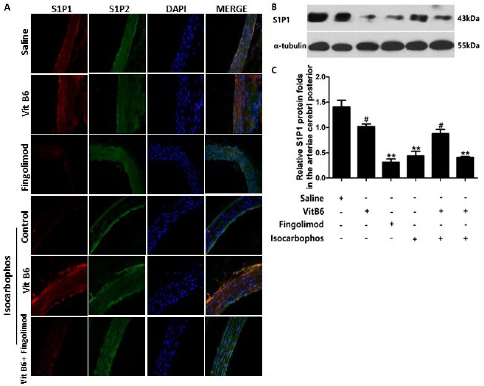
This corrigendum corrects errors in the original article 'Vitamin B6 prevents Isocarbophos-induced posterior cerebral artery injury in offspring rats through up-regulating S1P receptor expression' published in Acta Biochim Biophys Sin 2021, 53(12): 1691–1701. The errors were found in Figure 2B (Vit B6 + Fingolimod), Figure 5 (Saline), and Figure 7 (Isocarbophos/Control). The correct figures are shown. The authors apologize for the error. The corrigendum does not affect the interpretation of data and conclusions.

Corrigendum to: Vitamin B6 prevents Isocarbophos-induced posterior cerebral artery injury in offspring rats through up-regulating S1P receptor expression
Graphical Abstract