🧬 SinoBioData Academic Portal
🏛️ Indexed Academic JournalOriginal: 生物化学与生物物理学报

Acta Biochimica et Biophysica Sinica

Premier Chinese Biomedical Journal indexed in SinoBioData: Acta Biochimica et Biophysica Sinica (生物化学与生物物理学报).

Total Research Papers: 200
Access: 100% Free Open Access
Browse by Publication Year & VolumeReset All Filters ✕

Published Research PapersFiltered: Year 2025 • 57 • 6

Showing 4 of 200 peer-reviewed papers with full Graphical Abstracts.

Original ResearchVol. 57, Issue 6 • pp. 955-967DOI: 10.3724/abbs.2024235

Gastrodin inhibits reactive astrocyte-mediated inflammation in hypoxic-ischemic brain damage through S100B/RAGE-Smad3 signaling

Authors: Pengxiang Wang, Hanjun Zuo, Haolong Shi, Zhao Wang, Xueqi Ren, Jinsha Shi, Tao Guo, Xianfeng Kuang, Min Zhao, Jinghui Li, Juanjuan Li

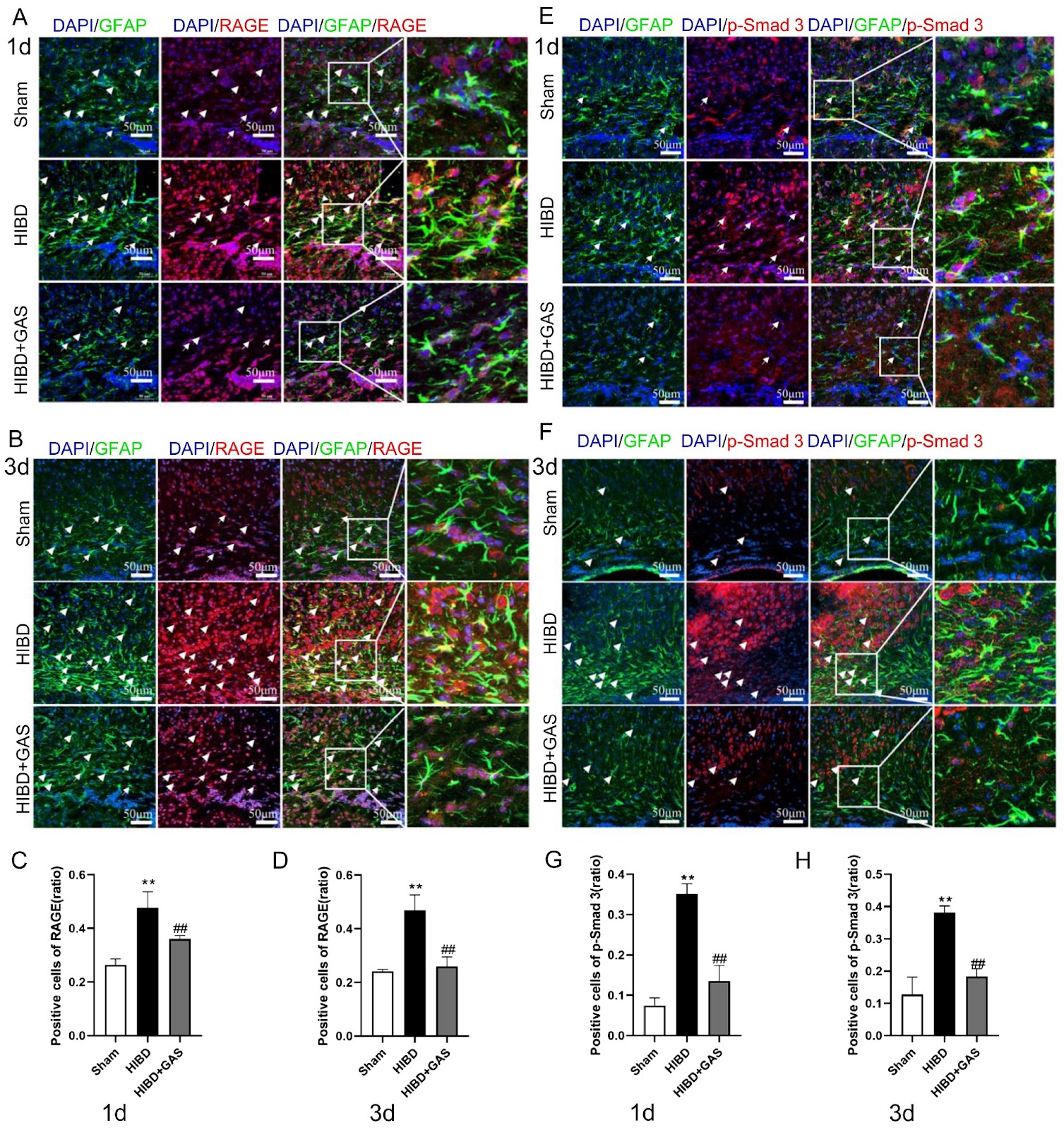
Activated astrocytes and their associated inflammatory responses play critical roles in the pathogenesis of hypoxic-ischemic brain damage (HIBD). Gastrodin (GAS), an anti-inflammatory herbal agent, is known to suppress microglial activation. Here, we investigate whether it exerts a similar effect on activated astrocytes and whether it acts through S100B/RAGE-Smad3 signaling. The expression changes of S100B/RAGE-Smad3 signaling pathway-related proteins, inflammatory factors and A1/A2 astrocyte markers were detected by ELISA, western blot analysis, immunofluorescence and immunohistochemistry. The results show that GAS decreases the expression of sRAGE in the brain tissue and S100B in the serum and brain tissue of HIBD mice. However, it promotes the expression of sRAGE in the serum of HIBD mice. Moreover, GAS inhibits the expressions of RAGE, p-Smad3, TNF-α, and C3 (A1 astrocyte marker), and promotes the expressions of S100A10 (A2 astrocyte marker) and BDNF in HIBD model mice, as well as in oxygen glucose deprivation (OGD)-treated TNC-1 astrocytes. The immunofluorescence and immunohistochemical results of RAGE and p-Smad3, as well as the immunofluorescence results of C3 and S100A10, reveal the same trend. Interestingly, FPS-ZM1 (a specific inhibitor of RAGE) inhibits the expressions of p-Smad3, TNF-α, C3, and S100A10, but promotes that of BDNF compared with those in the OGD group. The combination of GAS and FPS-ZM1 further decreases the expression of C3. These results indicate that GAS can inhibit the activation of Smad3 through S100B/RAGE signaling and regulate the expression of A1/A2-type astrocytes.

Gastrodin inhibits reactive astrocyte-mediated inflammation in hypoxic-ischemic brain damage through S100B/RAGE-Smad3 signaling
Graphical Abstract
Original ResearchVol. 57, Issue 6 • pp. 1029-1032DOI: 10.3724/abbs.2025040

Histone acetylases are required for iron homeostasis in yeast

Authors: Jian Zhang, Yong Xue, Xinya Zhang, Renjie Qi, Yaqi Zhang, Chen Lu, Zhidan Luo

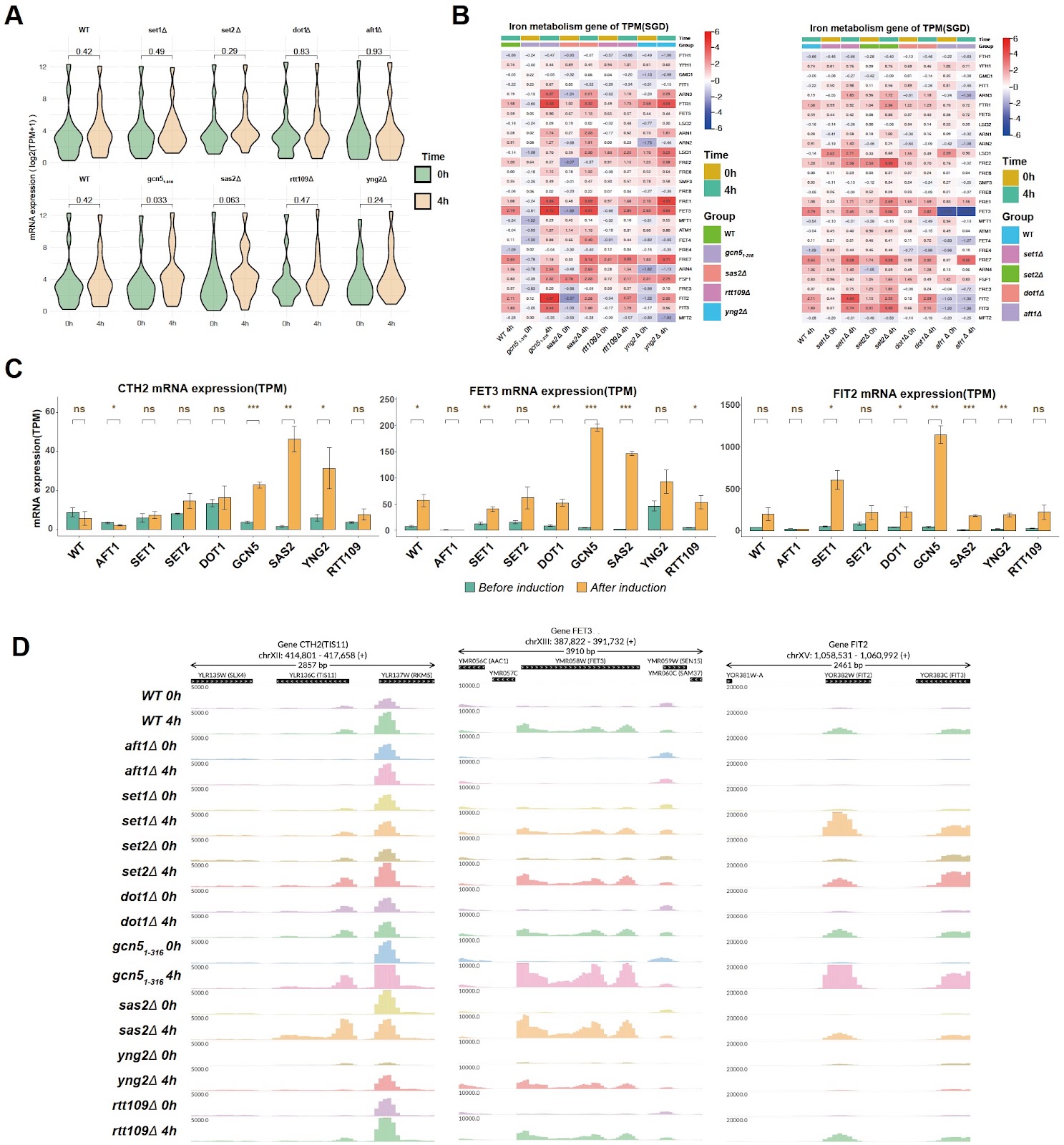
Iron, an ancient and essential transition metal, is involved in various biological functions, including oxygen transport, DNA synthesis, heme production, and iron-sulfur clusters, which participate in electron transport, DNA repair, and other cellular processes. However, excessive iron can lead to oxidative stress, lipid peroxidation, and cell damage. Thus, maintaining the iron content within an appropriate safe range and maintaining the balance of iron metabolism play crucial roles in both cellular function and human health [1]. An important aspect of maintaining the balance of iron homeostasis is the regulation of the iron uptake system. In Saccharomyces cerevisiae, cells can either obtain iron from the external environment via the non-reducing siderophore transport system or transport iron from the extracellular space to the intracellular space via the reducing iron transport system [2]. Iron uptake system-related genes are regulated mainly by the transcription factor Aft1p. During iron deficiency, Aft1p translocates into the nucleus, binds to genes involved in iron metabolism, and regulates the expressions of genes involved in iron uptake systems [3]. In addition, when there is a problem in the synthesis of iron-sulfur clusters in the mitochondria, such as the lack of the iron chaperone Yfh1p, which promotes the synthesis of iron-sulfur clusters, the transcription and nuclear entry of the transcription factor Aft1p are also activated, thereby regulating the expressions of iron metabolism-related genes [4]. In addition to transcription factors, gene expression is also regulated by histones and their modifications at the epigenetic level. For example, histone H3K4 methylation is related to gene activation, H3K36 methylation plays an important role in the elongation of transcription, and histone acetylation results in the loss of nucleosome structure and facilitates gene expression [5]. Therefore, histone modifications should also play important roles in the regulation of iron homeostasis. The relationship between histone modifications and iron homeostasis has been reported in the literature. For example, the DNA methylation-binding protein MBD5 can change histone acetylation in the promoter region of the ferritin gene by recruiting the histone acetylase KAT2A protein [6]. Histone acetylation has also been reported to be reduced in iron-deficient environments [7,8], and direct effects of histone acetylation on iron homeostasis gene loci have also been reported both in C. albicans [9] and mammals [10]. More recently, the histone H3-H4 tetramer was found to be a copper reductase enzyme, and H3-mediated Cu+ toxicity is a major determinant of the cellular functional pool of iron-sulfur (Fe-S) clusters [11,12]. However, information on the role of histone modifications in the regulation of iron homeostasis is limited. The mechanism by which and how histone modifications are involved in the transcriptional regulation of iron uptake-related genes or the iron deficiency response require further investigation. To determine whether histone acetylation and methylation are involved in the iron deficiency response, we first deleted the histone acetylase genes, including GCN5 (histones H2B and H3 N-terminal lysine acetylase, partial deletion of the ADA2 interaction sequence [13]), RTT109 (H3 lysine 9 and 56 acetylase), SAS2 (H4 lysine 16 acetylase), and YNG2 (subunit of the histone acetyltransferase complex NuA4 for acetylation of histone H4 or histone H2A) in the wild-type (WT) strain, as well as the histone methyltransferases SET1 (H3 lysine 4 methyltransferase), SET2 (H3 lysine 36 methyltransferase) and DOT1 (H3 lysine 79 methyltransferase). The genomic deletions were confirmed by colony PCR and genomic coverage analysis, as depicted in Supplementary Figures S1 and S2. The sensitivity of single histone modification enzyme mutants to iron deficiency induced by the iron chelator bathophenanthroline disulfonate (BPS) was tested. As shown in Supplementary Figure S3, the histone acetylation-related mutants gcn51–316, rtt109Δ, sas2Δ, and yng2Δ did not exhibit significant growth defects compared with the WT on the YPD + BPS plate. None of the histone methyltransferase knockout strains presented significant growth defects. As a positive control, the iron-responsive transcription factor gene AFT1 knockout strain grew slowly on YPD + BPS plates. It is possible that histone modifications do not have a strong effect on the equilibrium status of iron deficiency but still regulate transcription induction during the iron deficiency response. To investigate the role of histone modifications during the induction of the iron deficiency response, the expressions of iron response genes in the wild-type and mutant strains before and 4 h after BPS treatment were examined.
Histone acetylases are required for iron homeostasis in yeast
Graphical Abstract
Original ResearchVol. 57, Issue 6 • pp. 890-900DOI: 10.3724/abbs.2024202

Similarities and differences in the response and molecular characteristics of peripheral sensory neurons associated with pain and itch

Authors: Li Liu, Jiemin Yin, Youqiang Meng, Congrui Ye, Junhui Chen, Sa Wang, Wen Yin, Po Gao, Yingfu Jiao, Weifeng Yu, Yinghui Fan

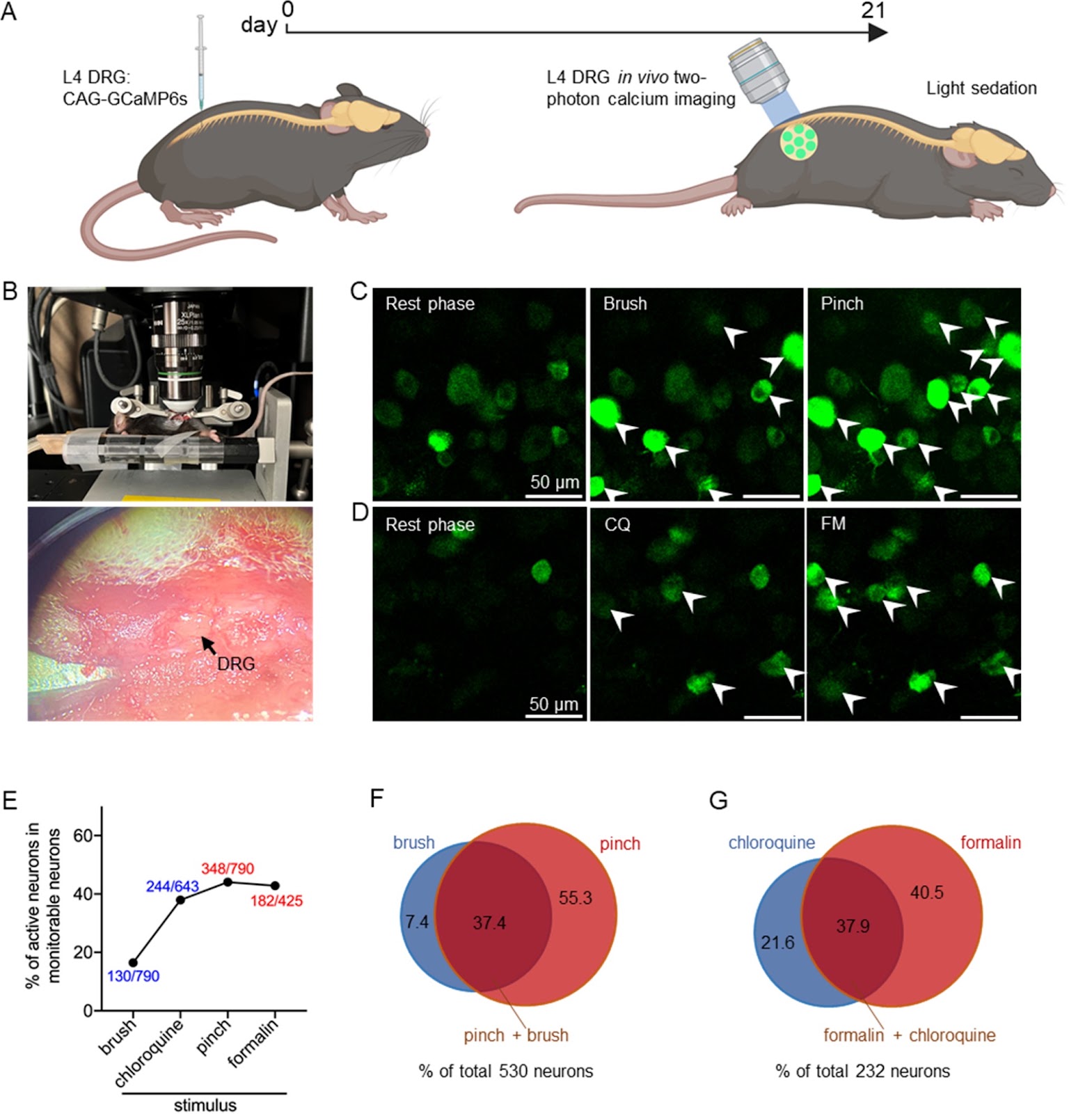
Dorsal root ganglion (DRG) neurons are responsible for the primary detection and transmission of peripheral noxious stimuli, mainly pain and itch. However, as two distinct noxious sensations, how DRG neurons respond differently to and code pain and itch is still an attractive topic. Here, we investigate the response and activation spectrum of DRG neurons under peripheral pain and itch stimuli using in vivo two-photon calcium imaging and find differences in the response intensity to pain and itch between multisensory neurons (both pain and itch) and single-sensory neurons (either pain or itch). In addition, single-cell RNA sequencing (scRNA-seq) is used to reveal the heterogeneity of distinct subpopulations on the basis of their expressions of pain- or itch-related marker genes and to determine the similarities and differences in their transcriptomic changes under chronic pain and itch. Our results show that primary sensory neurons with different sensory patterns respond differently to the same nociceptive stimuli. Additionally, distinct clusters of neurons exhibit unique transcriptomic changes in the development of chronic pain and itch, which may offer new insights for treating these conditions.

Similarities and differences in the response and molecular characteristics of peripheral sensory neurons associated with pain and itch
Graphical Abstract
Original ResearchVol. 57, Issue 6 • pp. 871-878DOI: 10.3724/abbs.2025011

Melanoma-derived versican reactivates tumor-associated macrophages by upregulating pyruvate carboxylase through TLR2-MyD88-RelB axis under normoxia

Authors: Yuxin Shu, Linmin Zhou, Jinqin Qian, Wei-Guo Zhu

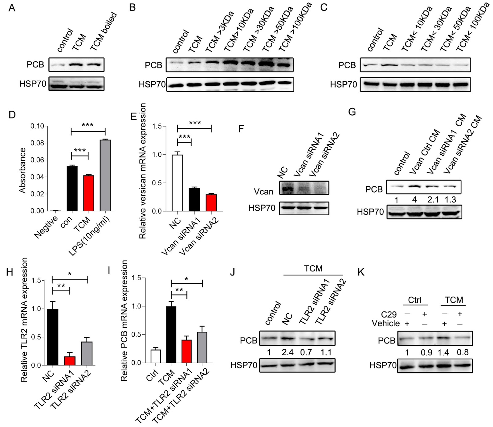
Relieving hypoxia in the tumor microenvironment (TME) promotes innate and adaptive immunity. Our previous research demonstrated that reoxygenation of the TME promotes the phagocytosis and tumor-killing functions of tumor-associated macrophages (TAMs) by upregulating pyruvate carboxylase (PCB). However, the mechanism remains obscure. In the present study, we find that versican derived from melanoma cells binds to TLR2 and activates the downstream transcription factor RelB, which transcribes PCB under normoxia. Blocking the versican-TLR2-MyD88-RelB axis not only reverses the upregulation of PCB in TAMs but also hinders the clearance of tumor cells by TAMs. Our work suggests a pathway that modulates the functions of TAMs under normoxia, which could be harnessed for strengthening anti-tumor immunity.

Melanoma-derived versican reactivates tumor-associated macrophages by upregulating pyruvate carboxylase through TLR2-MyD88-RelB axis under normoxia
Graphical Abstract