🧬 SinoBioData Academic Portal
🏛️ Indexed Academic JournalOriginal: 生物化学与生物物理学报

Acta Biochimica et Biophysica Sinica

Premier Chinese Biomedical Journal indexed in SinoBioData: Acta Biochimica et Biophysica Sinica (生物化学与生物物理学报).

Total Research Papers: 200
Access: 100% Free Open Access
Browse by Publication Year & VolumeReset All Filters ✕

Published Research PapersFiltered: Year 2026 • 58 • 5

Showing 9 of 200 peer-reviewed papers with full Graphical Abstracts.

Original ResearchVol. 58, Issue 5 • pp. 1155-1169DOI: 10.3724/abbs.2026055

Atractylenolide I mitigates Alzheimer’s disease pathology in ApoE–/– mice via ARG1/nNOS axis and lipid homeostasis regulation

Authors: Xun Zhou, Rui Wang, Jingsi Yan, Xiaolang Wu, Dongsheng Yuan, Qi Wang, Huilin Li, Wei Zhao

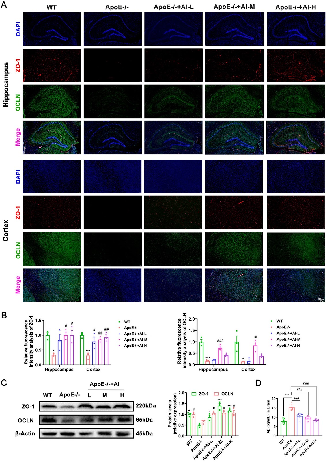
Apolipoprotein E (ApoE) serves as a critical molecular nexus between Alzheimer’s disease (AD) and atherosclerosis, two age-associated inflammatory disorders that share vascular pathology, amyloid-beta (Aβ) deposition, and lipid dysregulation. Atractylenolide I (AI), a promising therapeutic candidate derived from Atractylodes macrocephala Koidz., exhibits multimodal bioactivities with demonstrated anti-inflammatory and neuroprotective properties. To explore its therapeutic potential against AD pathology, we use high-fat diet (HFD)-fed ApoE knockout (ApoE–/–) mice treated with or without AI for 12 weeks. Integrated bioinformatics analyses and experimental validation reveal that AI treatment markedly attenuates systemic lipid dyshomeostasis, particularly cerebral lipid deposition, suppresses neuroinflammation via downregulation of M1 macrophage polarization markers, and restores cognitive function through neuronal preservation in hippocampal regions. Mechanistically, AI orchestrates cholesterol efflux by up-regulating ATP-binding cassette transporter A1 (ABCA1) and liver X receptor (LXR) expression, while concurrently modulating the abundance of arginine biosynthesis metabolites (urea, malic acid, and creatinine) to rebalance neurovascular homeostasis. Notably, western blot and RT-qPCR analyses reveal that AI differentially regulates key enzymes including arginase 1 (ARG1) and simultaneously upregulates the expression of neuronal nitric oxide synthase (nNOS). Further molecular docking and surface plasmon resonance (SPR) analyses confirm the direct binding of AI to ARG1, indicating a novel neuroprotective mechanism involving the modulation of arginine metabolism. These findings delineate the pleiotropic effects of AI against AD pathology and establish a preclinical foundation for the development of AI-based therapeutics targeting neurodegenerative-cardiovascular comorbidities.
Atractylenolide I mitigates Alzheimer’s disease pathology in ApoE–/– mice via ARG1/nNOS axis and lipid homeostasis regulation
Graphical Abstract
Original ResearchVol. 58, Issue 5 • pp. 1183-1186DOI: 10.3724/abbs.2025242

Expression characteristics of serum exosomal microRNAs in patients with liver injury induced by anti-tuberculosis drugs

Authors: Yinpeng Jin, Xiaofang Yu, Mingquan Guo, Li Li, Shuangshuang Sun, Liling Yang, Ying Yuan, Qingchun Fu, Rongfeng Shi, Meng Jin

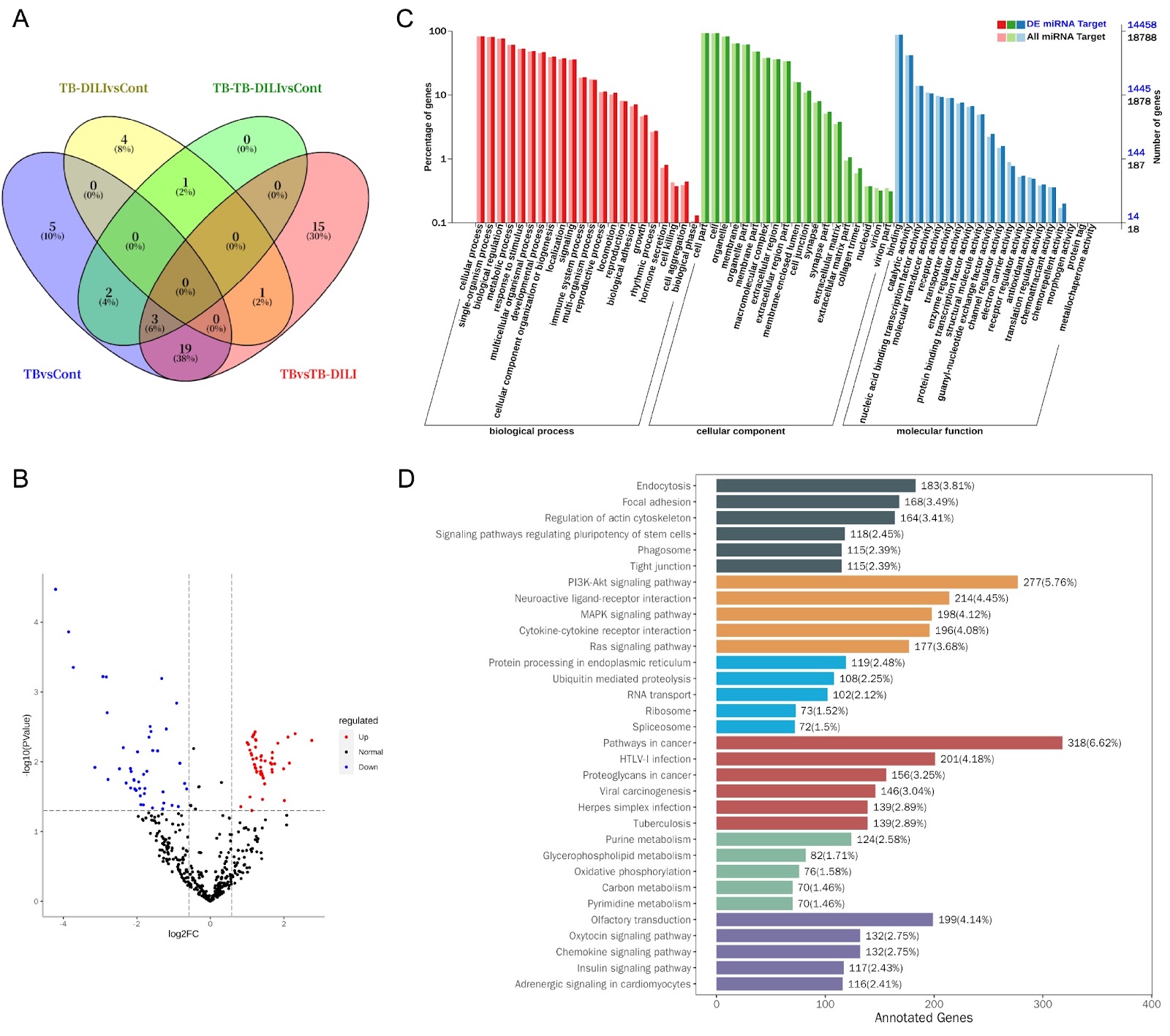
Drug-induced liver injury (DILI) caused by anti-tuberculosis drugs is a serious clinical problem that can lead to acute liver failure and even death. Current diagnosis relies on lagged indicators such as serum transaminase levels, which rise only 48–72 hours after liver injury. This study is the first to systematically analyze the microRNA expression profile of serum exosomes in patients with anti-tuberculosis drug-induced liver injury (TB-DILI) to discover early diagnostic markers. A total of 12 tuberculosis patients and 6 normal controls were included. Serum exosomes were isolated and characterized, and small RNA sequencing identified 701 miRNAs, with 128 differentially expressed between TB-DILI and TB groups. Notably, miR-122-5p was upregulated and has shown early warning value. Target gene prediction and enrichment analysis revealed involvement in GTPase activity regulation, cell migration, and BMP signaling. These findings suggest that exosomal miRNAs, particularly miR-122-5p, may serve as early diagnostic biomarkers for TB-DILI.

Expression characteristics of serum exosomal microRNAs in patients with liver injury induced by anti-tuberculosis drugs
Graphical Abstract
Original ResearchVol. 58, Issue 5 • pp. 975-988DOI: 10.3724/abbs.2025207

COCA-seq: genome-wide mapping of O-GlcNAc-associated open chromatin

Authors: Chang Ge, Ran Zhao, Hongyu Jiang, Qingbin Chen, Zhentao Yu, Hankai Yang, Xuan Jiang, Qile Ma, Lirui Han, Kairan Yu, Guofang Li, Huang Huang, Wei Wang, Yubo Liu, Qingyue Zhang, Xing Jin

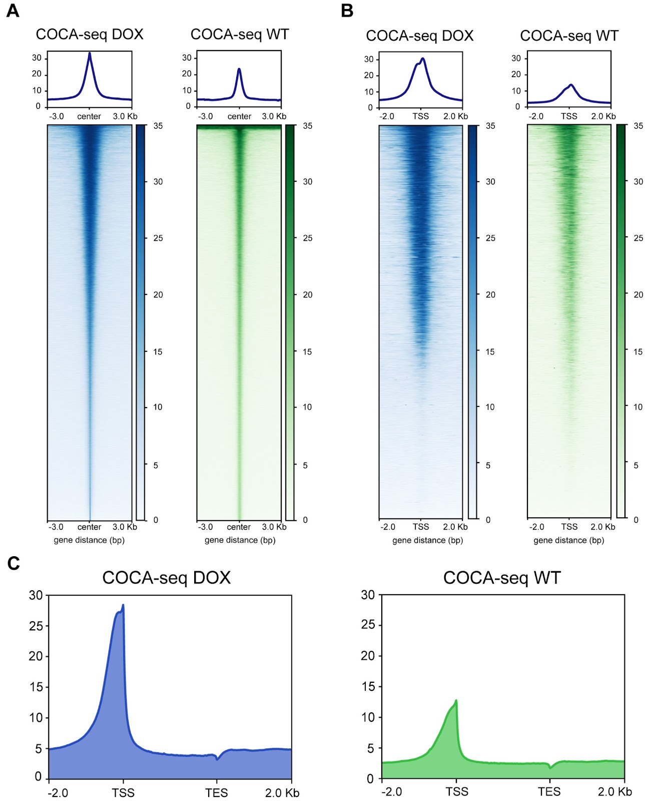
O-GlcNAcylation, a prevalent reversible post-translational modification, intricately alters non-histone proteins, influencing the organization of gene transcriptional regulation within the accessible chromatin environment. This nucleoplasmic landscape, characterized by histone-free regions, fundamentally enables O-GlcNAc-mediated modulation through dynamic accessibility. However, unraveling the O-GlcNAc-open chromatin interplay that governs sophisticated transcriptional regulatory networks remains constrained by current techniques, which lack the resolution to probe this spatiotemporal crosstalk. Here, we report a general strategy to systematically and chemoselectively profile O-GlcNAc-associated chromatin accessibility on a genome-wide scale (COCA-seq). Through comprehensive validation across low- and high-throughput levels, we demonstrate COCA-seq’s dual fidelity in both O-GlcNAc chemoselectivity and open chromatin specificity. We employ it to delve into doxorubicin resistance for breast cancer, scrutinizing pivotal regulatory genes and transcription factors implicated in this complex biological event. By integrating bulk RNA-seq with COCA-seq, we offer a multiomics perspective, shedding light on related biological processes and pathways like drug efflux and stress homeostasis, thereby uncovering potential mechanisms by which O-GlcNAc-associated open chromatin orchestrates tumor drug resistance. COCA-seq emerges as a general and versatile tool across various biological contexts, poised to reveal the landscape of O-GlcNAc-associated open chromatin regions across the genome and decipher the significance of glycosylation behind it.

COCA-seq: genome-wide mapping of O-GlcNAc-associated open chromatin
Graphical Abstract
Original ResearchVol. 58, Issue 5 • pp. 989-1007DOI: 10.3724/abbs.2025234

Cancer-specific bivalent promoters featuring low-level H3K27me3 signals favor active transcription and govern the cancer cell state transition

Authors: Fan Yang, Guanlan Fan, Jing Cao, Qiuyan Zhao, Kexiu Guo, Min Liu, Xin Yin, Hongying Zong, Feng Li, Fubing Wang, Jie Xiong

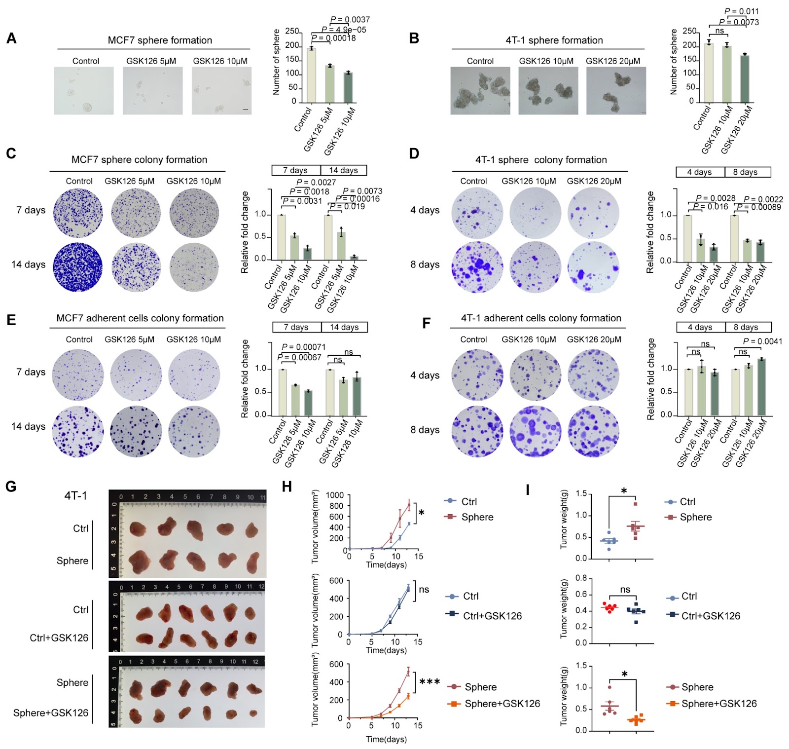
Bivalent chromatin maintains genes in low-expression, poised states in embryonic stem cells (ESCs). However, bivalent promoters correlate with the transcriptional activation of oncogenic programs in malignancies, a seemingly contradiction that remains to be resolved. Here, we identify a class of cancer-specific bivalent promoters (CSBPs) through the integration of a system-level longitudinal framework. Compared with ESCs, CSBPs are characterized by lower and narrower H3K27me3 deposition alongside abundant H3K4me3, thus permitting the persistent expression of genes critical for cancer stem cell (CSC) formation and maintenance, as exemplified by SOX9. The generation of CSBPs is essentially induced by the acquisition of H3K27me3 during cell state transition, which is mediated by specific binding of PRC2.1 and the de novo recruitment of PRC2.2. Notably, disrupting the bivalency of CSBPs significantly increases H3K4me3 levels, leading to hyperactivation of CSBPs and eventually inhibiting clonal expansion of CSCs and impairing tumorigenesis. Our study not only helps explain the puzzle of transcriptionally active bivalent genes in cancer but also provides insights into the development of therapies targeting phenotypic plasticity.

Cancer-specific bivalent promoters featuring low-level H3K27me3 signals favor active transcription and govern the cancer cell state transition
Graphical Abstract
Original ResearchVol. 58, Issue 5 • pp. 1102-1118DOI: 10.3724/abbs.2025136

Metabolic analysis and identification of potential biomarkers of early-stage melanoma lung metastasis

Authors: Chunying Gu, Hongyu Liu, Guangyu Jiang, Ying Lv, Jiafu Liu

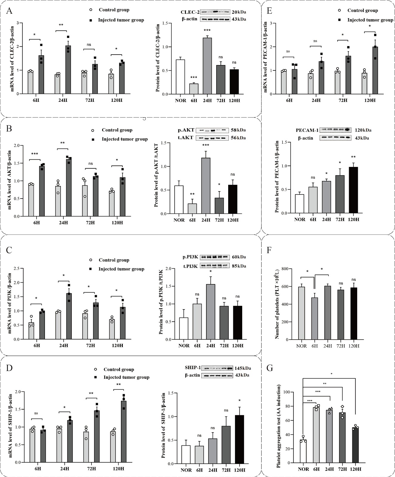
Tumor cells exhibit a notable ability to adapt to constantly changing microenvironments and possess distinct metabolic traits during metastasis. This study aims to establish a melanoma lung metastasis model in mice to elucidate the metabolic mechanisms involved in early-stage metastasis prior to treatment. The male C57BL/6 mice are divided into five groups based on time intervals of 6, 24, 72, and 120 h post-injection (SKCM-M groups) of melanoma cells, as well as a normal control group (NOR group). Our results demonstrate that platelet activation mainly occurs in the initial phases of metastasis to help tumor cells survive. NMR-based metabolomics analysis of mouse lung tissues identifies distinct metabolites and pathways associated with early-stage metastasis, revealing significant alterations in energy and amino acid metabolism during tumor progression. Further analysis indicates that methylxanthine and allantoin could serve as potential biomarkers for monitoring the early progression of tumor metastasis in cancer patients, providing novel insights into early diagnostic strategies for lung metastasis.

Metabolic analysis and identification of potential biomarkers of early-stage melanoma lung metastasis
Graphical Abstract
Original ResearchVol. 58, Issue 5 • pp. 1045-1054DOI: 10.3724/abbs.2025168

The DNA-PKcs-primary cilia axis maintains ionizing radiation-induced senescence in tumor cells

Authors: Xiuzhu Liu, Li Wei, Rong Zhang, Jiaxin Chen, Tongshan Zhang, Junrui Hua, Jufang Wang, Jinpeng He, Xiaodong Xie

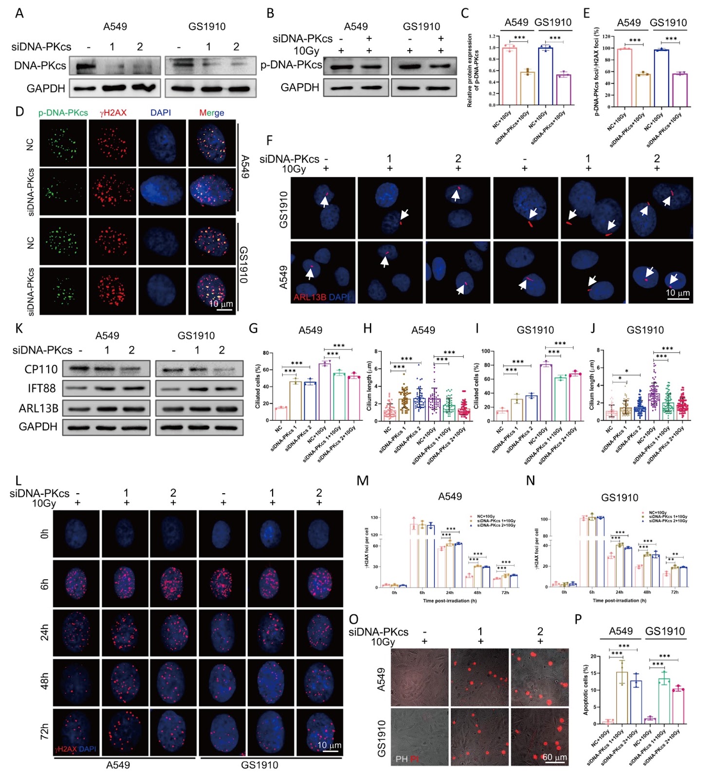
Senescence is a cellular response closely associated with genotoxic stress and plays a critical role in determining cell fate following irradiation exposure. Primary cilia, which are sensory organelles on the cell surface, detect and transmit diverse signaling cues. However, the relationship between primary cilia and senescence in long-term cell fate decisions after ionizing radiation remains poorly understood. Here, we show that the DNA-dependent protein kinase catalytic subunit (DNA-PKcs) colocalizes with centromeres during various stages of mitosis, whereas during interphase, phosphorylated DNA-PKcs (p-DNA-PKcs) is confined to the nucleus in tumor cells. Following irradiation exposure, primary cilia are formed and persistently maintained at high levels in senescent tumor cells. Inhibition of DNA-PKcs enhances primary cilia formation, whereas combined inhibition with siDNA-PKcs and irradiation reduces cilia generation. Moreover, chloral hydrate-induced primary cilia removal results in senescent cell death and decreases p-DNA-PKcs protein expression. Notably, treatment with the apoptosis inducer ABT263 also leads to increased cell death and decreased incidence of primary cilia. Inhibition of either primary cilia or DNA-PKcs further enhances the radiosensitivity of tumor cells. These findings suggest that DNA-PKcs contributes to primary cilia formation after irradiation and plays a critical role in both the induction and maintenance of cellular senescence.

The DNA-PKcs-primary cilia axis maintains ionizing radiation-induced senescence in tumor cells
Graphical Abstract
Original ResearchVol. 58, Issue 5 • pp. 1069-1082DOI: 10.3724/abbs.2025163

Myocyte enhancer factor 2A orchestrates vascular redox homeostasis via direct transcriptional activation of SIRT1

Authors: Benrong Liu, Lei Fang, Chunxia Miao, Xinyu Wen, Xiumiao Zheng, Minxing Xu, Junli Lin, Yujuan Xiong, Shi-Ming Liu

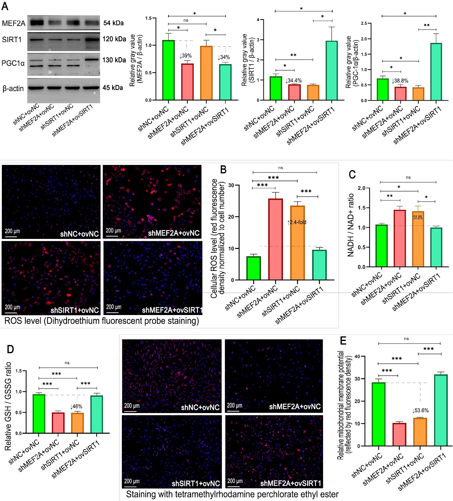
Myocyte enhancer factor 2A (MEF2A), a transcription factor implicated in coronary artery disease, remains unexplored in vascular redox regulation. To address this gap and overcome the limitations of current antioxidant therapies, we investigate the role of MEF2A in oxidative defense via human umbilical vein endothelial cells (HUVECs) and murine models. Adenoviral vectors encoding MEF2A-specific shRNAs or mRNAs are used to silence or overexpress MEF2A in HUVECs. For in vivo validation, endothelial-targeted MEF2A knockdown is achieved via AAV1-shRNA delivery in mice fed with a high-fat diet. Systemic redox status is assessed by measuring reactive oxygen species (ROS), glutathione homeostasis (GSH/GSSG ratio), the NADH/NAD+ balance, the mitochondrial membrane potential (ΔΨm), and 8-hydroxy-2′-deoxyguanosine (8-OHdG). Mechanistic insights are derived from immunofluorescence, qPCR, western blotting, and dual-luciferase reporter assays. MEF2A silencing induces redox imbalance, characterized by elevated ROS, a reduced GSH/GSSG ratio, and ΔΨm collapse. Conversely, MEF2A overexpression synergizes with SIRT1 to restore the glutathione pool, maintain NAD+ homeostasis, and suppress ROS under oxidative stress. Chromatin immunoprecipitation confirms that MEF2A directly binds to two cis-elements in the SIRT1 promoter, driving transcriptional activation. In vivo, MEF2A-deficient mice present increased vascular oxidative damage, as indicated by elevated DNA damage marker (8-OHdG) and ROS levels. The downregulation of SIRT1/PGC-1α in MEF2A-silenced cells is verified in vivo. Our findings establish MEF2A as a master regulator of endothelial redox defense via the SIRT1-PGC-1α axis, providing a mechanistic foundation for the treatment of oxidative cardiovascular disorders. This work suggests that pharmacological MEF2A activation is a novel strategy for precision antioxidant therapy in vascular medicine.

Myocyte enhancer factor 2A orchestrates vascular redox homeostasis via direct transcriptional activation of SIRT1
Graphical Abstract
Original ResearchVol. 58, Issue 5 • pp. 947-962DOI: 10.3724/abbs.2025212

Antibody-oligonucleotide conjugates for spatial proteomics: principles, applications, and challenges

Authors: Yinghui Qiu, Chunlan Li, Peiying Ye, Haiyun Zhang, Yanxiu Liu, Weiyan Ma, Chen Lin, Rongqin Ke

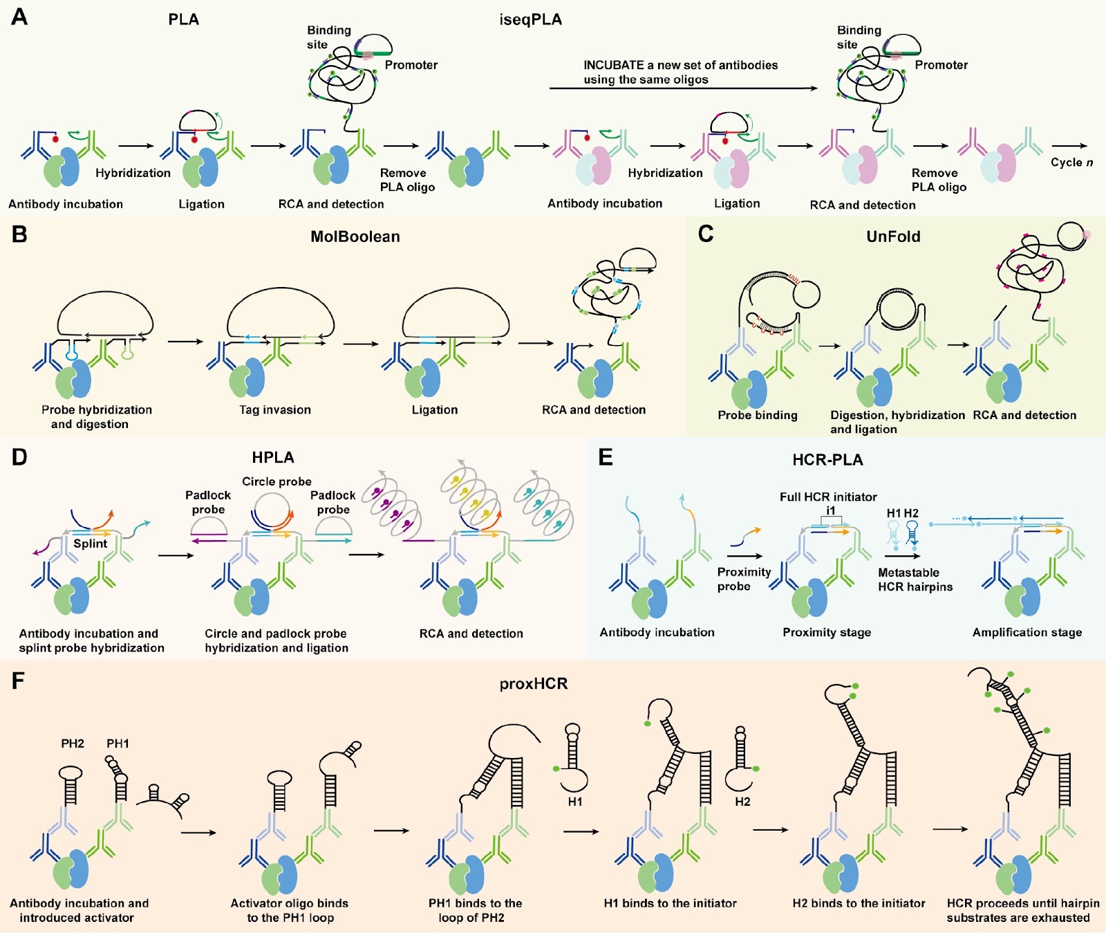
Spatial biology aims to elucidate cellular organization, function, and interactions within native tissue contexts, offering key insights into both normal physiology and disease. Spatial proteomics complements this by enabling high-resolution mapping of protein localization and abundance, directly reflecting functional cellular states. Unlike transcriptomics, which infers potential activity, proteomics captures actual molecular functions, including post-translational modifications and dynamic interactions. However, in situ protein profiling poses significant challenges, as proteins cannot be directly sequenced or easily targeted via nucleic acid hybridization. Antibody-oligonucleotide conjugates (AOCs) address this limitation by converting protein recognition into a DNA-based readout, thereby enabling sensitive and scalable detection. In this review, we outline the core principles of AOC-based spatial proteomic technologies, including multiplexed protein analysis, in situ protein-protein interactions, and integration with other biomolecular data. We highlight their applications in decoding tissue complexity and disease pathology and examine key technical challenges that remain. Overall, AOCs offer distinct advantages, including DNA-mediated signal amplification, spatially resolved proteomic profiling, and compatibility with multi-omics approaches, positioning them as powerful platforms in the advancement of spatial biology.

Antibody-oligonucleotide conjugates for spatial proteomics: principles, applications, and challenges
Graphical Abstract
Original ResearchVol. 58, Issue 5 • pp. 1023-1031DOI: 10.3724/abbs.2025183

Gut microbiota and bile acids changes in MASLD mice model with hepatic PLD1 knockout

Authors: Yushang Zhao, Huan Wang, Wanling Lin, Hui Wang, Lin-Lin Cao

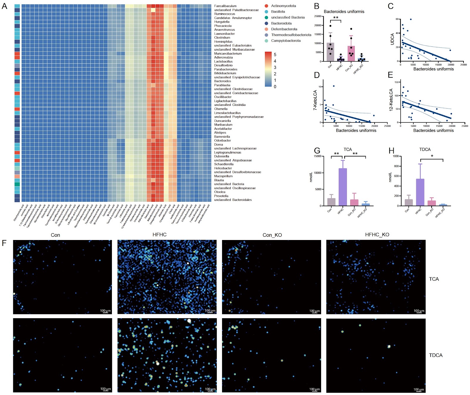
Hepatocyte phospholipase D1 (PLD1) knockout alleviates metabolic dysfunction-associated steatotic liver disease (MASLD) in mice, but the underlying mechanism is largely unknown. In this study, the mice are divided into four groups: Con (wild-type mice with normal control diet), HFHC (wild-type mice with high-fat diet), Con_KO (hepatocyte PLD1-knockout mice with normal control diet), and HFHC_KO (hepatocyte PLD1-knockout mice with high-fat diet). Intestinal contents of mice are analyzed via metagenomics and metabolomics, and the liver bile acids are assessed by mass spectrometry imaging. The results show that at the phylum level the abundance of Bacillota in the intestines of MASLD model mice is significantly increased, whereas that of Bacteroidota significantly is decreased. However, after the deletion of hepatocyte PLD1, Pseudomonadota and Candidatus Bathyarchaeota are significantly decreased in the MASLD model mice. At the species level, compared with that in the Con group, the abundance of Faecalibaculum rodentium is significantly increased in the HFHC group, whereas hepatocyte PLD1 knockout causes the abundances of Desulfovibrionaceae bacterium LT0009 and Lachnospiraceae bacterium 10-1 to be significantly decreased. In terms of intestinal bile acids, the levels of two bile acids (hyodeoxycholic acid and glycolithocholic acid) differ between the HFHC_KO group and the HFHC group. Association analysis shows that Faecalibaculum co-occurs with DCA, βMCA, ΩMCA and αMCA, while probiotic Bacteroides uniformis is significantly correlated with UDCA, 12-KetoLCA, and 7-KetoLCA. Finally, mass spectrometry imaging reveals that the TCA and TDCA contents in the liver are significantly decreased after PLD1 knockout in hepatocytes. These findings demonstrate that hepatocyte PLD1 knockout alters the gut microbiota and bile acids profiles, suggesting that PLD1 deficiency may modulate MASLD progression by changing intestinal microbiota-bile acid homeostasis.

Gut microbiota and bile acids changes in MASLD mice model with hepatic PLD1 knockout
Graphical Abstract