🧬 SinoBioData Academic Portal
🏛️ Indexed Academic JournalOriginal: 中国组织工程研究

Chinese Journal of Tissue Engineering Research

Premier Chinese Biomedical Journal indexed in SinoBioData: Chinese Journal of Tissue Engineering Research (中国组织工程研究).

Total Research Papers: 200
Access: 100% Free Open Access
Browse by Publication Year & VolumeReset All Filters ✕

Published Research PapersFiltered: Year 2026 • Vol 1895

Showing 5 of 200 peer-reviewed papers with full Graphical Abstracts.

Original ResearchVol 1895, Issue 23 • pp. 100-112DOI: 10.12307/2026.21206Jan 15, 2026

Chemokine receptor 7-bone marrow mesenchymal stem cells combined with porcine small intestinal submucosa promote skin repair in rats

Authors: Fan Meirong, Li Guangqi, Song Xumei, Yan Xin, Sui Ruizhi

BACKGROUND: The clinical outcomes of autologous and allogeneic skin transplants, which are commonly used for repairing skin lesions, are often suboptimal. In recent years, advancements in tissue engineering have provided new hope for skin repair. Nevertheless, the regeneration of blood vessels within skin tissue engineering remains a significant challenge. Porcine small intestinal submucosa and bone marrow-derived mesenchymal stem cells are widely utilized natural extracellular matrix biomaterials and seed cells in current tissue engineering research. Chemokine receptor 7 is a cytokine that can promote angiogenesis. OBJECTIVE: To observe the repair effect and angiogenesis ability of chemokine receptor 7-bone marrow mesenchymal stem cells-porcine small intestinal submucosa membrane on rat back skin damage. METHODS: (1) The adenovirus vector overexpressing chemokine receptor 7 was used to transfect bone marrow mesenchymal stem cells. The transfection efficiency was evaluated using western blot assay and RT-qPCR. (2) The bone marrow mesenchymal stem cells overexpressing chemokine receptor 7 were co-cultured with porcine small intestinal submucosa. Cytocompatibility was assessed through scanning electron microscopy and live/dead cell staining. (3) 12 SD rats were utilized to establish a skin defect animal experimental model, and chemokine receptor 7-bone marrow mesenchymal stem cells-porcine small intestinal submucosa (experimental group) and porcine small intestinal submucosa alone (control group) were applied to the skin defects. Wound healing was observed at 1, 3, 7, and 14 days post-modeling. At 7 and 14 days, western blot was used to detect vascular endothelial growth factor protein expression in wound healing tissue, and immunohistochemical staining was used to detect CD31 and proliferating cell nuclear antigen protein expression. RESULTS AND CONCLUSION: (1) Adenovirus-mediated overexpression of chemokine receptor 7 in bone marrow mesenchymal stem cells was successfully constructed. The protein and mRNA expression of chemokine receptor 7 in the transfection group was significantly upregulated compared with the control and empty vector groups (P < 0.001). (2) Scanning electron microscopy and live/dead cell staining showed that bone marrow mesenchymal stem cells overexpressing chemokine receptor 7 grew well on the porcine small intestinal submucosa membrane, indicating good cytocompatibility. (3) Compared with the control group, the wound area in the experimental group was significantly reduced (P < 0.05), and the protein expression of vascular endothelial growth factor, CD31, and proliferating cell nuclear antigen in the wound healing tissue was higher than that in the control group (P < 0.05), indicating that the chemokine receptor 7-bone marrow mesenchymal stem cells-porcine small intestinal submucosa membrane had a strong ability to promote wound angiogenesis.


Chemokine receptor 7-bone marrow mesenchymal stem cells combined with porcine small intestinal submucosa promote skin repair in rats
Graphical Abstract
Original ResearchVol 1895, Issue 23 • pp. 100-112DOI: 10.12307/2026.21207Jan 15, 2026

Function of human amniotic mesenchymal stem cell exosomes in repairing submandibular gland epithelial cells after radiation injury in SD rats

Authors: Zhang Ligang, Liu Tao, Yi Jie, Zhang Nini, Yao Li, Huang Guilin, Hu Xiaohua, Dai Min

BACKGROUND: Hypoxia preconditioning can increase extracellular vesicles, growth factors, anti-inflammatory and immunosuppressive factors in the parasecretory secretions of mesenchymal stem cells. Exosomes derived from human amniotic mesenchymal stem cells preconditioned with hypoxia are expected to play a better therapeutic role in tissue damage repair. OBJECTIVE: To observe the repair effect of exosomes from human amniotic mesenchymal stem cells preconditioned with hypoxia on radiation-induced submandibular gland epithelial cell damage. METHODS: The passage 3 human amniotic mesenchymal stem cells were divided into two groups: hypoxia and normoxia. They were pretreated with hypoxia (1% O2 by volume) and normoxia (20% O2 by volume) for 48 hours, respectively. Then, normoxic and hypoxic human amniotic mesenchymal stem cell exosomes were extracted from the culture supernatant of human amniotic mesenchymal stem cells by modified ultrahigh-speed centrifugation. The submandibular gland epithelial cells of SD newborn rats were divided into blank control group, radiation control group, normoxia-treated group, and hypoxia-treated group. The submandibular gland epithelial cells were treated with 5 Gy radiation to construct a radiation-induced injury model. The normoxia-treated and hypoxia-treated groups were co-cultured with normoxic or hypoxic exosomes for 3 days. CCK-8 assay was used to detect the proliferation activity of rat submandibular gland epithelial cells; ELISA was used to measure the content of α-amylase in the supernatant; RT-qPCR was used to detect the expression of aquaporin 5 mRNA. RESULTS AND CONCLUSION: The cell proliferation activity in the radiation control group was significantly lower than that in the blank control group (P < 0.05). The cell proliferation activity in the normoxia-treated and hypoxia-treated groups on days 2 and 3 was significantly higher than that in the radiation control group (P < 0.05). The cell proliferation activity in the hypoxia-treated group on day 1 was higher than that in the normoxia-treated group, and showed an increasing trend on days 2 and 3, but there was no statistical difference compared with the normoxia-treated group (P > 0.05). The α-amylase content in the radiation control group was significantly lower than that in the blank control group (P < 0.05). The α-amylase content in the normoxia-treated and hypoxia-treated groups was significantly higher than that in the radiation control group (P < 0.05). Compared with the normoxia-treated group, the α-amylase content in the hypoxia-treated group showed an increasing trend, but there was no statistical difference (P > 0.05). The expression of aquaporin 5 mRNA in the radiation control group was significantly lower than that in the blank control group (P < 0.05). The expression of aquaporin 5 mRNA in the normoxia-treated and hypoxia-treated groups was significantly higher than that in the radiation control group (P < 0.05). The expression of aquaporin 5 mRNA in the hypoxia-treated group was slightly higher than that in the normoxia-treated group, but there was no statistical difference (P > 0.05). The results indicate that both normoxic and hypoxic preconditioned human amniotic mesenchymal stem cell exosomes have a repair effect on radiation-induced submandibular gland epithelial cell damage, but hypoxic exosomes have no obvious advantage.

Function of human amniotic mesenchymal stem cell exosomes in repairing submandibular gland epithelial cells after radiation injury in SD rats
Graphical Abstract
Original ResearchVol 1895, Issue 23 • pp. 100-112DOI: 10.12307/2026.21205Jan 15, 2026

Isolation, cultivation, identification, and induction of M1/M2 polarization in bone marrow-derived macrophages from C57BL/6 mice

Authors: Tan Yuhang, Li Bo, Tang Minghong, Sun Zeyu, Luo Xu

BACKGROUND: Macrophage polarization demonstrates significant potential in disease treatment, particularly in areas such as cancer, inflammation, and autoimmune diseases. Establishing standardized in vitro models can lay the groundwork for in-depth research into the mechanisms of macrophage polarization. OBJECTIVE: To observe the in vitro growth characteristics of bone marrow-derived macrophages from C57BL/6 mice and to establish a standardized in vitro model for M1 and M2 macrophage polarization. METHODS: Femurs and tibias of C57BL/6 mice were aseptically separated, and the contents of the bone marrow cavity were collected. After filtering through a mesh and lysing erythrocytes, the contents were resuspended in high-glucose DMEM containing 20 ng/mL macrophage colony-stimulating factor and inoculated in 6-well plates according to experimental requirements. On day 7, they were differentiated into mature mouse bone marrow-derived macrophages (M0 type). Then, 100 ng/mL lipopolysaccharide was used to induce polarization to M1 type, and 20 ng/mL interleukin-4 was used to induce polarization to M2 type. Flow cytometry and RT-qPCR were used to detect the expression of corresponding markers in macrophages under different polarization states, and Western blot was used to detect the expression of M1 macrophage marker pathway proteins p-STAT1, STAT1 and M2 macrophage marker pathway proteins p-STAT6, STAT6. RESULTS AND CONCLUSION: (1) After stimulation with 20 ng/mL macrophage colony-stimulating factor for 7 days, flow cytometry showed that the positive rate of macrophage surface marker F4/80 reached 98.1%. (2) After stimulation with 100 ng/mL lipopolysaccharide for 6 h, the positive rates of F4/80 and CD86 were about 35%, and RT-qPCR showed that the mRNA expression of M1 macrophage markers inducible nitric oxide synthase, interleukin-6, macrophage inflammatory protein 1α, and monocyte chemoattractant protein 1 were significantly higher than those in the control group (P < 0.01). (3) After stimulation with 20 ng/mL interleukin-4 for 24 h, the mean fluorescence intensity of CD206 was significantly increased, and RT-qPCR showed that the mRNA expression of M2 macrophage markers Chi3l3 (Ym1), interleukin-10, and arginase 1 were significantly higher than those in the control group (P < 0.01). (4) Western blot results showed that lipopolysaccharide-induced M1 macrophage marker pathway protein p-STAT1 was significantly activated; interleukin-4-induced M2 macrophage marker pathway protein p-STAT6 was significantly activated. These results indicate that lipopolysaccharide and interleukin-4 effectively induced polarization of bone marrow-derived macrophages to M1 and M2 types, respectively.

Isolation, cultivation, identification, and induction of M1/M2 polarization in bone marrow-derived macrophages from C57BL/6 mice
Graphical Abstract
Original ResearchVol 1895, Issue 23 • pp. 100-112DOI: 10.12307/2026.21204Jan 15, 2026

Extracellular matrix stiffness affects the proliferation activity of bone marrow stromal stem cells

Authors: GAO Feng, WANG Jiliang, WANG Hongbo, YANG Yongsheng, LIU Yuan, FU Su

BACKGROUND: In tissue engineering bone construction, the physical properties of the scaffold can directly affect the activity and repair effect of seed cells, among which extracellular matrix hardness is a key factor affecting seed cell proliferation activity. Primary cilia and YAP proteins have been shown to be classical mechanoreceptors and downstream transduction factors, which may directly mediate this mechanism. OBJECTIVE: To investigate the regulatory effect of extracellular matrix hardness on the proliferation activity of bone marrow stromal stem cells and the related mechanisms. METHODS: Bone marrow stromal stem cells were passaged and seeded under different hardness of polydimethylsiloxane extracellular matrix conditions (soft, median, and rigid) for culture. Cell proliferation activity was detected using CCK-8 assay. Transcriptional activity of proliferation genes c-myc and CCND1 was measured using qRT-PCR. Activation of Wnt/β-catenin pathway was evaluated using western blot assay. Primary cilia and YAP protein expression levels were evaluated by acetylated α-tubulin and YAP immunofluorescence staining. After passage, bone marrow stromal stem cells were inoculated on polydimethylsiloxane-based membranes of different hardness (soft and hard) for culture. Then siRNA was used to interfere with YAP protein expression. Western blot assay was used to detect YAP, phosphorylated GSK-3β, and β-catenin protein expression. qRT-PCR was used to detect the transcriptional activity of c-myc and CCND1. The length of primary cilia was analyzed after immunofluorescence staining of acetylated α-tubulin. RESULTS AND CONCLUSION: The cell proliferation activity, c-myc and CCND1 transcriptional activity under rigid polydimethylsiloxane conditions were significantly higher than those under soft and median hardness, and the activation of Wnt/β-catenin pathway was stronger. Immunofluorescence staining showed that rigid polydimethylsiloxane induced shortening of primary cilia and increased YAP-positive cells. After siRNA interference of YAP expression, the differences in YAP, phosphorylated GSK-3β, β-catenin protein expression, and c-myc and CCND1 transcriptional activity between groups disappeared, accompanied by the disappearance of primary cilia length differences. The results indicate that extracellular matrix stiffness regulates the proliferation activity of bone marrow stromal stem cells through a novel YAP protein/primary cilia mechanism.

Extracellular matrix stiffness affects the proliferation activity of bone marrow stromal stem cells
Graphical Abstract
Original ResearchVol 1895, Issue 23 • pp. 100-112DOI: 10.12307/2026.21203Jan 15, 2026

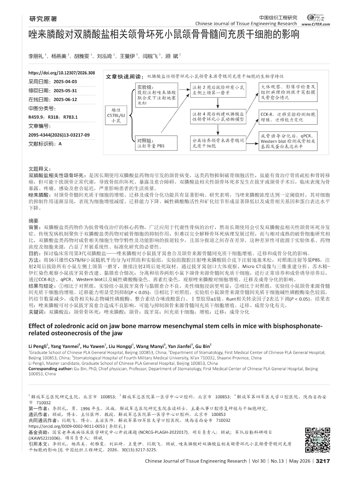
Effect of zoledronic acid on jaw bone marrow mesenchymal stem cells in mice with bisphosphonate-related osteonecrosis of the jaw

Authors: LI Pengli, YANG Yanmei, HU Yawen, LIU Hongqi, WANG Manyi, YAN Jianfei, GU Bin

BACKGROUND: Bisphosphonates, as the core drugs of anti-bone resorption therapy, are widely used in the treatment of metabolic bone diseases. However, long-term use can cause the complications of bisphosphonate related osteonecrosis of the jaw. The traditional pathogenesis focuses on the inhibitory effect of bisphosphonates on osteoclasts, but it is difficult to fully explain the pathological development of osteonecrosis. Compared with the relatively mature osteoclast research, there are fewer reports on the effects of bisphosphonates on the biological characteristics and functions of osteoblast-related cells, and there are differences between some reports. This difference may be due to the experimental system, drug concentration and cell source, highlighting the necessity of conducting systematic and standardized research. OBJECTIVE: To investigate the effect of the third-generation bisphosphonate-zoledronic acid commonly used in clinical practice on the healing of tooth extraction sockets and the proliferation, migration and osteogenic differentiation of bone marrow mesenchymal stem cells derived from the jaw in mice. METHODS: Sixteen male C57BL/6J mice were randomly divided into control and experimental groups. The experimental group received intraperitoneal injection of zoledronic acid combined with subcutaneous injection of dexamethasone, while the control group received an equal volume of PBS. After 2 weeks of injection, the left maxillary first molars of all mice were extracted, and after another 2 weeks of injection, the mice were sacrificed. The healing of extraction sockets was evaluated by gross observation, Micro CT imaging and three-dimensional reconstruction, and hematoxylin-eosin staining. Jaw bone marrow mesenchymal stem cells were isolated and cultured from both groups. After normal culture and osteogenic induction, cell proliferation, migration, and osteogenic differentiation were assessed by CCK-8 assay, qPCR, Western blot, alkaline phosphatase staining, and alizarin red staining. RESULTS AND CONCLUSION: Compared with the control group, the experimental group showed poor healing of extraction sockets with more inflammatory cell infiltration. The proliferation and migration abilities of jaw bone marrow mesenchymal stem cells were significantly inhibited in the experimental group (P < 0.05). Alkaline phosphatase staining was weaker, calcium nodule formation was reduced, and the expression of osteogenic markers (alkaline phosphatase, integrin-binding sialoprotein, collagen type I alpha 1 chain, Runt-related transcription factor 2) was downregulated in the experimental group (P < 0.05). These results indicate that zoledronic acid can adversely affect extraction socket healing, possibly by inhibiting the proliferation, migration, and osteogenic differentiation of jaw bone marrow mesenchymal stem cells.

Effect of zoledronic acid on jaw bone marrow mesenchymal stem cells in mice with bisphosphonate-related osteonecrosis of the jaw
Graphical Abstract