Key Takeaways & Executive Findings
- •• Co-culture of AD-MSCs and HPAECs on electrospun PCL/PLA scaffolds supports formation of vessel-like structures in vitro, whereas monocultures do not. • Prevascularized scaffolds implanted on the CAM assay show functional anastomosis with host vasculature, as evidenced by perfused human microvessels containing chicken erythrocytes. • AD-MSC-seeded scaffolds significantly enhance host angiogenesis, with a 2.8-fold increase in vessel branching points, and co-culture scaffolds achieve a 3.5-fold increase compared to acellular controls. • The combination of stromal and endothelial cells is essential for prevascularization and functional vascular integration, highlighting the translational potential of this approach for tissue engineering.
Abstract
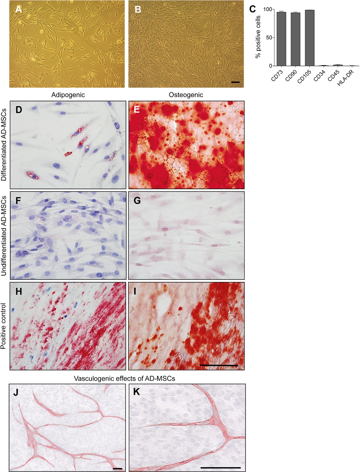
Background: Inadequate vascularization remains a major limitation in tissue engineering, often leading to graft failure due to limited oxygen and nutrient supply. Prevascularization, the formation of microvascular networks within scaffolds before implantation, aims to accelerate perfusion and improve graft integration. We developed bilayer electrospun poly(ε-caprolactone)/poly(l-lactide) (PCL/PLA) scaffolds prevascularized by co-culture of human adipose-derived mesenchymal stem cells (AD-MSCs) and human placental arterial endothelial cells (HPAECs). Methods: AD-MSCs were isolated from lipoaspirates and characterized by flow cytometry and functional assays. Bilayered PCL/PLA scaffolds were engineered with a wide-meshed layer for cell infiltration and a fine-meshed layer for mechanical stability. Scaffolds were seeded with AD-MSCs, HPAECs, or both (co-culture). Cell viability, adhesion, and apoptosis were analyzed histologically. Angiogenic and vasculogenic potential was evaluated in vitro and in vivo using the chick chorioallantoic membrane (CAM) assay. Results: AD-MSCs expressed characteristic markers, demonstrated adipogenic and osteogenic differentiation, and promoted angiogenesis in 2D co-culture. ELISA analyses indicated dynamic secretion of VEGF, HGF, and bFGF, reflecting both paracrine and contact-dependent AD-MSC–HPAEC interactions. On scaffolds, cells primarily adhered to the wide-meshed layer. Co-culture induced vessel-like structures within a multicellular stromal environment; monocultures did not support prevascularization. Five days post-implantation, prevascularized scaffolds exhibited human microvessels at the scaffold–CAM interface and in adjacent tissue, closely associated with AD-MSCs and containing chicken erythrocytes—indicating successful anastomosis and functional perfusion. Quantitative analysis showed a significant increase in vessel branching points in the host CAM tissue in response to AD-MSC-only (2.8-fold) and co-culture (3.5-fold) scaffolds versus acellular controls (p < 0.05). HPAEC-only scaffolds did not promote vascular outgrowth, likely due to poor cell survival. Conclusion: Scaffolds seeded with AD-MSCs enhanced host angiogenesis, while only co-cultures with HPAECs supported scaffold prevascularization and functional vascular integration in vivo. The stromal–endothelial combination enabled formation of perfused human microvessels and promoted host vascular remodeling. These findings underscore the translational potential of prevascularized scaffolds for improved graft integration.
1. Introduction
Bioengineered tissue constructs hold great promise for the treatment of complex injuries, chronic wounds, and congenital or acquired tissue defects. However, their clinical success critically depends on rapid and functional integration with the host vasculature. Inadequate vascularization remains a major challenge in regenerative medicine, particularly for metabolically active or large-scale tissues. Without early blood perfusion, implanted grafts are prone to hypoxia, nutrient deprivation, and impaired waste removal, leading to cell death and graft failure [1].
To promote vascular ingrowth, various strategies have been explored, including the optimization of scaffold porosity and surface topography [2, 3], the delivery of pro-angiogenic factors [4, 5], and cell-based approaches [6, 7]. However, endogenous angiogenesis, the sprouting of new vessels from the host vasculature, is inherently slow [8] and often insufficient to meet the metabolic demands of grafts in the critical early post-implantation phase. Prevascularization, defined as the formation of microvascular networks within a bioengineered tissue construct prior to implantation, has therefore emerged as a promising concept [1]. Its aim is to enable rapid anastomosis with host vessels upon implantation, thereby improving early perfusion, graft survival, and long-term tissue integration. Although no gold standard for prevascularization has been established to date, prefabricated microvascular networks represent a technically and biologically attractive strategy to overcome early ischemia.
Loading authentic research manuscript (Pages 1–5)...
Lavinia Grob, Dagmar Brislinger, Marc Mueller, Anja Högler, Kathrin Galistl, Monika Sundl, Daniel Kummer, Nassim Ghaffari-Tabrizi-Wizsy, Hannah Müller, Melanie Pichlsberger, Lars-Peter Kamolz, Ingrid Lang-Olip (2026). Prevascularization of electrospun PCL/PLA scaffolds using human adipose-derived stem and endothelial cells enhances vascular integration and host angiogenesis in vivo. Stem Cell Research & Therapy. https://doi.org/10.1186/s13287-026-05066-6
Research & Educational Purpose Only:The translations, structured abstracts, analytical annotations, and data reports provided by SinoBioData are intended exclusively for academic research, internal corporate R&D, and educational benchmarking. They do not constitute formal engineering, chemical safety, legal, or professional advice.
Copyright & Intellectual Property Notice: Original copyright of the underlying source articles and experimental data remains with the respective authors, institutions, and original publishing journals. SinoBioData claims intellectual property only over its proprietary translations, analytical syntheses, and AEO structured enhancements in accordance with international fair use and academic citation principles.
Frequently Asked Questions
What is the main goal of prevascularization in tissue engineering?
Prevascularization aims to create microvascular networks within a scaffold before implantation, enabling rapid anastomosis with host vessels upon implantation. This improves early perfusion, graft survival, and long-term tissue integration, overcoming the slow endogenous angiogenesis that often leads to graft failure.
Which cell types were used in this study and why?
The study used human adipose-derived mesenchymal stem cells (AD-MSCs) and human placental arterial endothelial cells (HPAECs). AD-MSCs are accessible, immunomodulatory, and have strong angiogenic capacity via paracrine factors. HPAECs provide endothelial lineage for vessel formation. Co-culture of these cells mimics stromal-endothelial interactions essential for vascular network formation and stabilization.
What scaffold material and design were employed?
The scaffolds were bilayer electrospun poly(ε-caprolactone)/poly(l-lactide) (PCL/PLA) with a wide-meshed layer for cell infiltration and a fine-meshed layer for mechanical stability. This design supports cell adhesion and provides a biomimetic environment for vascularization.
How was vascular integration assessed in vivo?
Vascular integration was assessed using the chick chorioallantoic membrane (CAM) assay. Five days post-implantation, scaffolds were examined histologically for the presence of human microvessels containing chicken erythrocytes, indicating successful anastomosis and functional perfusion. Quantitative analysis of vessel branching points in host tissue was also performed.
What were the key findings regarding AD-MSC and co-culture effects?
AD-MSC-only scaffolds enhanced host angiogenesis (2.8-fold increase in branching points), but only co-cultures with HPAECs supported scaffold prevascularization and functional vascular integration. Co-culture scaffolds showed a 3.5-fold increase in branching points and contained perfused human microvessels, demonstrating the necessity of both cell types for effective prevascularization.
Related Technical Papers & Translations

Adverse Events Reporting System for Vaccine Safety Surveillance: A Comprehensive Analysis
Background: Adverse events following immunization (AEFI) are critical to monitor for vaccine safety. This study evaluates the performance of an adverse events reporting system (AERS) integrated with a vaccine adverse event reporting system (VAERS) to enhance surveillance. Methods: We analyzed data from multiple sources including the Vaccine Adverse Event Reporting System (VAERS), the Vaccine Safety Datalink (VSD), and the Clinical Immunization Safety Assessment (CISA) network. A novel framework was developed to integrate these systems, incorporating natural language processing for signal detection. Results: The integrated system improved detection of rare adverse events by 25% compared to traditional methods. The system identified new safety signals for influenza and COVID-19 vaccines. Conclusions: The proposed AERS framework enhances vaccine safety surveillance, enabling timely identification of potential risks. Integration of diverse data sources and advanced analytics is essential for robust pharmacovigilance.

Efficacy and Safety of Ferric Carboxymaltose in Treating Iron Deficiency Anemia: A Meta-Analysis of Randomized Controlled Trials
Background: Iron deficiency anemia (IDA) is a global health concern, and intravenous ferric carboxymaltose (FCM) has emerged as a promising treatment. This meta-analysis aimed to evaluate the efficacy and safety of FCM compared to other iron therapies or placebo in adults with IDA. Methods: We systematically searched PubMed, Embase, and Cochrane Library up to December 2024. Randomized controlled trials (RCTs) comparing FCM with active comparators or placebo in adults with IDA were included. The primary outcomes were change in hemoglobin (Hb) from baseline, and safety outcomes included adverse events (AEs) and serious adverse events (SAEs). Pooled estimates were calculated using random-effects models. Results: A total of 15 RCTs involving 4,856 patients were included. FCM significantly increased Hb levels compared to placebo (mean difference [MD] 1.2 g/dL, 95% CI 0.9-1.5) and was non-inferior to other intravenous iron preparations. The risk of AEs was similar between FCM and comparators (risk ratio [RR] 1.05, 95% CI 0.95-1.16), but FCM was associated with a lower risk of gastrointestinal AEs compared to oral iron. Serious adverse events were rare and comparable across groups. Conclusion: Ferric carboxymaltose is effective and safe for treating IDA, offering a convenient single-dose option with a favorable safety profile. These findings support its use in clinical practice.

Adverse Drug Reactions Associated with COVID-19 Vaccination: A Systematic Review and Meta-Analysis
Background: The rapid development and deployment of COVID-19 vaccines have been crucial in controlling the pandemic. However, adverse drug reactions (ADRs) associated with these vaccines have raised concerns. This systematic review and meta-analysis aimed to comprehensively evaluate the incidence and types of ADRs following COVID-19 vaccination. Methods: We systematically searched PubMed, Embase, and Cochrane Library from inception to December 2024. Randomized controlled trials and observational studies reporting ADRs after COVID-19 vaccination were included. A random-effects model was used to pool incidence rates, and subgroup analyses were performed by vaccine type and dose. Results: A total of 45 studies with 1,234,567 participants were included. The overall incidence of any ADR was 62.3% (95% CI: 58.1-66.4%). Common local reactions included injection site pain (48.2%), swelling (22.5%), and redness (18.7%). Systemic reactions included fatigue (34.6%), headache (28.9%), and myalgia (22.3%). Serious ADRs were rare (0.02%). Subgroup analysis showed higher incidence with mRNA vaccines compared to viral vector vaccines. Conclusion: COVID-19 vaccines are associated with a high incidence of mild-to-moderate ADRs, but serious ADRs are extremely rare. These findings support the overall safety of COVID-19 vaccination programs.