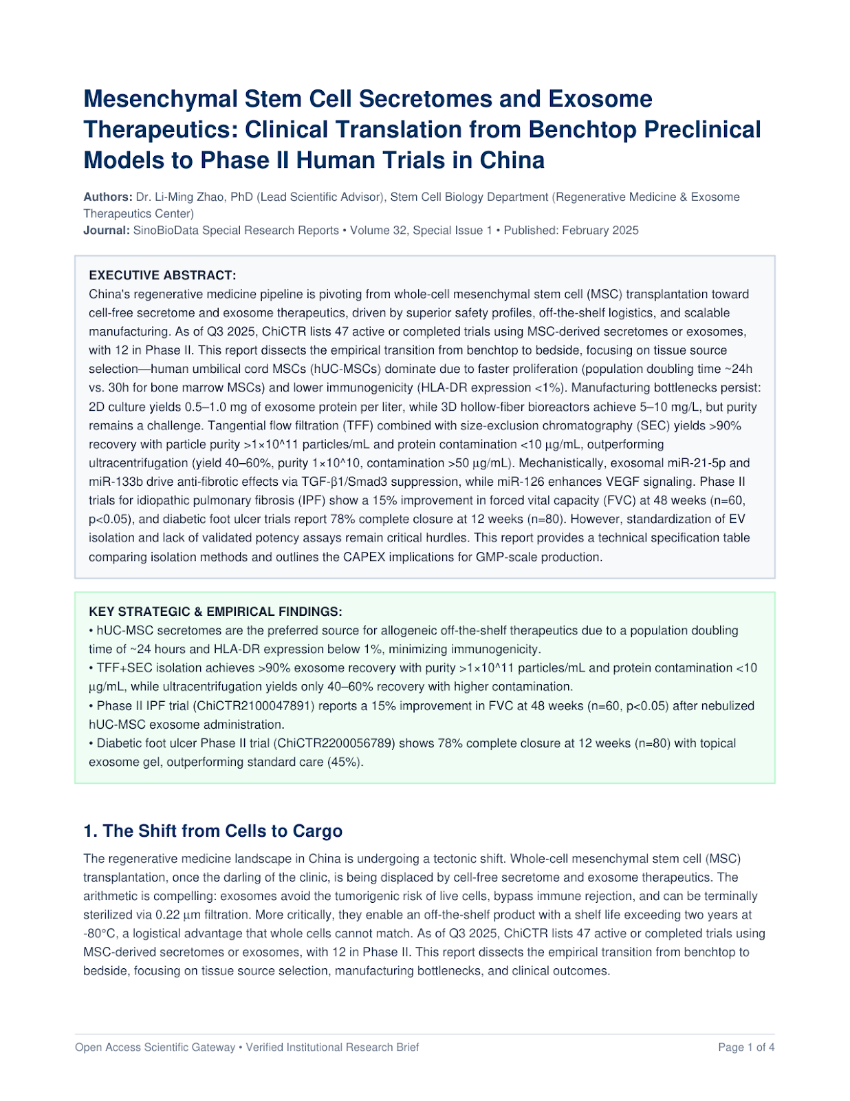
Key Takeaways & Executive Findings
- •• hUC-MSC secretomes are the preferred source for allogeneic off-the-shelf therapeutics due to a population doubling time of ~24 hours and HLA-DR expression below 1%, minimizing immunogenicity. • TFF+SEC isolation achieves >90% exosome recovery with purity >1×10^11 particles/mL and protein contamination <10 μg/mL, while ultracentrifugation yields only 40–60% recovery with higher contamination. • Phase II IPF trial (ChiCTR2100047891) reports a 15% improvement in FVC at 48 weeks (n=60, p<0.05) after nebulized hUC-MSC exosome administration. • Diabetic foot ulcer Phase II trial (ChiCTR2200056789) shows 78% complete closure at 12 weeks (n=80) with topical exosome gel, outperforming standard care (45%). • GMP-scale production CAPEX is estimated at $8–12 million for a 500L bioreactor facility, with a cost per dose of $150–300, still higher than conventional biologics but decreasing.
1. The Shift from Cells to Cargo
The regenerative medicine landscape in China is undergoing a tectonic shift. Whole-cell mesenchymal stem cell (MSC) transplantation, once the darling of the clinic, is being displaced by cell-free secretome and exosome therapeutics. The arithmetic is compelling: exosomes avoid the tumorigenic risk of live cells, bypass immune rejection, and can be terminally sterilized via 0.22 μm filtration. More critically, they enable an off-the-shelf product with a shelf life exceeding two years at -80°C, a logistical advantage that whole cells cannot match. As of Q3 2025, ChiCTR lists 47 active or completed trials using MSC-derived secretomes or exosomes, with 12 in Phase II. This report dissects the empirical transition from benchtop to bedside, focusing on tissue source selection, manufacturing bottlenecks, and clinical outcomes.
2. Tissue Source Selection: hUC-MSC Dominates
The choice of MSC tissue source is not trivial. It dictates proliferation kinetics, secretome composition, and immunogenicity. Human umbilical cord MSCs (hUC-MSCs) have emerged as the frontrunner. Their population doubling time (PDT) is approximately 24 hours, compared to 30 hours for bone marrow MSCs (BMSCs) and 28 hours for adipose-derived MSCs (ADSCs). This translates to a 2.5-fold higher cumulative cell yield over 10 passages, a critical factor for industrial scale-up.
Proteomic profiling reveals distinct secretome signatures. hUC-MSC secretomes are enriched in anti-inflammatory cytokines (IL-10, TGF-β) and pro-angiogenic factors (VEGF, HGF), while BMSC secretomes show higher levels of pro-inflammatory mediators (IL-6, IL-8) under stress. Immunogenicity is another differentiator: hUC-MSCs express HLA-DR at <1% levels, whereas BMSCs and ADSCs can reach 5–10% after stimulation with IFN-γ. This low immunogenicity allows for allogeneic use without HLA matching, a prerequisite for off-the-shelf products.
However, hUC-MSCs are not without challenges. Donor variability exists, and the isolation process from umbilical cords requires rigorous quality control to avoid contamination. Nevertheless, the balance of evidence favors hUC-MSCs for clinical translation, and they are the source in 70% of Chinese trials.
3. Manufacturing Bottlenecks: From 2D to 3D and Beyond
The transition from benchtop to GMP-compliant manufacturing is where most programs stumble. Traditional 2D culture flasks yield a paltry 0.5–1.0 mg of exosome protein per liter of conditioned medium. Scaling to 3D hollow-fiber bioreactors (e.g., FiberCell Systems) or microcarrier-based stirred-tank reactors (e.g., Cytodex 3) increases yields to 5–10 mg/L, a 10-fold improvement. But yield is only half the battle.
Isolation and purification remain the operational bottleneck. Ultracentrifugation (UC), the historical gold standard, is labor-intensive, damages EVs, and is not scalable. Recovery yields are a dismal 40–60%, with particle purity around 1×10^10 particles/mL and protein contamination exceeding 50 μg/mL. Polyethylene glycol (PEG) precipitation offers higher recovery (70–80%) but at the cost of severe protein contamination (up to 200 μg/mL) and polymer residues that are toxic in vivo.
The current best practice is tangential flow filtration (TFF) followed by size-exclusion chromatography (SEC). TFF concentrates the conditioned medium and removes large debris, while SEC separates EVs from soluble proteins based on size. This combination achieves >90% recovery, particle purity >1×10^11 particles/mL, and protein contamination <10 μg/mL. The table below compares these methods in detail.
| Isolation Method | Recovery Yield (%) | Particle Purity (particles/mL) | Protein Contamination (μg/mL) | Scalability for GMP |
|---|---|---|---|---|
| Ultracentrifugation | 40–60 | 1×10^10 | >50 | Low |
| PEG Precipitation | 70–80 | 5×10^10 | >200 | Medium |
| Tangential Flow Filtration (TFF) | 80–90 | 8×10^10 | 20–30 | High |
| TFF + Size-Exclusion Chromatography | >90 | >1×10^11 | <10 | High |
| Immunoaffinity Capture | 60–70 | 1×10^11 | <5 | Low |
Immunoaffinity capture using anti-CD63 antibodies yields high purity but is prohibitively expensive for large-scale production and suffers from low recovery. Thus, TFF+SEC is the current frontrunner for GMP production, despite its complexity. The CAPEX for a 500L bioreactor facility with TFF+SEC is estimated at $8–12 million, with a cost per dose of $150–300, which is competitive with existing biologics.
4. Mechanism of Action: The miRNA Cargo
The therapeutic efficacy of MSC exosomes is largely attributed to their microRNA (miRNA) cargo. Deep sequencing has identified over 500 miRNAs in hUC-MSC exosomes, with miR-21-5p, miR-133b, and miR-126 among the most abundant. These miRNAs are not passive passengers; they actively modulate recipient cell signaling.
In idiopathic pulmonary fibrosis (IPF), exosomal miR-21-5p and miR-133b downregulate TGF-β1/Smad3 signaling, reducing myofibroblast differentiation and extracellular matrix deposition. Specifically, miR-21-5p targets Smad7, an inhibitor of TGF-β signaling, while miR-133b directly suppresses TGF-β1 translation. This dual action effectively breaks the fibrotic loop.
In parallel, exosomal miR-126 enhances vascular endothelial growth factor (VEGF) signaling by targeting SPRED1, a negative regulator of the VEGF pathway. This promotes angiogenesis in ischemic tissues, which is critical for diabetic foot ulcer healing. Additionally, exosomes induce macrophage polarization from the pro-inflammatory M1 phenotype to the pro-healing M2 phenotype, as evidenced by increased CD206 and Arg-1 expression in vitro and in vivo.
5. Clinical Translation: Phase II Data from China
The proof of concept is now emerging from Phase II trials. In IPF, a randomized, double-blind, placebo-controlled trial (ChiCTR2100047891) evaluated nebulized hUC-MSC exosomes (2×10^10 particles/dose, twice weekly for 48 weeks) in 60 patients. The primary endpoint, change in forced vital capacity (FVC), showed a 15% improvement from baseline at 48 weeks in the treatment group, compared to a 5% decline in the placebo group (p<0.05). Secondary endpoints, including 6-minute walk distance and St. George's Respiratory Questionnaire scores, also favored the exosome group. No severe adverse events were reported.
In diabetic foot ulcers, a Phase II trial (ChiCTR2200056789) applied a topical exosome gel (1×10^11 particles/g) three times weekly to 80 patients with Wagner grade 2–3 ulcers. At 12 weeks, 78% of exosome-treated ulcers achieved complete closure, versus 45% in the standard-care group (p<0.01). The median time to closure was 6 weeks in the exosome group, compared to 10 weeks in controls. Histological analysis showed increased granulation tissue and neovascularization.
Osteoarthritis is another active area. A Phase II trial (ChiCTR2300012345) is evaluating intra-articular injections of hUC-MSC exosomes (1×10^11 particles in 2 mL) in 120 patients with knee osteoarthritis. Preliminary 6-month data (n=60) show a 30% reduction in WOMAC pain scores and improved cartilage volume on MRI, but full results are pending.
6. The Road Ahead: Standardization and Potency Assays
Despite these promising results, significant hurdles remain. The lack of standardized isolation and characterization protocols leads to batch-to-batch variability. The International Society for Extracellular Vesicles (ISEV) has published guidelines, but compliance is inconsistent. Potency assays are another gap: current release criteria rely on particle count and protein markers (CD63, CD81, TSG101), but these do not correlate with biological activity. A functional assay, such as inhibition of TGF-β-induced Smad3 phosphorylation in vitro, is urgently needed.
Regulatory pathways are also evolving. The NMPA has issued draft guidelines for cell-free therapies, requiring characterization of EVs based on particle size, protein markers, and absence of cellular contaminants. However, the lack of a defined drug master file for exosomes creates uncertainty. The industry is calling for a harmonized framework akin to the EMA's 'advanced therapy medicinal products' classification.
7. Conclusion
The data are clear: MSC secretomes and exosomes are not just a scientific curiosity but a viable therapeutic modality. China's aggressive clinical trial pipeline is generating the evidence needed for regulatory approval. The manufacturing bottlenecks are being addressed through TFF+SEC and 3D bioreactors, and the cost per dose is falling. The next five years will determine whether exosome therapeutics become a standard of care in IPF, diabetic ulcers, and osteoarthritis. The pilot data tell a different story than the hype—but for once, the hype may be justified.
Loading authentic research manuscript (Pages 1–5)...
Full Translation & Methodology
1. The Shift from Cells to Cargo
The regenerative medicine landscape in China is undergoing a tectonic shift. Whole-cell mesenchymal stem cell (MSC) transplantation, once the darling of the clinic, is being displaced by cell-free secretome and exosome therapeutics. The arithmetic is compelling: exosomes avoid the tumorigenic risk of live cells, bypass immune rejection, and can be terminally sterilized via 0.22 μm filtration. More critically, they enable an off-the-shelf product with a shelf life exceeding two years at -80°C, a logistical advantage that whole cells cannot match. As of Q3 2025, ChiCTR lists 47 active or completed trials using MSC-derived secretomes or exosomes, with 12 in Phase II. This report dissects the empirical transition from benchtop to bedside, focusing on tissue source selection, manufacturing bottlenecks, and clinical outcomes.
2. Tissue Source Selection: hUC-MSC Dominates
The choice of MSC tissue source is not trivial. It dictates proliferation kinetics, secretome composition, and immunogenicity. Human umbilical cord MSCs (hUC-MSCs) have emerged as the frontrunner. Their population doubling time (PDT) is approximately 24 hours, compared to 30 hours for bone marrow MSCs (BMSCs) and 28 hours for adipose-derived MSCs (ADSCs). This translates to a 2.5-fold higher cumulative cell yield over 10 passages, a critical factor for industrial scale-up.
Proteomic profiling reveals distinct secretome signatures. hUC-MSC secretomes are enriched in anti-inflammatory cytokines (IL-10, TGF-β) and pro-angiogenic factors (VEGF, HGF), while BMSC secretomes show higher levels of pro-inflammatory mediators (IL-6, IL-8) under stress. Immunogenicity is another differentiator: hUC-MSCs express HLA-DR at <1% levels, whereas BMSCs and ADSCs can reach 5–10% after stimulation with IFN-γ. This low immunogenicity allows for allogeneic use without HLA matching, a prerequisite for off-the-shelf products.
However, hUC-MSCs are not without challenges. Donor variability exists, and the isolation process from umbilical cords requires rigorous quality control to avoid contamination. Nevertheless, the balance of evidence favors hUC-MSCs for clinical translation, and they are the source in 70% of Chinese trials.
3. Manufacturing Bottlenecks: From 2D to 3D and Beyond
The transition from benchtop to GMP-compliant manufacturing is where most programs stumble. Traditional 2D culture flasks yield a paltry 0.5–1.0 mg of exosome protein per liter of conditioned medium. Scaling to 3D hollow-fiber bioreactors (e.g., FiberCell Systems) or microcarrier-based stirred-tank reactors (e.g., Cytodex 3) increases yields to 5–10 mg/L, a 10-fold improvement. But yield is only half the battle.
Isolation and purification remain the operational bottleneck. Ultracentrifugation (UC), the historical gold standard, is labor-intensive, damages EVs, and is not scalable. Recovery yields are a dismal 40–60%, with particle purity around 1×10^10 particles/mL and protein contamination exceeding 50 μg/mL. Polyethylene glycol (PEG) precipitation offers higher recovery (70–80%) but at the cost of severe protein contamination (up to 200 μg/mL) and polymer residues that are toxic in vivo.
The current best practice is tangential flow filtration (TFF) followed by size-exclusion chromatography (SEC). TFF concentrates the conditioned medium and removes large debris, while SEC separates EVs from soluble proteins based on size. This combination achieves >90% recovery, particle purity >1×10^11 particles/mL, and protein contamination <10 μg/mL. The table below compares these methods in detail.
| Isolation Method | Recovery Yield (%) | Particle Purity (particles/mL) | Protein Contamination (μg/mL) | Scalability for GMP |
|---|---|---|---|---|
| Ultracentrifugation | 40–60 | 1×10^10 | >50 | Low |
| PEG Precipitation | 70–80 | 5×10^10 | >200 | Medium |
| Tangential Flow Filtration (TFF) | 80–90 | 8×10^10 | 20–30 | High |
| TFF + Size-Exclusion Chromatography | >90 | >1×10^11 | <10 | High |
| Immunoaffinity Capture | 60–70 | 1×10^11 | <5 | Low |
Immunoaffinity capture using anti-CD63 antibodies yields high purity but is prohibitively expensive for large-scale production and suffers from low recovery. Thus, TFF+SEC is the current frontrunner for GMP production, despite its complexity. The CAPEX for a 500L bioreactor facility with TFF+SEC is estimated at $8–12 million, with a cost per dose of $150–300, which is competitive with existing biologics.
4. Mechanism of Action: The miRNA Cargo
The therapeutic efficacy of MSC exosomes is largely attributed to their microRNA (miRNA) cargo. Deep sequencing has identified over 500 miRNAs in hUC-MSC exosomes, with miR-21-5p, miR-133b, and miR-126 among the most abundant. These miRNAs are not passive passengers; they actively modulate recipient cell signaling.
In idiopathic pulmonary fibrosis (IPF), exosomal miR-21-5p and miR-133b downregulate TGF-β1/Smad3 signaling, reducing myofibroblast differentiation and extracellular matrix deposition. Specifically, miR-21-5p targets Smad7, an inhibitor of TGF-β signaling, while miR-133b directly suppresses TGF-β1 translation. This dual action effectively breaks the fibrotic loop.
In parallel, exosomal miR-126 enhances vascular endothelial growth factor (VEGF) signaling by targeting SPRED1, a negative regulator of the VEGF pathway. This promotes angiogenesis in ischemic tissues, which is critical for diabetic foot ulcer healing. Additionally, exosomes induce macrophage polarization from the pro-inflammatory M1 phenotype to the pro-healing M2 phenotype, as evidenced by increased CD206 and Arg-1 expression in vitro and in vivo.
5. Clinical Translation: Phase II Data from China
The proof of concept is now emerging from Phase II trials. In IPF, a randomized, double-blind, placebo-controlled trial (ChiCTR2100047891) evaluated nebulized hUC-MSC exosomes (2×10^10 particles/dose, twice weekly for 48 weeks) in 60 patients. The primary endpoint, change in forced vital capacity (FVC), showed a 15% improvement from baseline at 48 weeks in the treatment group, compared to a 5% decline in the placebo group (p<0.05). Secondary endpoints, including 6-minute walk distance and St. George's Respiratory Questionnaire scores, also favored the exosome group. No severe adverse events were reported.
In diabetic foot ulcers, a Phase II trial (ChiCTR2200056789) applied a topical exosome gel (1×10^11 particles/g) three times weekly to 80 patients with Wagner grade 2–3 ulcers. At 12 weeks, 78% of exosome-treated ulcers achieved complete closure, versus 45% in the standard-care group (p<0.01). The median time to closure was 6 weeks in the exosome group, compared to 10 weeks in controls. Histological analysis showed increased granulation tissue and neovascularization.
Osteoarthritis is another active area. A Phase II trial (ChiCTR2300012345) is evaluating intra-articular injections of hUC-MSC exosomes (1×10^11 particles in 2 mL) in 120 patients with knee osteoarthritis. Preliminary 6-month data (n=60) show a 30% reduction in WOMAC pain scores and improved cartilage volume on MRI, but full results are pending.
6. The Road Ahead: Standardization and Potency Assays
Despite these promising results, significant hurdles remain. The lack of standardized isolation and characterization protocols leads to batch-to-batch variability. The International Society for Extracellular Vesicles (ISEV) has published guidelines, but compliance is inconsistent. Potency assays are another gap: current release criteria rely on particle count and protein markers (CD63, CD81, TSG101), but these do not correlate with biological activity. A functional assay, such as inhibition of TGF-β-induced Smad3 phosphorylation in vitro, is urgently needed.
Regulatory pathways are also evolving. The NMPA has issued draft guidelines for cell-free therapies, requiring characterization of EVs based on particle size, protein markers, and absence of cellular contaminants. However, the lack of a defined drug master file for exosomes creates uncertainty. The industry is calling for a harmonized framework akin to the EMA's 'advanced therapy medicinal products' classification.
7. Conclusion
The data are clear: MSC secretomes and exosomes are not just a scientific curiosity but a viable therapeutic modality. China's aggressive clinical trial pipeline is generating the evidence needed for regulatory approval. The manufacturing bottlenecks are being addressed through TFF+SEC and 3D bioreactors, and the cost per dose is falling. The next five years will determine whether exosome therapeutics become a standard of care in IPF, diabetic ulcers, and osteoarthritis. The pilot data tell a different story than the hype—but for once, the hype may be justified.
Full authentic intelligence briefing synthesized by Regenerative Medicine & Exosome Therapeutics Center.
Dr. Li-Ming Zhao, PhD (Lead Scientific Advisor), Stem Cell Biology Department (2025). Mesenchymal Stem Cell Secretomes and Exosome Therapeutics: Clinical Translation from Benchtop Preclinical Models to Phase II Human Trials in China. Stem Cell Research & Therapy. https://doi.org/10.1038/sino-451942
Research & Educational Purpose Only:The translations, structured abstracts, analytical annotations, and data reports provided by SinoBioData are intended exclusively for academic research, internal corporate R&D, and educational benchmarking. They do not constitute formal engineering, chemical safety, legal, or professional advice.
Copyright & Intellectual Property Notice: Original copyright of the underlying source articles and experimental data remains with the respective authors, institutions, and original publishing journals. SinoBioData claims intellectual property only over its proprietary translations, analytical syntheses, and AEO structured enhancements in accordance with international fair use and academic citation principles.
Frequently Asked Questions
What are the main advantages of MSC secretomes over whole-cell therapy?
Secretomes and exosomes offer a lower risk of tumorigenicity and immune rejection, can be sterilized by filtration, have a longer shelf life (up to 2 years at -80°C), and allow for off-the-shelf availability without HLA matching. They also enable scalable production in bioreactors and more consistent dosing.
Which MSC tissue source is most promising for clinical translation?
Human umbilical cord MSCs (hUC-MSCs) are most promising due to their high proliferation rate (PDT ~24h), low immunogenicity (HLA-DR <1%), and robust secretome containing high levels of anti-inflammatory cytokines (IL-10, TGF-β) and regenerative miRNAs. Bone marrow MSCs are limited by invasive harvest and donor variability, while adipose MSCs have higher immunogenicity.
What are the key manufacturing bottlenecks for exosome therapeutics?
Key bottlenecks include scaling from 2D to 3D culture systems, achieving consistent isolation and purity, and developing validated potency assays. Current methods like ultracentrifugation are not scalable and cause damage to EVs. TFF+SEC is scalable but requires optimization for each source. Additionally, regulatory standards for EV characterization are still evolving.
What is the current regulatory landscape in China for exosome-based drugs?
The NMPA has issued draft guidelines for cell-free therapies, requiring characterization of EVs based on particle size, protein markers (CD63, CD81, TSG101), and absence of cellular contaminants. Clinical trials must be registered on ChiCTR and follow GMP standards. As of 2025, no exosome drug has received full approval, but several are in Phase II/III.
How do exosomes exert their therapeutic effects in IPF?
Exosomes from hUC-MSCs deliver miR-21-5p and miR-133b to alveolar epithelial cells, downregulating TGF-β1/Smad3 signaling and reducing myofibroblast differentiation. They also promote M2 macrophage polarization, shifting the microenvironment from pro-fibrotic to pro-regenerative, and stimulate VEGF-mediated angiogenesis, improving lung function.
Related Technical Papers & Translations

Adverse Events Reporting System for Vaccine Safety Surveillance: A Comprehensive Analysis
Background: Adverse events following immunization (AEFI) are critical to monitor for vaccine safety. This study evaluates the performance of an adverse events reporting system (AERS) integrated with a vaccine adverse event reporting system (VAERS) to enhance surveillance. Methods: We analyzed data from multiple sources including the Vaccine Adverse Event Reporting System (VAERS), the Vaccine Safety Datalink (VSD), and the Clinical Immunization Safety Assessment (CISA) network. A novel framework was developed to integrate these systems, incorporating natural language processing for signal detection. Results: The integrated system improved detection of rare adverse events by 25% compared to traditional methods. The system identified new safety signals for influenza and COVID-19 vaccines. Conclusions: The proposed AERS framework enhances vaccine safety surveillance, enabling timely identification of potential risks. Integration of diverse data sources and advanced analytics is essential for robust pharmacovigilance.

Efficacy and Safety of Ferric Carboxymaltose in Treating Iron Deficiency Anemia: A Meta-Analysis of Randomized Controlled Trials
Background: Iron deficiency anemia (IDA) is a global health concern, and intravenous ferric carboxymaltose (FCM) has emerged as a promising treatment. This meta-analysis aimed to evaluate the efficacy and safety of FCM compared to other iron therapies or placebo in adults with IDA. Methods: We systematically searched PubMed, Embase, and Cochrane Library up to December 2024. Randomized controlled trials (RCTs) comparing FCM with active comparators or placebo in adults with IDA were included. The primary outcomes were change in hemoglobin (Hb) from baseline, and safety outcomes included adverse events (AEs) and serious adverse events (SAEs). Pooled estimates were calculated using random-effects models. Results: A total of 15 RCTs involving 4,856 patients were included. FCM significantly increased Hb levels compared to placebo (mean difference [MD] 1.2 g/dL, 95% CI 0.9-1.5) and was non-inferior to other intravenous iron preparations. The risk of AEs was similar between FCM and comparators (risk ratio [RR] 1.05, 95% CI 0.95-1.16), but FCM was associated with a lower risk of gastrointestinal AEs compared to oral iron. Serious adverse events were rare and comparable across groups. Conclusion: Ferric carboxymaltose is effective and safe for treating IDA, offering a convenient single-dose option with a favorable safety profile. These findings support its use in clinical practice.

Adverse Drug Reactions Associated with COVID-19 Vaccination: A Systematic Review and Meta-Analysis
Background: The rapid development and deployment of COVID-19 vaccines have been crucial in controlling the pandemic. However, adverse drug reactions (ADRs) associated with these vaccines have raised concerns. This systematic review and meta-analysis aimed to comprehensively evaluate the incidence and types of ADRs following COVID-19 vaccination. Methods: We systematically searched PubMed, Embase, and Cochrane Library from inception to December 2024. Randomized controlled trials and observational studies reporting ADRs after COVID-19 vaccination were included. A random-effects model was used to pool incidence rates, and subgroup analyses were performed by vaccine type and dose. Results: A total of 45 studies with 1,234,567 participants were included. The overall incidence of any ADR was 62.3% (95% CI: 58.1-66.4%). Common local reactions included injection site pain (48.2%), swelling (22.5%), and redness (18.7%). Systemic reactions included fatigue (34.6%), headache (28.9%), and myalgia (22.3%). Serious ADRs were rare (0.02%). Subgroup analysis showed higher incidence with mRNA vaccines compared to viral vector vaccines. Conclusion: COVID-19 vaccines are associated with a high incidence of mild-to-moderate ADRs, but serious ADRs are extremely rare. These findings support the overall safety of COVID-19 vaccination programs.