🧬 SinoBioData Academic Portal
πŸ›οΈ Indexed Academic JournalOriginal: η”Ÿη‰©εŒ–ε­¦δΈŽη”Ÿη‰©η‰©η†ε­¦ζŠ₯

Acta Biochimica et Biophysica Sinica

Premier Chinese Biomedical Journal indexed in SinoBioData: Acta Biochimica et Biophysica Sinica (η”Ÿη‰©εŒ–ε­¦δΈŽη”Ÿη‰©η‰©η†ε­¦ζŠ₯).

Total Research Papers: 200
Access: 100% Free Open Access
Browse by Publication Year & VolumeReset All Filters βœ•

Published Research PapersFiltered: Year 2024 β€’ 56 β€’ 12

Showing 3 of 200 peer-reviewed papers with full Graphical Abstracts.

Original ResearchVol. 56, Issue 12 β€’ pp. 1733-1747DOI: 10.3724/abbs.2024130

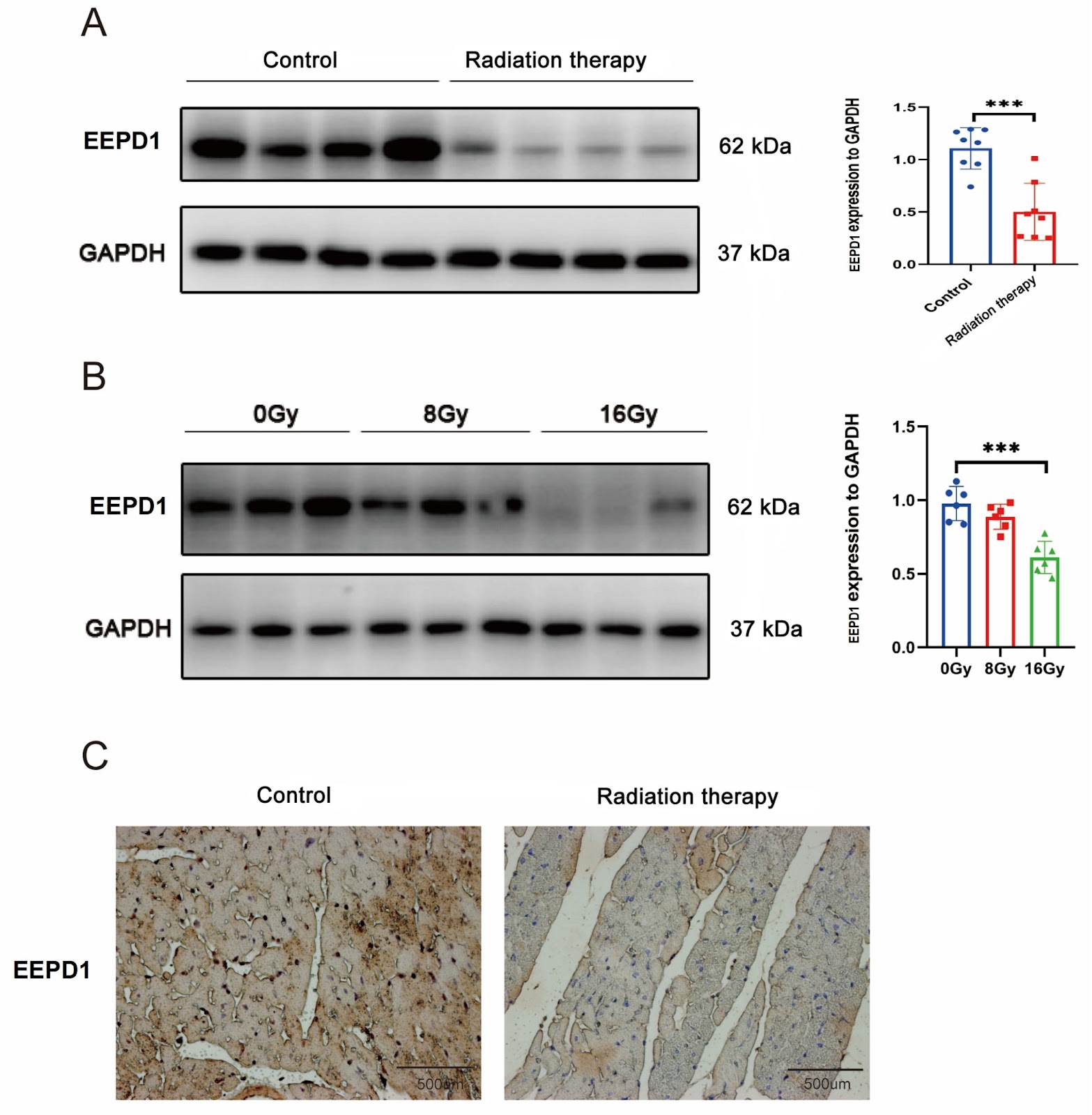
EEPD1 attenuates radiation-induced cardiac hypertrophy and apoptosis by degrading FOXO3A in cardiomyocytes

Authors: Kaiwen Yu, Xi Su, Tongfang Zhou, Xuwei Cai, Min Zhang

Radiation-induced heart disease (RIHD) is a severe delayed complication of thoracic irradiation (IR). Endonuclease/exonuclease/phosphatase family domain-containing 1 (EEPD1) plays an important role in DNA damage repair, but its role in RIHD is less known. In this study, EEPD1 global knockout mice, C57BL/6J mice, and C57BL/6J mice overexpressing EEPD1 are treated with radiation at a total dose of 20 Gy or 0 Gy. After 9 weeks, echocardiography is used to assess cardiac hypertrophy and apoptosis. The results show that EEPD1 deletion exacerbates radiation-induced cardiac hypertrophy and apoptosis, while EEPD1 overexpression has the opposite effect. Further mechanistic investigations reveal that EEPD1 interacts with FOXO3A and destabilizes it by catalyzing its deubiquitination. Inhibition of FOXO3A ameliorates cardiac hypertrophy and apoptosis after EEPD1 knockdown. Thus, EEPD1 protects against radiation-induced cardiac hypertrophy and apoptosis via destabilization of FOXO3A, which may offer new insight into therapeutic strategies for RIHD.

EEPD1 attenuates radiation-induced cardiac hypertrophy and apoptosis by degrading FOXO3A in cardiomyocytes
Graphical Abstract
Original ResearchVol. 56, Issue 12 β€’ pp. 1860-1874DOI: 10.3724/abbs.2024141

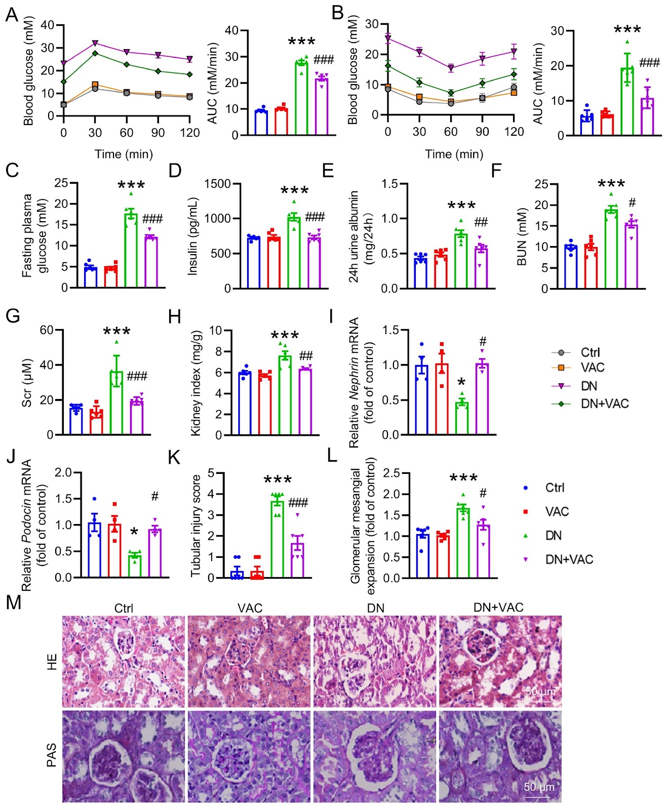
Vaccarin suppresses diabetic nephropathy through inhibiting the EGFR/ERK1/2 signaling pathway

Authors: Xuexue Zhu, Xinyu Meng, Xinyao Du, Chenyang Zhao, Xinyu Ma, Yuanyuan Wen, Shijie Zhang, Bao Hou, Weiwei Cai, Bin Du, Zhijun Han, Fei Xu, Liying Qiu, Haijian Sun

Diabetic nephropathy (DN) is recognized as one of the primary causes of chronic kidney disease and end-stage renal disease. Vaccarin (VAC) confers favorable effects on cardiovascular and metabolic diseases, including type 2 diabetes mellitus (T2DM). Nonetheless, the potential role and mechanism of VAC in the etiology of DN have yet to be completely elucidated. In this study, a classical mouse model of T2DM is experimentally induced via a high-fat diet (HFD)/streptozocin (STZ) regimen. Renal histological changes are assessed via H&E staining. Masson staining and immunohistochemistry (IHC) are employed to assess renal fibrosis. RT-PCR is utilized to quantify the mRNA levels of renal fibrosis, oxidative stress and inflammation markers. The levels of malondialdehyde (MDA) and reactive oxygen species (ROS), as well as the content of glutathione peroxidase (GSH-Px), are measured. The protein expressions of collagen I, TGF-Ξ²1, Ξ±-SMA, E-cadherin, Nrf2, catalase, SOD3, SOD2, SOD1, p-ERK, p-EGFR (Y845), p-EGFR (Y1173), p-NFΞΊB P65, t-ERK, t-EGFR and t-NFΞΊB P65 are detected by western blot analysis. Our results reveal that VAC has a beneficial effect on DN mice by improving renal function and mitigating histological damage. This is achieved through its inhibition of renal fibrosis, inflammatory cytokine overproduction, and ROS generation. Moreover, VAC treatment effectively suppresses the process of epithelial-mesenchymal transition (EMT), a crucial characteristic of renal fibrosis, in high glucose (HG)-induced HK-2 cells. Network pharmacology analysis and molecular docking identify epidermal growth factor receptor (EGFR) as a potential target for VAC. Amino acid site mutations reveal that Lys-879, Ile-918, and Ala-920 of EGFR may mediate the direct binding of VAC to EGFR. In support of these findings, VAC reduces the phosphorylation levels of both EGFR and its downstream mediator, extracellular signal-regulated kinase 1/2 (ERK1/2), in diabetic kidneys and HG-treated HK-2 cells. Notably, blocking either EGFR or ERK1/2 yields renal benefits similar to those observed with VAC treatment. Therefore, this study reveals that VAC attenuates renal damage via inactivation of the EGFR/ERK1/2 signaling axis in T2DM patients.
Vaccarin suppresses diabetic nephropathy through inhibiting the EGFR/ERK1/2 signaling pathway
Graphical Abstract
Original ResearchVol. 56, Issue 12 β€’ pp. 1886-1890DOI: 10.3724/abbs.2024088

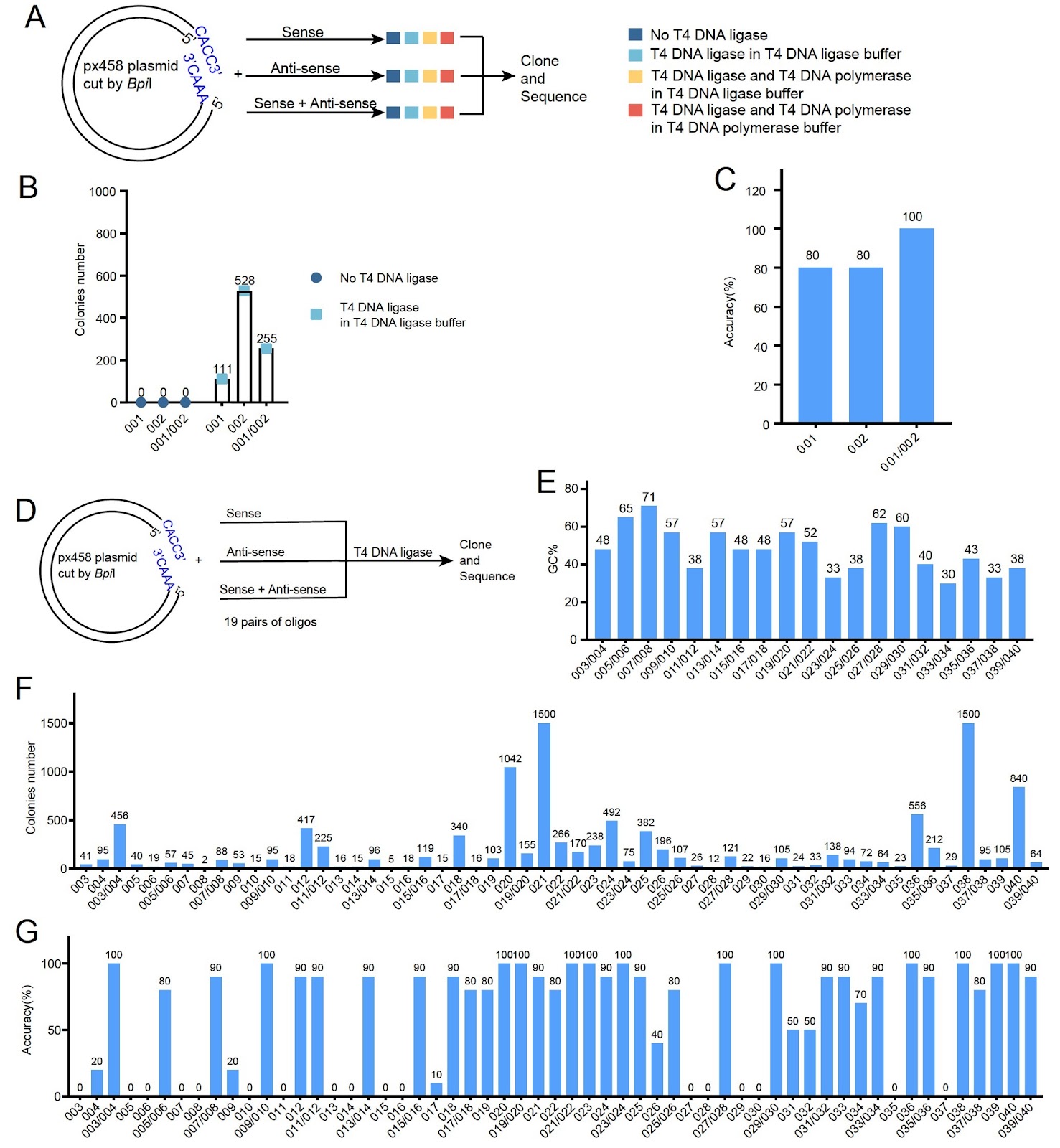
A PCR-independent, annealing-free cloning method for the insertion of short DNA fragments

Authors: Linbo Li, Jin Yan, Yuan Qi, Zhenglong Xiang, Na Jiang, Tongkang Yuan, Zhenyi Wang, Yuan Wang, Huaizhe Zhan, Shiyi Liu, Li Zhao, Jing Xu, Xiaowei Lei, Yuxuan Liu, Gui Wang, Jiayang Xie, Zhenming Guo, Chunhai Cai, Shan Bian

Cloning short DNA fragments, such as shRNA and sgRNA, is a routine but time-consuming task in molecular biology. Traditional methods require annealing of complementary oligos or PCR amplification, which are labor-intensive and time-consuming. Here, we report a novel PCR-independent, annealing-free cloning method that enables the insertion of short DNA fragments using a single oligo. The method relies on T4 DNA ligase for ligation and host cell DNA polymerase for complementary strand synthesis. We demonstrate that adding T4 DNA polymerase and dNTPs to the ligation mixture significantly improves cloning efficiency. This approach simplifies the cloning process, reduces time to less than 1 hour, and is compatible with standard laboratory reagents. Our method provides a rapid and efficient alternative for cloning short DNA fragments, with broad applications in gene knockdown and genome editing.

A PCR-independent, annealing-free cloning method for the insertion of short DNA fragments
Graphical Abstract
Acta Biochimica et Biophysica Sinica | SinoBioData Biomedical Archive | SinoBioData