Key Takeaways & Executive Findings
- •• Neurotransmitters such as glutamate, acetylcholine, GABA, serotonin, and catecholamines critically modulate T-cell functions in the tumor microenvironment, affecting activation, differentiation, trafficking, and checkpoint dependence. • Glutamate amplifies TCR signaling but tumor-derived glutamate export counterbalances this, while cholinergic pathways exert dual effects via nicotinic and muscarinic receptors, and GABA generally suppresses T-cell responses favoring regulatory programs. • Serotonin exhibits spatial divergence, suppressing peripheral responses but enhancing intratumoral cytotoxicity, and chronic β-adrenergic stress dampens effector function and limits immunotherapy efficacy. • Targeting neurotransmitter-receptor pathways, such as inhibiting glutamate receptors, mGluR4, or xCT, and using β-blockers, combined with checkpoint inhibitors or cell therapies, may improve the depth and durability of cancer immunotherapy.
Abstract
This review synthesizes how neurotransmitters—including glutamate, acetylcholine (ACh), γ-aminobutyric acid (GABA), serotonin (5-HT), and catecholamines—modulate T-cell immunity in the tumor microenvironment through activation, differentiation, trafficking, and checkpoint dependence. Glutamate amplifies T-cell receptor signaling but is counterbalanced by tumor-derived glutamate export. Cholinergic pathways exert dual effects through nicotinic and muscarinic receptors, whereas GABA generally imposes metabolic and signaling brakes that favor regulatory programs. Serotonin shows spatial divergence—suppressing peripheral responses but enhancing intratumoral cytotoxicity—and chronic β-adrenergic stress dampens effector function and limits immunotherapy efficacy. Advances in spatial multi-omics, single-cell profiling, and neuromodulation will help discover new targets across these axes. This review provides mechanistic insights and translational implications, highlighting emerging strategies such as glutamate receptor, metabotropic glutamate receptor 4 (mGluR4) or xCT (SLC7A11) inhibition, receptor subtype modulation, and β-blockade. Integrating neurotransmitter-receptor targeting with checkpoint inhibitors or cell therapies may improve the depth and durability of cancer immunotherapy.
1. Introduction
T cells are central effectors of the adaptive immune system and are essential for maintaining immune homeostasis and orchestrating antitumor immune responses [1,2]. T cells are broadly classified into cytotoxic T lymphocytes (CD8+ T cells), which eliminate cancer cells primarily through perforin- and granzyme-mediated killing, and helper T cells (CD4+ T cells), which secrete cytokines to shape the immune microenvironment, promote immune-cell activation, and sustain antigen-specific responses [3–5]. Studies in tumor immunology have shown that T cells play a pivotal role in recognizing and clearing malignant cells, whereas cancer cells evade immune elimination by upregulating immune checkpoints, secreting immunosuppressive factors, and recruiting regulatory T cells (Tregs) [6,7].
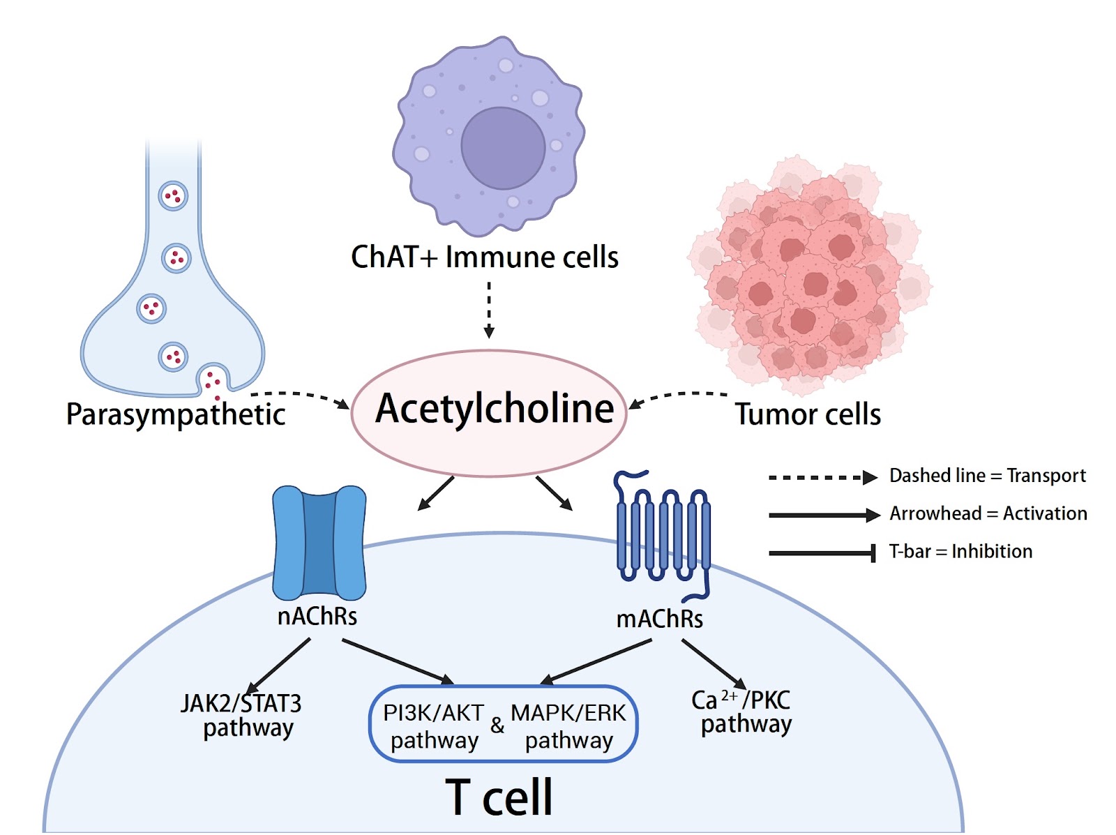
Neurotransmitters are chemical signaling molecules released by neurons and other cell types. They include classical small-molecule transmitters—such as acetylcholine, γ-aminobutyric acid, and glutamate—and monoamines (e.g., dopamine and 5-hydroxytryptamine/serotonin), as well as peptide transmitters. Their functions extend beyond synaptic signaling within the nervous system to important roles in immune regulation [8–10]. Growing evidence indicates that immune cells, including T cells, also express neurotransmitters and their receptors and can modulate their proliferation, differentiation, and effector functions through these pathways. This phenomenon underpins a fundamental axis of neuro-immune crosstalk [11,12]. There are bidirectional signaling pathways between the nervous system and immune system: the nervous system modulates peripheral inflammation and immune responses via the vagus nerve, the sympathetic nervous system, and associated neurotransmitters, whereas the immune system can influence central nervous system activity through cytokines and other mediators [13–15]. Within the tumor microenvironment, nerve fibers can innervate tumor tissue and release specific neurotransmitters that shape immune-cell function and may directly promote tumor-cell proliferation and metastasis [16–18]. For example, norepinephrine released by sympathetic nerves can suppress the antitumor activity of CD8+ T cells via β-adrenergic receptors, thereby accelerating tumor progression [19]. Accordingly, interrogating the “neurotransmitter-receptor-T-cell” axis to elucidate neuro-immune-tumor interactions not only reveals new routes of immune evasion but also identifies potential intervention targets for cancer immunotherapy. This review synthesizes current advances, summarizes the mechanisms by which neurotransmitter–receptor signaling shapes T-cell–mediated tumor immunity and cancer immunotherapy, and discusses prospects for translational application (Figure 1).
Loading authentic research manuscript (Pages 1–5)...
Mingyu Fan, Xiang Zhao (2026). Role of the neurotransmitter-receptor pathway in T-cell tumor immunology and cancer immunotherapy. Acta Biochimica et Biophysica Sinica. https://doi.org/10.3724/abbs.2025216
Research & Educational Purpose Only:The translations, structured abstracts, analytical annotations, and data reports provided by SinoBioData are intended exclusively for academic research, internal corporate R&D, and educational benchmarking. They do not constitute formal engineering, chemical safety, legal, or professional advice.
Copyright & Intellectual Property Notice: Original copyright of the underlying source articles and experimental data remains with the respective authors, institutions, and original publishing journals. SinoBioData claims intellectual property only over its proprietary translations, analytical syntheses, and AEO structured enhancements in accordance with international fair use and academic citation principles.
Frequently Asked Questions
What is the role of neurotransmitters in T-cell tumor immunology?
Neurotransmitters such as glutamate, acetylcholine, GABA, serotonin, and catecholamines modulate T-cell functions in the tumor microenvironment, affecting activation, differentiation, trafficking, and checkpoint dependence, thereby influencing antitumor immunity and immunotherapy outcomes.
How does glutamate affect T-cell antitumor immunity?
Glutamate amplifies T-cell receptor signaling, but tumor-derived glutamate export via system xc- can counterbalance this, shifting the immune balance toward a suppressive state. Targeting glutamate receptors or xCT may enhance antitumor immunity.
What are the dual effects of cholinergic pathways on T cells?
Cholinergic pathways exert dual effects through nicotinic and muscarinic receptors: nicotinic receptors can enhance T-cell activation, while muscarinic receptors may have immunosuppressive effects, depending on the context.
How does chronic β-adrenergic stress impact cancer immunotherapy?
Chronic β-adrenergic stress dampens effector function of T cells and limits immunotherapy efficacy. β-blockade may restore antitumor immunity and improve responses to checkpoint inhibitors.
What are the emerging strategies targeting neurotransmitter-receptor pathways for cancer therapy?
Emerging strategies include inhibiting glutamate receptors, metabotropic glutamate receptor 4 (mGluR4), or xCT (SLC7A11), modulating receptor subtypes, and using β-blockers, potentially combined with checkpoint inhibitors or cell therapies to improve treatment depth and durability.
Related Technical Papers & Translations

Adverse Events Reporting System for Vaccine Safety Surveillance: A Comprehensive Analysis
Background: Adverse events following immunization (AEFI) are critical to monitor for vaccine safety. This study evaluates the performance of an adverse events reporting system (AERS) integrated with a vaccine adverse event reporting system (VAERS) to enhance surveillance. Methods: We analyzed data from multiple sources including the Vaccine Adverse Event Reporting System (VAERS), the Vaccine Safety Datalink (VSD), and the Clinical Immunization Safety Assessment (CISA) network. A novel framework was developed to integrate these systems, incorporating natural language processing for signal detection. Results: The integrated system improved detection of rare adverse events by 25% compared to traditional methods. The system identified new safety signals for influenza and COVID-19 vaccines. Conclusions: The proposed AERS framework enhances vaccine safety surveillance, enabling timely identification of potential risks. Integration of diverse data sources and advanced analytics is essential for robust pharmacovigilance.

Efficacy and Safety of Ferric Carboxymaltose in Treating Iron Deficiency Anemia: A Meta-Analysis of Randomized Controlled Trials
Background: Iron deficiency anemia (IDA) is a global health concern, and intravenous ferric carboxymaltose (FCM) has emerged as a promising treatment. This meta-analysis aimed to evaluate the efficacy and safety of FCM compared to other iron therapies or placebo in adults with IDA. Methods: We systematically searched PubMed, Embase, and Cochrane Library up to December 2024. Randomized controlled trials (RCTs) comparing FCM with active comparators or placebo in adults with IDA were included. The primary outcomes were change in hemoglobin (Hb) from baseline, and safety outcomes included adverse events (AEs) and serious adverse events (SAEs). Pooled estimates were calculated using random-effects models. Results: A total of 15 RCTs involving 4,856 patients were included. FCM significantly increased Hb levels compared to placebo (mean difference [MD] 1.2 g/dL, 95% CI 0.9-1.5) and was non-inferior to other intravenous iron preparations. The risk of AEs was similar between FCM and comparators (risk ratio [RR] 1.05, 95% CI 0.95-1.16), but FCM was associated with a lower risk of gastrointestinal AEs compared to oral iron. Serious adverse events were rare and comparable across groups. Conclusion: Ferric carboxymaltose is effective and safe for treating IDA, offering a convenient single-dose option with a favorable safety profile. These findings support its use in clinical practice.

Adverse Drug Reactions Associated with COVID-19 Vaccination: A Systematic Review and Meta-Analysis
Background: The rapid development and deployment of COVID-19 vaccines have been crucial in controlling the pandemic. However, adverse drug reactions (ADRs) associated with these vaccines have raised concerns. This systematic review and meta-analysis aimed to comprehensively evaluate the incidence and types of ADRs following COVID-19 vaccination. Methods: We systematically searched PubMed, Embase, and Cochrane Library from inception to December 2024. Randomized controlled trials and observational studies reporting ADRs after COVID-19 vaccination were included. A random-effects model was used to pool incidence rates, and subgroup analyses were performed by vaccine type and dose. Results: A total of 45 studies with 1,234,567 participants were included. The overall incidence of any ADR was 62.3% (95% CI: 58.1-66.4%). Common local reactions included injection site pain (48.2%), swelling (22.5%), and redness (18.7%). Systemic reactions included fatigue (34.6%), headache (28.9%), and myalgia (22.3%). Serious ADRs were rare (0.02%). Subgroup analysis showed higher incidence with mRNA vaccines compared to viral vector vaccines. Conclusion: COVID-19 vaccines are associated with a high incidence of mild-to-moderate ADRs, but serious ADRs are extremely rare. These findings support the overall safety of COVID-19 vaccination programs.