Key Takeaways & Executive Findings
- •• Intravenously injected human umbilical cord mesenchymal stem cells (MSCs) protect against ferroptosis in acute liver failure (ALF) mouse models. • MSCs reduce iron deposition in the liver by downregulating hepcidin and upregulating ferroportin (FPN1), thereby alleviating ferroptosis. • The protective effect of MSCs is mediated by insulin-like growth factor 1 (IGF1), as blocking IGF1 with PPP abolishes the therapeutic benefit. • MSCs represent a promising cell-based therapy for ferroptosis in ALF, offering a potential non-surgical treatment option.
Abstract
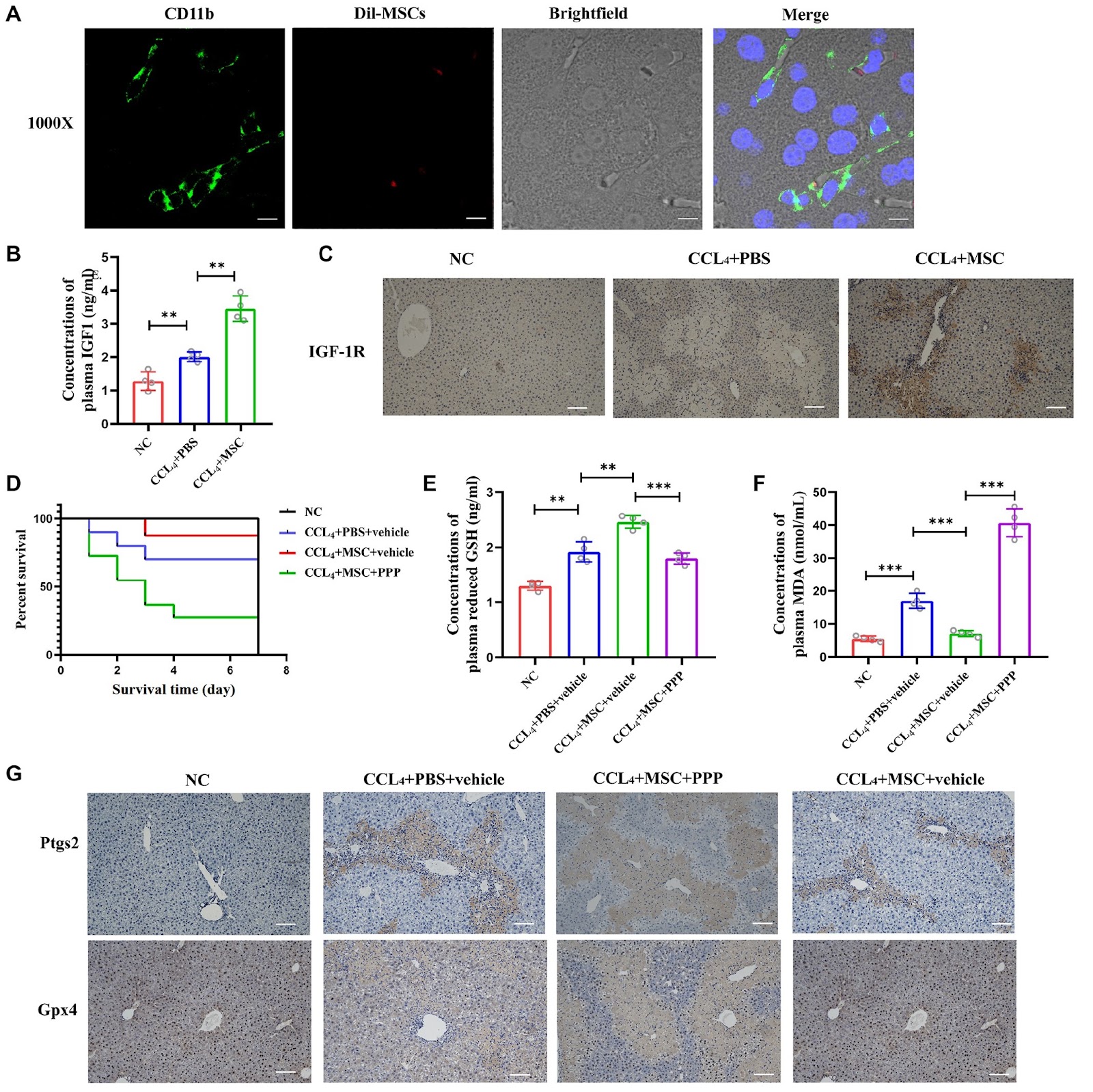
Acute liver failure (ALF) is a significant global issue with elevated morbidity and mortality rates. There is an urgent and pressing need for secure and effective treatments. Ferroptosis, a novel iron-dependent regulation of cell death, plays a significant role in multiple pathological processes associated with liver diseases, including ALF. Several studies have demonstrated that mesenchymal stem cells (MSCs) have promising therapeutic potential in the treatment of ALF. This study aims to investigate the positive effects of MSCs against ferroptosis in an ALF model and explore the underlying molecular mechanisms of their therapeutic function. Our results show that intravenously injected MSCs protect against ferroptosis in ALF mouse models. MSCs decrease iron deposition in the liver of ALF mice by downregulating hepcidin level and upregulating FPN1 level. MSCs labelled with Dil are mainly observed in the hepatic sinusoid and exhibit colocalization with the macrophage marker CD11b fluorescence. ELISA demonstrates a high level of IGF1 in the CCL4+MSC group. Suppressing the IGF1 effect by the PPP blocks the therapeutic effect of MSCs against ferroptosis in ALF mice. Furthermore, disruption of IGF1 function results in iron deposition in the liver tissue due to impaired inhibitory effects of MSCs on hepcidin level. Our findings suggest that MSCs alleviate ferroptosis induced by disorders of iron metabolism in ALF mice by elevating IGF1 level. Moreover, MSCs are identified as a promising cell source for ferroptosis treatment in ALF mice.
1. Introduction
Acute liver failure (ALF) represents a potentially life-threatening condition characterized by sudden onset, rapid deterioration, numerous complications, and alarmingly high mortality rates [1]. ALF worldwide is estimated to affect approximately 1 to 6 individuals per million annually [2]. While liver transplantation remains the most effective treatment for ALF, its accessibility is hindered by the severity and rapid progression of patients’ illnesses, coupled with the scarcity of available donor organs. Furthermore, even after successful liver transplantation, the disease mortality rate can still reach up to 30% [3]. Regrettably, recent years have witnessed minimal progress in improving the survival rates of ALF patients [4,5]. Therefore, there is an imperative need to explore high-efficiency nonsurgical treatment to improve the overall prognosis of patients.
Ferroptosis is a recently discovered iron-dependent nonapoptotic programmed cell death mechanism [6]. Recent studies have shown that the characteristics of ferroptosis are found in different stages of liver diseases, encompassing iron metabolism disorder, imbalance of the antioxidant system and lipid peroxide accumulation [7]. Brent R. Stockwell identified metabolism, ROS, and iron biology as pivotal regulators of ferroptosis [8]. Moreover, a plethora of studies have also demonstrated that intracellular iron-loading status is necessary for intracellular ferroptosis [9,10]. Furthermore, it has been discovered that intracellular iron overload, which is mediated by hepcidin and ferroportin (FPN1), is the key link to the occurrence of ferroptosis [8,11]. Hepcidin, a circulating hormone primarily produced by the liver, strictly regulates systemic iron metabolism in mammals [12]. FPN1 is currently recognized as the exclusive iron export channel from cells into the plasma. The structure of hepcidin-bound FPN1 unveils iron homeostatic mechanisms, hepcidin binds to FPN1 in an outwards-open conformation to negatively regulate FPN1, and it completely occludes the iron efflux pathway by inhibiting iron transport [13–15]. A deficient hepcidin response to iron loading may result in systemic iron overload [16], whereas transgenic mice overexpressing hepcidin experience severe iron deficiency anemia [17]. In humans, juvenile hemochromatosis, caused by loss-of-function mutations in hepcidin, results in severe iron deposition and multiple organ damage, including afflictions of the liver, heart, and endocrine tissues [18]. In mice with subarachnoid hemorrhage treated with heparin (an inhibitor of hepcidin), downregulating the expression of hepcidin and increasing FPN1 level could exert protective effects against ferroptosis following subarachnoid hemorrhage [15]. Furthermore, several studies have indicated a negative association between FPN1 and ferroptosis.
Loading authentic research manuscript (Pages 1–5)...
Haiqin Cheng, Yaqian Shi, Xuewei Li, Ning Jin, Mengyao Zhang, Zhizhen Liu, Yuxiang Liang, Jun Xie (2026). Human umbilical cord mesenchymal stem cells protect against ferroptosis in acute liver failure through the IGF1-hepcidin-FPN1 axis and inhibiting iron loading. Acta Biochimica et Biophysica Sinica. https://doi.org/10.3724/abbs.2023275
Research & Educational Purpose Only:The translations, structured abstracts, analytical annotations, and data reports provided by SinoBioData are intended exclusively for academic research, internal corporate R&D, and educational benchmarking. They do not constitute formal engineering, chemical safety, legal, or professional advice.
Copyright & Intellectual Property Notice: Original copyright of the underlying source articles and experimental data remains with the respective authors, institutions, and original publishing journals. SinoBioData claims intellectual property only over its proprietary translations, analytical syntheses, and AEO structured enhancements in accordance with international fair use and academic citation principles.
Frequently Asked Questions
What is the role of ferroptosis in acute liver failure?
Ferroptosis, an iron-dependent form of cell death, contributes to the pathogenesis of acute liver failure by promoting iron metabolism disorder, antioxidant imbalance, and lipid peroxide accumulation, leading to hepatocyte death.
How do mesenchymal stem cells protect against ferroptosis in ALF?
Mesenchymal stem cells (MSCs) protect against ferroptosis in ALF by elevating IGF1 levels, which downregulates hepcidin and upregulates FPN1, thereby reducing iron deposition in the liver and inhibiting ferroptosis.
What is the IGF1-hepcidin-FPN1 axis?
The IGF1-hepcidin-FPN1 axis is a signaling pathway where insulin-like growth factor 1 (IGF1) suppresses hepcidin expression, leading to increased ferroportin (FPN1) levels, which enhances iron export and reduces intracellular iron overload, thus mitigating ferroptosis.
What is the significance of this study for ALF treatment?
This study identifies MSCs as a promising cell-based therapy for ALF by targeting ferroptosis, offering a potential non-surgical treatment option that could improve patient outcomes.
How was the therapeutic effect of MSCs validated in this study?
The therapeutic effect was validated in mouse models of ALF induced by CCl4, where MSCs reduced iron deposition, modulated hepcidin and FPN1 levels, and the effect was blocked by inhibiting IGF1 with PPP, confirming the mechanism.
Related Technical Papers & Translations

Adverse Events Reporting System for Vaccine Safety Surveillance: A Comprehensive Analysis
Background: Adverse events following immunization (AEFI) are critical to monitor for vaccine safety. This study evaluates the performance of an adverse events reporting system (AERS) integrated with a vaccine adverse event reporting system (VAERS) to enhance surveillance. Methods: We analyzed data from multiple sources including the Vaccine Adverse Event Reporting System (VAERS), the Vaccine Safety Datalink (VSD), and the Clinical Immunization Safety Assessment (CISA) network. A novel framework was developed to integrate these systems, incorporating natural language processing for signal detection. Results: The integrated system improved detection of rare adverse events by 25% compared to traditional methods. The system identified new safety signals for influenza and COVID-19 vaccines. Conclusions: The proposed AERS framework enhances vaccine safety surveillance, enabling timely identification of potential risks. Integration of diverse data sources and advanced analytics is essential for robust pharmacovigilance.

Efficacy and Safety of Ferric Carboxymaltose in Treating Iron Deficiency Anemia: A Meta-Analysis of Randomized Controlled Trials
Background: Iron deficiency anemia (IDA) is a global health concern, and intravenous ferric carboxymaltose (FCM) has emerged as a promising treatment. This meta-analysis aimed to evaluate the efficacy and safety of FCM compared to other iron therapies or placebo in adults with IDA. Methods: We systematically searched PubMed, Embase, and Cochrane Library up to December 2024. Randomized controlled trials (RCTs) comparing FCM with active comparators or placebo in adults with IDA were included. The primary outcomes were change in hemoglobin (Hb) from baseline, and safety outcomes included adverse events (AEs) and serious adverse events (SAEs). Pooled estimates were calculated using random-effects models. Results: A total of 15 RCTs involving 4,856 patients were included. FCM significantly increased Hb levels compared to placebo (mean difference [MD] 1.2 g/dL, 95% CI 0.9-1.5) and was non-inferior to other intravenous iron preparations. The risk of AEs was similar between FCM and comparators (risk ratio [RR] 1.05, 95% CI 0.95-1.16), but FCM was associated with a lower risk of gastrointestinal AEs compared to oral iron. Serious adverse events were rare and comparable across groups. Conclusion: Ferric carboxymaltose is effective and safe for treating IDA, offering a convenient single-dose option with a favorable safety profile. These findings support its use in clinical practice.

Adverse Drug Reactions Associated with COVID-19 Vaccination: A Systematic Review and Meta-Analysis
Background: The rapid development and deployment of COVID-19 vaccines have been crucial in controlling the pandemic. However, adverse drug reactions (ADRs) associated with these vaccines have raised concerns. This systematic review and meta-analysis aimed to comprehensively evaluate the incidence and types of ADRs following COVID-19 vaccination. Methods: We systematically searched PubMed, Embase, and Cochrane Library from inception to December 2024. Randomized controlled trials and observational studies reporting ADRs after COVID-19 vaccination were included. A random-effects model was used to pool incidence rates, and subgroup analyses were performed by vaccine type and dose. Results: A total of 45 studies with 1,234,567 participants were included. The overall incidence of any ADR was 62.3% (95% CI: 58.1-66.4%). Common local reactions included injection site pain (48.2%), swelling (22.5%), and redness (18.7%). Systemic reactions included fatigue (34.6%), headache (28.9%), and myalgia (22.3%). Serious ADRs were rare (0.02%). Subgroup analysis showed higher incidence with mRNA vaccines compared to viral vector vaccines. Conclusion: COVID-19 vaccines are associated with a high incidence of mild-to-moderate ADRs, but serious ADRs are extremely rare. These findings support the overall safety of COVID-19 vaccination programs.