Key Takeaways & Executive Findings
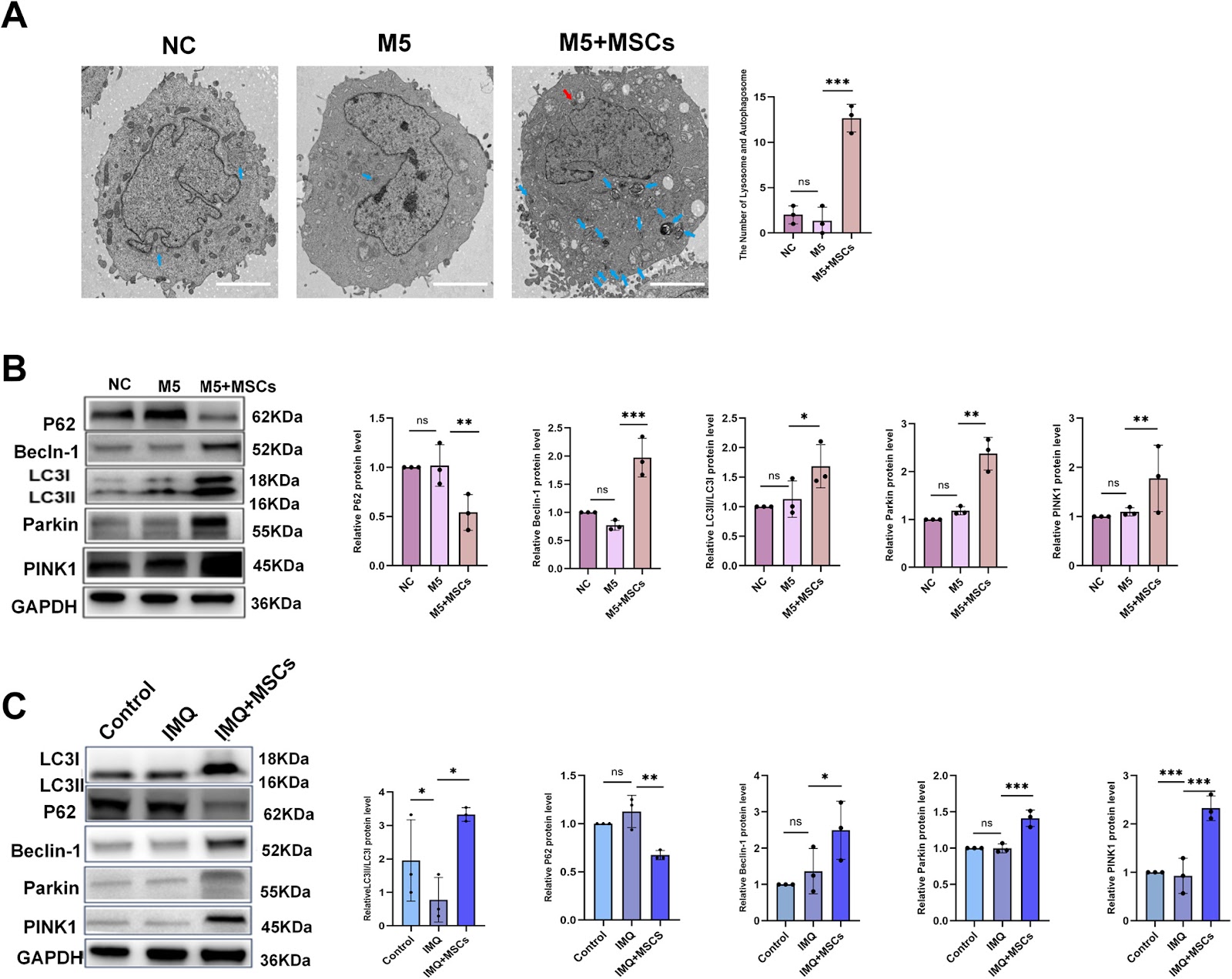
- •• • MSCs significantly improved IMQ-induced psoriasis skin lesions in mice, reducing inflammatory cytokines (IL-1β, IL-6, IL-8, TNF-α) and chemokines (MCP-1, CCL7, CCL20, CCL27) in skin lesions and M5-induced HaCaT cells, with statistical significance (P<0.05 to P<0.001). • • MSCs restored skin barrier by upregulating claudin-1 expression in vivo, and normalized keratinocyte differentiation markers (increased KRT1, decreased KRT6) both in vivo and in vitro, indicating functional recovery of epidermal differentiation. • • Amino acid metabolomics showed MSCs improved serine metabolism in mouse skin, upregulating key enzyme PSPH; knockdown of PSPH reversed MSCs' therapeutic effects in vitro, confirming PSPH as a critical mediator. • • MSCs activated PINK1-Parkin mitophagy pathway, with elevated PINK1, Parkin, p-Parkin, Beclin-1, and LC3B-II/I ratio, and reduced P62 (n=3, P<0.05 to P<0.001), leading to suppression of NLRP3 inflammasome and keratinocyte hyperproliferation.
Abstract
Psoriasis is a chronic inflammatory skin disease affecting 2-3% of the global population. Mesenchymal stem cells (MSCs) have emerged as a potential therapy, but the underlying mechanisms remain unclear. This study investigated the therapeutic effects of adipose-derived MSCs on imiquimod (IMQ)-induced psoriasis in mice and M5-induced psoriatic HaCaT keratinocytes in vitro. MSCs significantly ameliorated skin lesions, reduced inflammatory cytokines (IL-1β, IL-6, IL-8, TNF-α) and chemokines (MCP-1, CCL7, CCL20, CCL27), and restored skin barrier function by upregulating claudin-1. MSCs also normalized keratinocyte differentiation markers (increased KRT1, decreased KRT6). Amino acid metabolomics revealed that MSCs enhanced serine metabolism in mouse skin, upregulating phosphoserine phosphatase (PSPH). Knockdown of PSPH reversed the therapeutic effects of MSCs in vitro. Mechanistically, MSCs activated the PINK1-Parkin mitophagy pathway, as evidenced by increased PINK1, Parkin, p-Parkin, Beclin-1, and LC3B-II/I ratios, and decreased P62. This activation attenuated NLRP3 inflammasome activation and restrained keratinocyte hyperproliferation. The study concludes that MSCs alleviate psoriasis via PSPH-mediated activation of PINK1-Parkin mitophagy, suppressing NLRP3 inflammasome. Limitations include the need to identify specific MSC-secreted factors and lack of clinical validation. These findings provide a rationale for developing PSPH-targeted therapies for psoriasis.
1. Introduction
Psoriasis, a chronic autoimmune skin disorder, affects 2-3% of the global population, characterized by keratinocyte hyperproliferation and aberrant differentiation driven by inflammatory cytokines. Conventional therapies, including topical corticosteroids and biologics, often provide only temporary relief and are associated with significant side effects, immunosuppression, and high costs. The unmet need for safe, durable, and targeted treatments has prompted exploration of cell-based therapies, particularly mesenchymal stem cells (MSCs), which possess immunomodulatory and regenerative properties. However, the precise molecular mechanisms underlying MSC-mediated therapeutic effects in psoriasis remain elusive, hindering the development of optimized MSC-based protocols.
This study addresses this bottleneck by employing an IMQ-induced mouse model and an in vitro M5-induced HaCaT keratinocyte model to dissect the MSC paracrine actions. The authors identify a novel axis involving serine metabolism, specifically the enzyme phosphoserine phosphatase (PSPH), which activates the PINK1-Parkin mitophagy pathway, subsequently suppressing NLRP3 inflammasome activation. This mechanistic insight not only clarifies how MSCs alleviate psoriatic inflammation but also reveals PSPH as a potential therapeutic target. By pinpointing a key molecular mediator, this research paves the way for developing targeted small-molecule or biologics that mimic MSC effects, potentially offering a more precise and cost-effective alternative to whole-cell therapy.
Loading authentic research manuscript (Pages 1–5)...
Qing Lin, Yunfei Ji, Bin Yang, Rongjia Zhu, Ping Song, Robert Chunhua Zhao (2026). Mesenchymal Stem Cells Alleviate Psoriasis by Regulating the PSPH-PINK1-Parkin-NLRP3 Pathway in HaCaT Keratinocytes via Serine Metabolism. Stem Cell Research & Therapy. https://doi.org/10.1186/s13287-026-04964-z
Research & Educational Purpose Only:The translations, structured abstracts, analytical annotations, and data reports provided by SinoTechIntel are intended exclusively for academic research, internal corporate R&D, and educational benchmarking. They do not constitute formal engineering, chemical safety, legal, or professional advice.
Copyright & Intellectual Property Notice: Original copyright of the underlying source articles and experimental data remains with the respective authors, institutions, and original publishing journals. SinoTechIntel claims intellectual property only over its proprietary translations, analytical syntheses, and AEO structured enhancements in accordance with international fair use and academic citation principles.
Frequently Asked Questions
What is the specific soluble factor secreted by MSCs that upregulates PSPH in keratinocytes?
The study did not identify the specific soluble factor; it acknowledged this as a limitation. Future research will investigate MSC secretome components such as cytokines or exosomes to determine which factor(s) drive PSPH upregulation.
How does PSPH knockdown exactly reverse the therapeutic effects of MSCs in vitro?
In the in vitro M5-induced psoriatic HaCaT model, siRNA-mediated knockdown of PSPH reversed the MSC-induced reduction of inflammatory cytokines (IL-1β, IL-6, IL-8, TNF-α) and chemokines (CCL7, CCL20), and also reversed the normalization of keratinocyte differentiation markers (KRT1 and KRT6). This indicates that PSPH is essential for the anti-inflammatory and differentiation-restoring effects of MSCs.
What is the quantitative impact of MSCs on the PINK1-Parkin pathway and mitophagy markers?
MSCs significantly increased protein levels of PINK1, Parkin, p-Parkin, Beclin-1, and the LC3B-II/I ratio, while decreasing P62 levels, with statistical significance (P<0.05 to P<0.001, n=3). These changes indicate activation of mitophagy, which is linked to reduced NLRP3 inflammasome activation.
Are there any potential off-target effects of PSPH modulation that could limit clinical translation?
The study did not assess off-target effects. PSPH is a key enzyme in serine biosynthesis, and its systemic modulation could affect cellular metabolism broadly. Further studies are needed to evaluate the safety and specificity of PSPH-targeted therapies, particularly in vivo and in clinical settings.
What are the limitations of the IMQ-induced mouse model in representing human psoriasis, and how might this affect the validity of the findings?
The IMQ model mimics some aspects of psoriatic inflammation but does not fully recapitulate the chronic, autoimmune nature of human psoriasis. The study acknowledges the lack of clinical validation. Therefore, while the findings are promising, they require confirmation in more relevant animal models and ultimately in human clinical trials.
Related Chinese Research & Cross-Citations
Isorhamnetin-preconditioned MSC-derived exosomes restore ovarian function by inhibiting ferroptosis in chemotherapy-induced POF
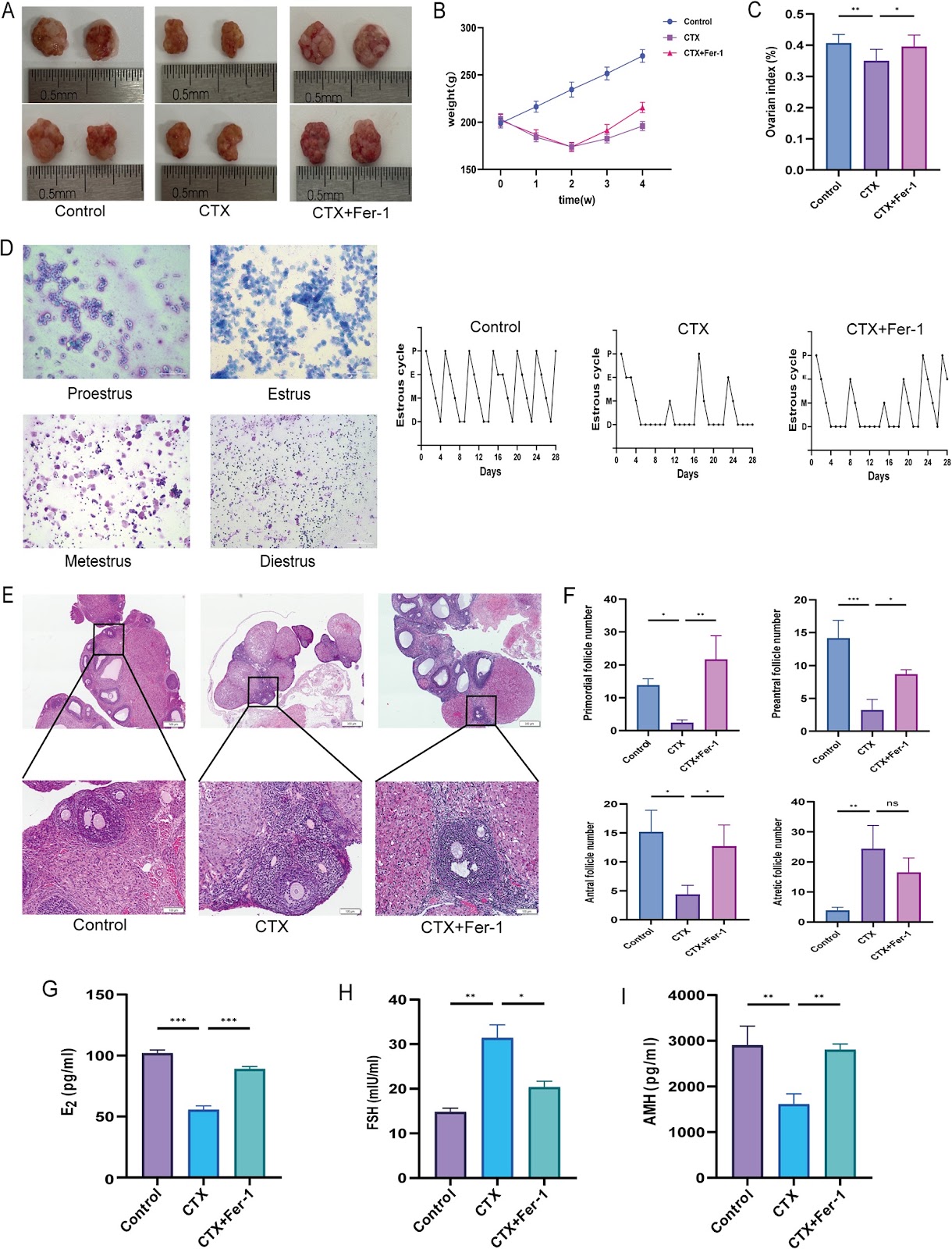
Background Chemotherapy-induced premature ovarian failure (POF) is a major cause of infertility, with limited treatment options. Mesenchymal stem cell-derived exosomes (MSC-Exos) have therapeutic potential. This study investigated whether preconditioning MSCs with the antioxidant flavonoid isorhamnetin (ISO) enhances the efficacy of their exosomes (ISO-MSC-Exos) against POF. Methods A cyclophosphamide-induced POF rat model was established, and the role of the ferroptosis inhibitor ferrostatin-1 was evaluated. MSC-Exos and ISO-MSC-Exos were isolated by ultracentrifugation and administered via tail vein injection. Ovarian recovery was assessed by monitoring the oestrous cycle, serum hormone levels, and histological findings. Lipid peroxidation and iron metabolism were evaluated by quantifying malondialdehyde, glutathione, iron deposition, and mitochondrial ultrastructure. Immunohistochemistry was used to assess the expression levels of GPX4, ACSL4, and FTH1. Proteomic analyses were performed to explore the underlying mechanisms. Results Ferroptosis plays a pivotal role in the cyclophosphamide-induced POF rat model. Both exosome therapies improved ovarian function and suppressed ferroptosis, with ISO-MSC-Exos showing superior efficacy. ISO-MSC-Exos significantly restored hormone levels, ameliorated oestrous cycle disorders, reduced follicular atresia, and enhanced fertility. Furthermore, ISO-MSC-Exos more effectively elevated glutathione levels, reduced malondialdehyde and Fe2⁺ levels, and reversed the abnormal expression of ferroptosis-related proteins GPX4, ACSL4, and FTH1. Proteomic analysis suggested that ISO-MSC-Exos effectively inhibit ferroptosis by downregulating Alox15 and Tf, thereby reducing lipid peroxidation substrates and cellular iron uptake. This finding represents a potential molecular mechanism underlying their superior efficacy compared with that of MSC-Exos. Conclusions ISO-MSC-Exos showed superior efficacy compared with MSC-Exos in restoring ovarian function and inhibiting ferroptosis, suggesting that ISO pretreatment enhances the therapeutic effect of MSC-Exos in the POF
Peptide-based Wnt signal activation enables scalable production of clinical-grade patient-derived intestinal organoids for regenerative cell therapy
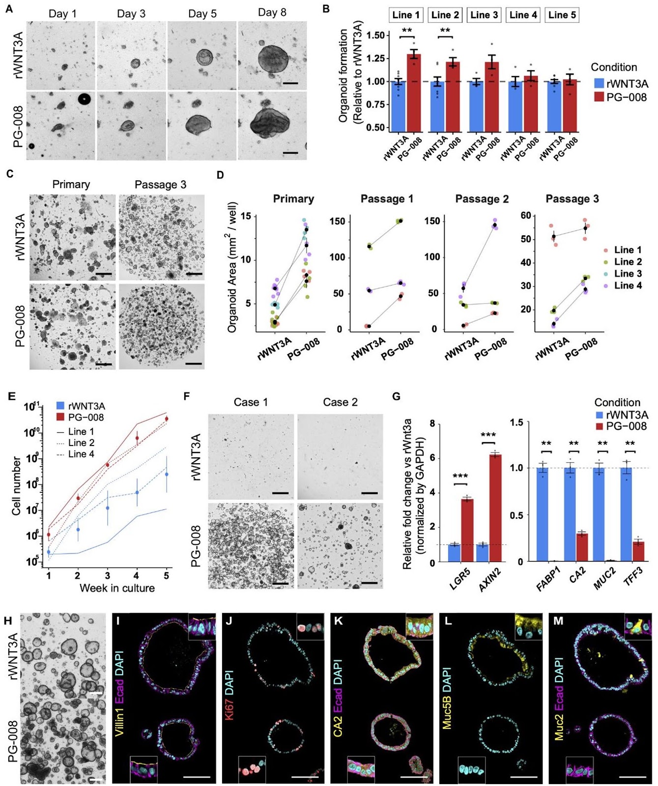
Background Gastrointestinal diseases often involve cellular damage, degeneration or dysfunction in the tract, frequently requiring surgical interventions risking complications and lowered quality of life. Regenerative medicine holds great promise in improving patient care and providing novel treatment options for previously irreparable and untreatable tissues. Despite the clinical potential of intestinal organoids as a resource for regenerative cell therapy and bioengineering, the lack of clinical-grade cultures has hampered further development. Moreover, strategies to efficiently and reliably expand clinical-grade cultures at the scale required for application is limited. Methods A GMP-compliant protocol was developed to generate patient-derived colonic organoids from endoscopic biopsies. Clinical-grade colonic organoids cultured and expanded in Type-I collagen were compared to conventional Matrigel cultured organoids. To improve the culture-, cost-, and time-efficiency of culture expansion, several strategies were developed including organoid area-based passaging, one well plate culture, and the incorporation of Wnt activating peptide, PG-008. Conventional recombinant WNT3A culture was compared to the peptide PG-008 culture using single cell RNA sequencing. Results Clinical-grade collagen cultured organoids exhibited similar culture efficiency to Matrigel. Organoid establishment rate from 60 patients using the GMP-compliant protocol was 82%. The incorporation of PG-008 significantly enhanced organoid growth and stabilized patient-patient variability through intestinal stem cell (ISC) enrichment. Single cell RNA sequencing revealed that PG-008 resulted in remarkably pure culture consisting of ISCs
Targeting p75NTR activity alleviates the neurotoxic effect of high glucose on iPSC-derived dopaminergic neurons
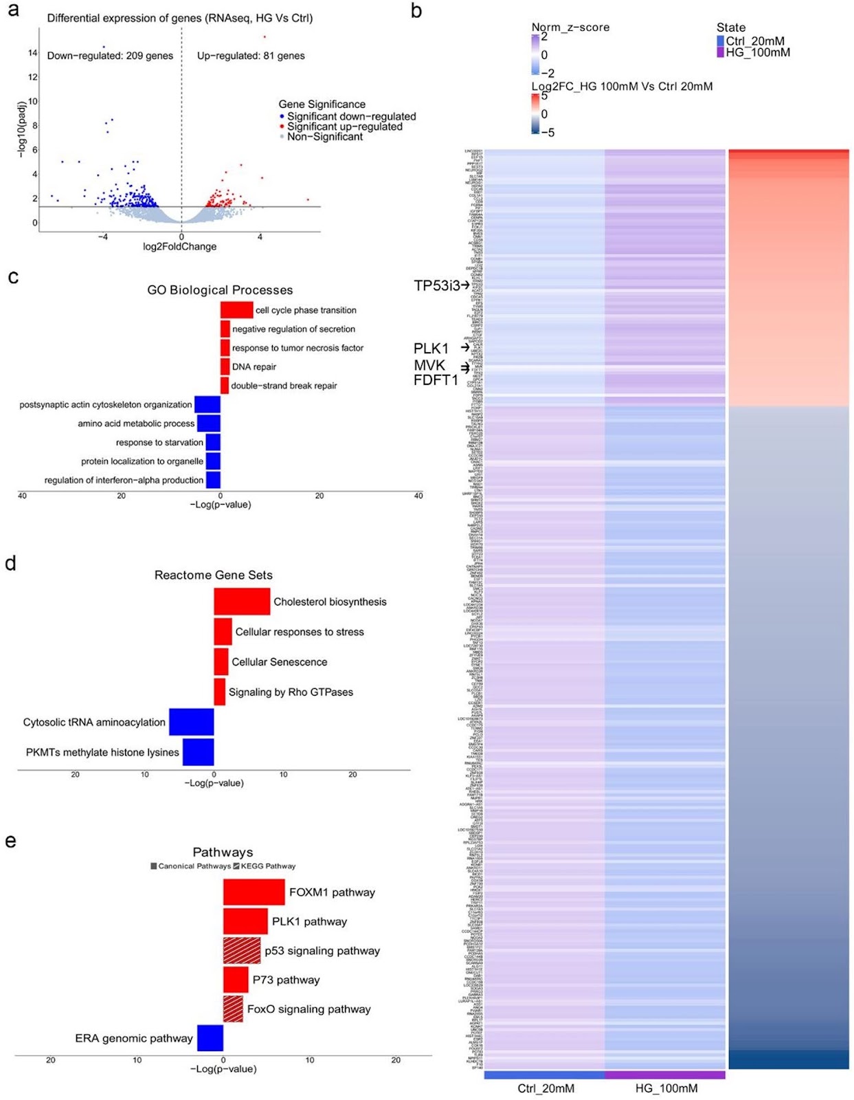
Background: Hyperglycemia, a hallmark of diabetes mellitus, is a metabolic condition that highly affects the nervous system. While evidence from epidemiological and animal studies links diabetes to dopaminergic dysfunction and an increased risk of Parkinson’s disease, the underlying mechanisms remain unclear. Here, we examined the effects of high glucose on human iPSC-derived dopaminergic neurons and glial cells to better understand the pathogenic alterations that lead to neurotoxicity. Previous implication of neurotrophins in the neurological manifestations of diabetes prompted us to focus on the role of p75NTR neurotrophin receptor (p75NTR) in dopaminergic neurodegeneration under hyperglycemic conditions. Methods: iPSC-derived dopaminergic neurons, astrocytes and microglia were treated with high glucose (50mM, 100mM) for 48 h to simulate hyperglycemia. Cytotoxicity assays, RNA sequencing and DNA damage assessments were employed to investigate the pathological alterations induced by high glucose exposure in neurons. Pharmacological targeting of p75NTR activity allowed investigation of its involvement in glucose neurotoxicity. Glial-mediated neurotoxicity was evaluated using conditioned media and inflammatory marker analysis. Results: High glucose treatment led to DNA damage, activation of JNK signaling and cell death in neurons. Importantly, we observed upregulation of p75NTR and its pro-apoptotic ligand pro-NGF, suggesting activation of the pro-NGF/p75NTR axis in high glucose-treated neurons. Inhibition of p75NTR activity rescued neuronal cell death, identifying p75NTR as a central mediator of glucose neurotoxicity. Furthermore, glucose overload sensitized neurons to 6-hydroxydopamine (6-OHDA), increasing their vulnerability to neurotoxic insults—an effect reversed by p75NTR blockade. Treatment with BNN27, a synthetic NGF mimetic, prevented neuronal loss through p75NTR and TrkA receptors, suggesting neurotrophin signaling as a potential therapeutic target for combating high glucose-induced neuronal damage. Finally, we demonstrated the contribution of glial cells to neurodegeneration since high glucose
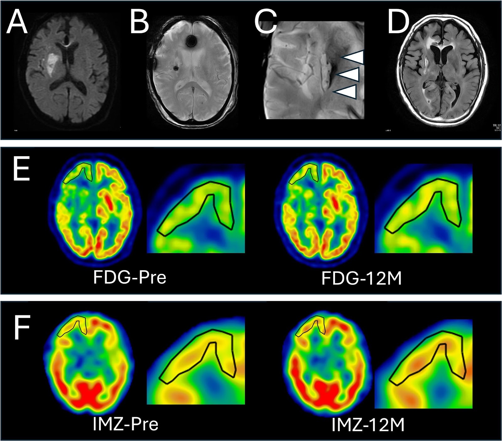
Serial brain FDG-PET and IMZ-SPECT following intracerebral MSC transplantation in patients with subacute ischemic stroke
Ischemic stroke is a leading cause of mortality and long-term neurological disability worldwide, and cell-based therapies represent a promising approach. Although clinical studies have reported favorable outcomes following cell transplantation, the effects on host neuronal integrity remain incompletely understood. This study investigated temporal and spatial changes in fluorodeoxyglucose positron emission tomography (FDG-PET) and 123I-iomazenil single-photon emission computed tomography (IMZ-SPECT) after intracerebral cell transplantation in patients with subacute ischemic stroke and examined their relationship with functional recovery. Seven adults with severe post-stroke disability underwent autologous mesenchymal stromal cell (HUNS001-01) transplantation 47–64 days after stroke onset. Brain FDG-PET and IMZ-SPECT were performed preoperatively and at 1, 3, and 12 months post-transplantation. Regions of interest were first manually set in the ipsilateral cortex where the 12-month postoperative-to-preoperative standard uptake value ratio seems increased, and followed by quantitative measurement. Five of seven patients demonstrated 5% or more increase of FDG-PET and/or IMZ-SPECT uptake in peri-infarct cortical regions, predominantly within the frontal or temporal cortex. Transplanted cells localized either within metabolically enhanced regions or in anatomically remote areas. FDG-PET and IMZ-SPECT changes were strongly interacted in each other and were associated with functional improvement. Overall, improvement of glucose metabolism and synaptic density/viability were observed in patient with subacute ischemic stroke, which may have been attributable to cell transplantation. Trial registration: UMIN000026130.
Beyond conventional PRP: a rationale for bioengineered, growth-factor-defined platelet mimetics in alopecia—the precision re-engineered efficacy optimization framework
Autologous platelet-rich plasma (PRP) is widely used for alopecia, but outcomes are often inconsistent due to procedural differences and patient-to-patient biological variability, including platelet yield, leukocyte content, and the mixed presence of stimulatory and inhibitory mediators. This commentary outlines a rationale for moving from variable autologous PRP toward defined PRP-inspired, growth-factor-based platelet mimetic formulations with batch-specified concentrations and relative proportions to enable more reproducible dosing and clearer clinical evaluation, aligned with Precision Re-Engineered Efficacy Optimization as a framework for standardizing potency, composition, and performance. Such formulations may improve consistency and scalability, but should be viewed as controlled reconstructions of selected PRP-associated signals rather than complete replicas of platelet releasate. Their translational value will depend on careful formulation characterization, staged proof-of-concept testing, and controlled clinical studies to establish safety, dosing, and comparative effectiveness.
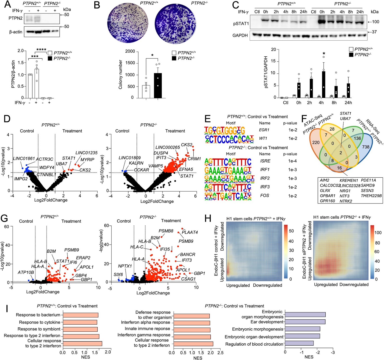
PTPN2 deficiency amplifies inflammatory signalling and impairs functional maturation of human stem cell-derived islets
Background Protein tyrosine phosphatases (PTPs) play key roles in β-cell function and diabetes development. PTPN2 is a candidate gene for type 1 diabetes (T1D) that negatively regulates JAK/STAT signalling. However, the impact of PTPN2 deficiency on the differentiation and functionality of human stem cell-derived somatic metabolic cells remains unclear. Methods PTPN2 expression in β cells from T1D organ donors and during the differentiation of human stem cell-derived islets (SC-islets) was evaluated using single-cell RNA-Sequencing (scRNA-Seq) datasets. We differentiated CRISPR-Cas12a genome-edited PTPN2-deficient H1 human embryonic stem cells (H1-hESCs) into SC-islets, and scRNA-Seq was performed. The maturation and functionality of PTPN2-deficient SC-islets were assessed by implantation under the kidney capsule of NOD-SCID mice. Results scRNA-Seq analysis showed that PTPN2 expression was increased in β cells from recently diagnosed T1D and decreased in long-standing T1D organ donors compared with controls. Conversely, we found that PTPN2 expression was decreased at the early stages of SC-islet differentiation and reconstituted at the later stages, suggesting a developmental dynamic. PTPN2 deficiency exacerbated interferon-induced inflammatory signalling in stem cells and differentiated somatic metabolic cells. Interestingly, PTPN2 deficiency increased hedgehog signalling and reduced SC-islet differentiation efficiency in vitro. In addition, PTPN2-knockout SC-islets exhibited reduced glycaemic control after implantation in vivo, mediated by reduced endocrine cell identity and enhanced interferon signalling. Conclusions Our study postulates a key role of PTPN2 in preserving β-cell function during inflammatory and metabolic stress in SC-islets.