Key Takeaways & Executive Findings
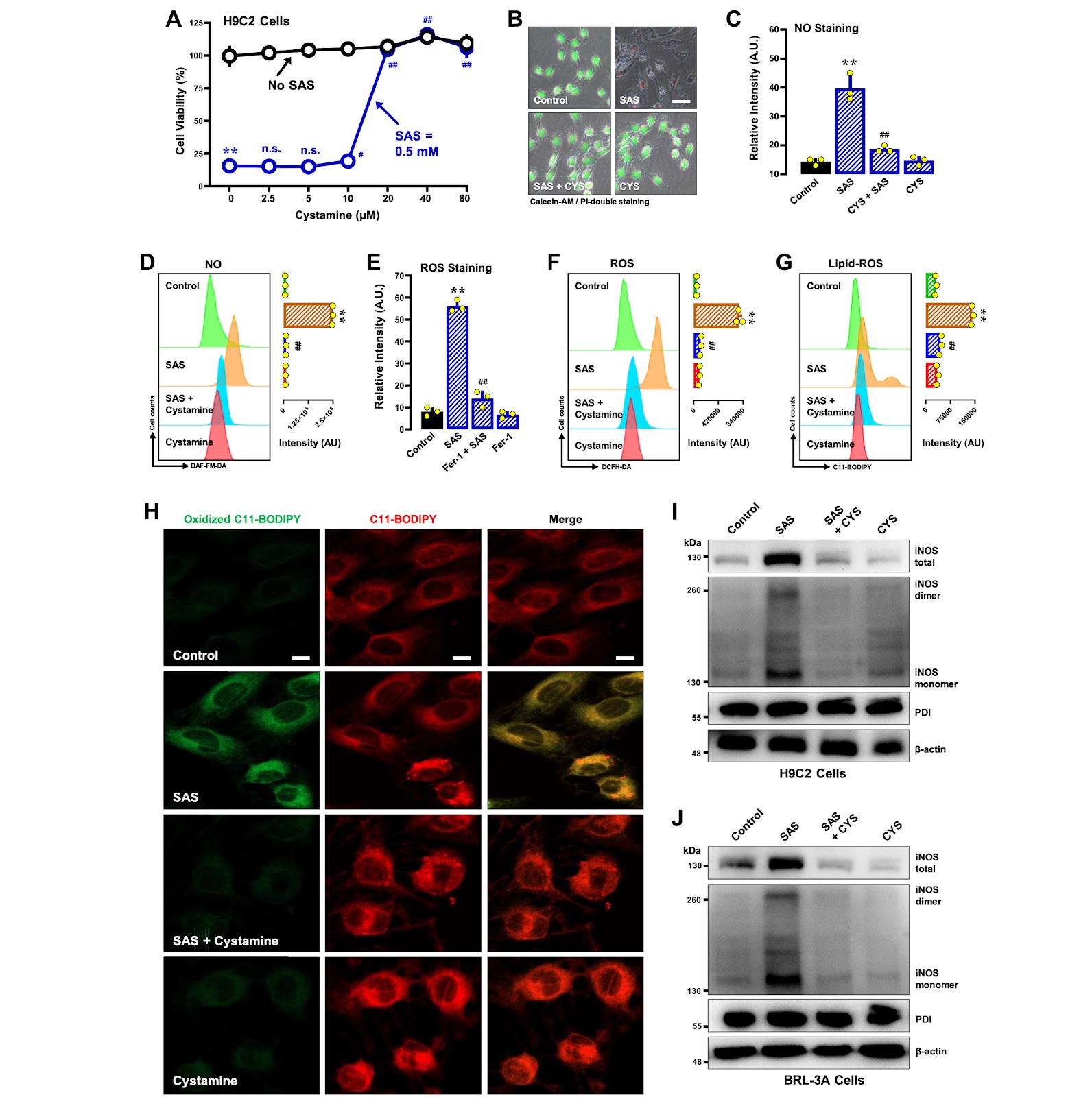
- •• • SAS induces ferroptosis in H9C2 and BRL-3A cells with a time-dependent sequential increase in NO, ROS, and lipid-ROS; this cascade identifies NO as an early mediator, offering a therapeutic window for intervention with NO scavengers or iNOS inhibitors. • • PDI knockdown or pharmacological inhibition abrogates SAS-induced iNOS dimerization and prevents accumulation of NO, ROS, and lipid-ROS, conferring strong protection against ferroptotic cell death; this validates PDI as a druggable target for mitigating SAS toxicity in normal tissues. • • PDI activation by TrxR1 inhibitors sensitizes cells to SAS-induced ferroptosis, suggesting a combination strategy to enhance SAS efficacy in cancer therapy by promoting oxidative cell death. • • SAS upregulates iNOS protein levels, contributing to elevated NO production; this identifies iNOS as a potential biomarker for SAS responsiveness and a target for modulating ferroptosis in inflammatory and malignant conditions.
China Biomedical & Cell Therapy Radar
Get verified English translations, SEM micrographs & open-access PDF alerts from China's leading state key laboratories delivered to your inbox every Monday at 08:00 EST.
Abstract
Sulfasalazine (SAS), a clinically utilized anti-inflammatory drug, induces ferroptosis by inhibiting system Xc− and depleting glutathione. This study characterizes the biochemical and cellular mechanisms of SAS-induced ferroptosis in H9C2 rat cardiomyocytes and BRL-3A rat hepatocytes, focusing on protein disulfide isomerase (PDI). SAS induced ferroptosis with sequential increases in cellular nitric oxide (NO), reactive oxygen species (ROS), and lipid-ROS. SAS activated PDI-mediated dimerization of inducible NO synthase (iNOS) and NO accumulation, followed by ROS and lipid-ROS buildup. SAS also upregulated iNOS protein levels. PDI knockdown or pharmacological inhibition suppressed iNOS dimerization, abrogated NO, ROS, and lipid-ROS accumulation, and prevented ferroptosis. Conversely, PDI activation via TrxR1 inhibitors sensitized cells to SAS-induced ferroptosis. These findings support a pivotal role of the PDI-NOS-NO axis in SAS-induced cytotoxicity, leading to oxidative cell death. The study provides mechanistic insights and suggests strategies for sensitizing cancer cells to SAS-induced ferroptosis.
1. Introduction
Sulfasalazine (SAS) is a well-established anti-inflammatory drug for Crohn's disease and ulcerative colitis, but its clinical utility is limited by dose-dependent toxicity, including ferroptosis induction via system Xc− inhibition and glutathione depletion. Existing ferroptosis inducers lack specificity, and SAS's mechanism involves a poorly understood cascade that hampers the development of safer or more effective analogs.
This study addresses the bottleneck by elucidating the PDI-NOS-NO axis in SAS-induced ferroptosis. Using H9C2 cardiomyocytes and BRL-3A hepatocytes, the authors demonstrate that PDI activation mediates iNOS dimerization and NO accumulation, preceding ROS and lipid-ROS buildup. Genetic and pharmacological interventions confirm PDI's pivotal role, and TrxR1 inhibition sensitizes cells to SAS. These findings provide a mechanistic framework for optimizing SAS-based therapies and mitigating off-target toxicity.
Loading authentic research manuscript (Pages 1–5)...
JIA Yi-Chen, ZHONG Jia-Ling, HAO Xiangyu, ZHU Bao Ting (2025). Characterization of the mechanisms underlying sulfasalazine-induced ferroptotic cell death: role of protein disulfide isomerase-mediated NOS activation and NO accumulation. Acta Biochimica et Biophysica Sinica. https://doi.org/10.3724/abbs.2025100
Research & Educational Purpose Only: The translations, structured abstracts, analytical annotations, and data reports provided by SinoBioDataare intended exclusively for academic research, internal corporate R&D, and educational benchmarking. They do not constitute formal engineering, chemical safety, legal, or professional advice.
Copyright & Intellectual Property Notice: Original copyright of the underlying source articles and experimental data remains with the respective authors, institutions, and original publishing journals. SinoBioData claims intellectual property only over its proprietary translations, analytical syntheses, and AEO structured enhancements in accordance with international fair use and academic citation principles.
Frequently Asked Questions
What is the temporal sequence of NO, ROS, and lipid-ROS accumulation in SAS-induced ferroptosis, and how does it inform intervention timing?
The study reports a sequential increase: NO accumulation precedes ROS and lipid-ROS buildup. This order suggests that early intervention with NO scavengers or iNOS inhibitors could block the downstream oxidative cascade, potentially preventing ferroptosis more effectively than targeting later events.
How does PDI inhibition compare to genetic knockdown in preventing SAS-induced ferroptosis, and what are the translational implications?
Both PDI knockdown and pharmacological inhibition effectively suppress iNOS dimerization, abrogate NO, ROS, and lipid-ROS accumulation, and prevent ferroptosis. Pharmacological inhibition offers a more feasible therapeutic strategy, but off-target effects and bioavailability must be optimized for clinical use.
What is the role of TrxR1 inhibitors in sensitizing cells to SAS-induced ferroptosis, and what are the potential clinical applications?
TrxR1 inhibitors activate PDI, accelerating iNOS dimerization and ferroptosis. This sensitization could be exploited in cancer therapy to enhance SAS efficacy, particularly in tumors with high TrxR1 expression, but requires careful dosing to avoid systemic toxicity.
Does SAS directly activate PDI, and what are the implications for drug design?
The study rules out direct PDI activation by SAS; instead, PDI oxidation occurs due to glutathione depletion. This indirect mechanism suggests that drugs modulating glutathione levels could influence SAS-induced ferroptosis, offering a target for combination therapies.
What are the limitations of using H9C2 and BRL-3A cell lines to model SAS-induced ferroptosis, and how might they affect clinical translation?
These rat cell lines provide mechanistic insights but may not fully replicate human tissue responses. Species-specific differences in PDI, iNOS, and glutathione metabolism could affect translation. Further validation in human primary cells or animal models is necessary to confirm the PDI-NOS-NO axis as a therapeutic target.
Related Chinese Research & Cross-Citations
miR-199a-3p suppresses Vldlr expression to promote cardiomyocyte proliferation
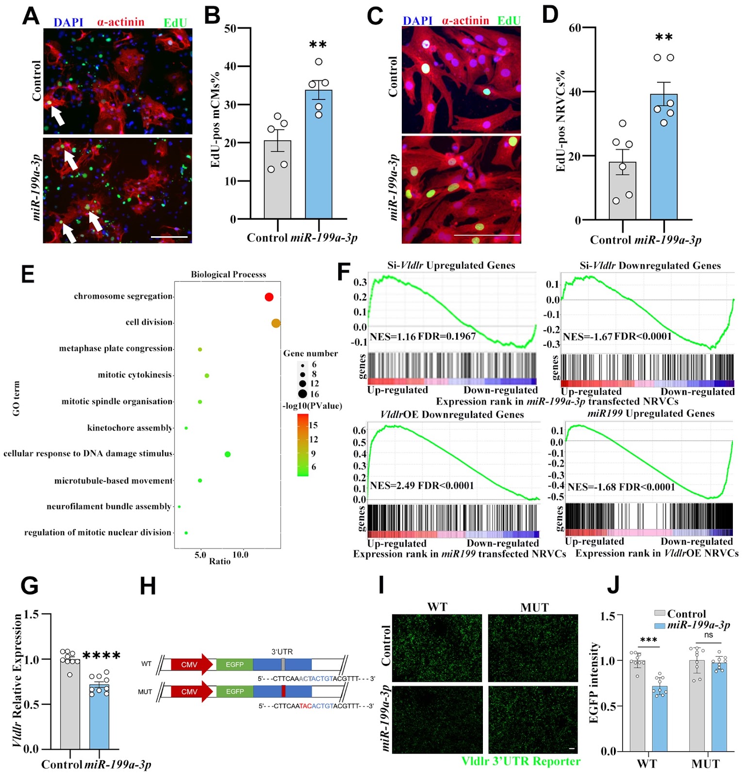
The proliferative capacity of cardiomyocytes is limited in adult mammals, and replacing lost tissue following acute ischemic injury is challenging. Previous studies have demonstrated that miR-199a-3p can promote cardiomyocyte proliferation, but the exact mechanism by which this occurs remains unclear, although multiple targets of miR-199a-3p have been identified. We recently showed that very-low-density-lipoprotein receptor (Vldlr) inhibits cardiomyocyte proliferation, and in this study we aim to test whether Vldlr is a functional target gene of miR-199a-3p. 3′UTR reporter assays demonstrate that miR-199a-3p directly binds to the 3′UTR of Vldlr and inhibits its translation. Overexpressing Vldlr blunts the pro-proliferative effect of miR-199a-3p on cardiomyocytes, suggesting that Vldlr is indeed a functional target of miR-199a-3p. Mechanistically, Vldlr reduces S807/811 phosphorylation of RB1, and inhibiting CDK4/6 to prevent RB1 phosphorylation can block the pro-proliferative effect of both Vldlr knockdown and miR-199a-3p, suggesting that RB1 phosphorylation is required for the cardiomyocyte proliferation induced by miR-199a-3p and Vldlr knockdown. The findings of this study reveal Vldlr as a novel functional target of miR-199a-3p in cardiomyocytes and identify RB1 as a downstream effector of cardiomyocyte proliferation. The identification of the role of the miR-199a-3p-Vldlr-RB1 axis in cardiomyocyte proliferation may provide potential therapeutic targets for cardiac regenerative medicine.
A positive feedback loop between FOSB and miR-133b controls colon cancer cell proliferation
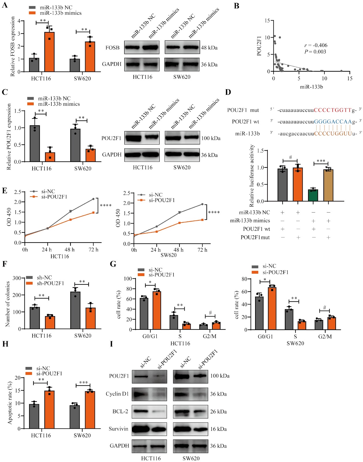
FOSB, a member of the FOS gene family, forms heterodimers with JUN family proteins to engage in diverse cellular processes. Its biological impacts vary among different types of tumors, yet its specific function in colon cancer (CC) remains ambiguous. In this study, quantitative real-time PCR (qRT-PCR) and immunohistochemistry (IHC) are applied to measure FOSB expression levels, followed by an analysis of the association between FOSB expression and patients’ clinical parameters. In vitro experiments are performed to assess cell proliferation, including growth rate, cell cycle distribution, and apoptosis. A subcutaneous xenograft model in nude mice is utilized to monitor tumor growth in vivo. Additionally, chromatin immunoprecipitation (ChIP) and luciferase reporter assays are conducted to dissect the interactions among FOSB, miR-133b, and POU2F1. The results indicate that FOSB expression is downregulated in CC tissues relative to normal controls. Overexpression of FOSB suppresses proliferation and promotes apoptosis in CC cells. Mechanistically, FOSB binds to the promoter region of miR-133b, enhancing its transcription and subsequently repressing POU2F1 expression. Notably, decreased POU2F1 expression also alleviates the transcriptional repression of the FOSB promoter region, establishing a FOSB-miR-133b-POU2F1 feedback loop that inhibits CC proliferation. In summary, our findings suggest that FOSB acts as a tumor suppressor gene in CC and may exert its inhibitory effects on CC growth via the FOSB-miR-133b-POU2F1 feedback loop.
Reduced expression of the PER2 protein contributes to β1-AA-induced cardiac autophagy rhythm disorders
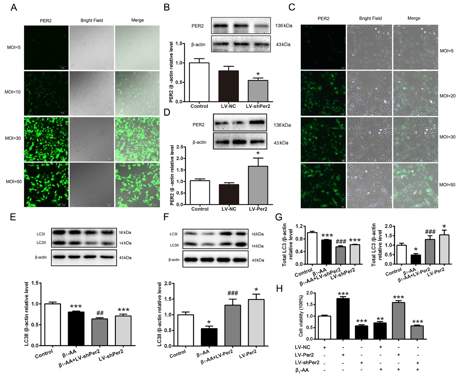
Heart failure may be linked to fluctuations in the rhythm of autophagy in cardiomyocytes throughout the day. Circadian rhythms depend on the regulation of core biological clock proteins, with PER2 playing a crucial role. Our previous research confirmed that the presence of β1-adrenergic receptor autoantibodies (β1-AAs) could inhibit myocardial autophagy, leading to cell death and heart failure. However, it remains unclear whether β1-AA induces cardiac autophagy rhythm disorders by affecting PER2 expression. In this study, we find that β1-AA disrupts the autophagy rhythm in cardiomyocytes, which is primarily indicated by decreased expression of the autophagy marker protein LC3. β1-AA disrupts the rhythmic expression of the PER2 protein in myocardial cells, which is manifested mainly by a decrease in PER2 protein expression. Metoprolol is used to verify that the β1-adrenergic receptor contributes to the reduction in the Per2 protein caused by β1-AA. Knockdown of Per2 with lentivirus reduces the inhibition of LC3 expression caused by β1-AA, whereas overexpression of Per2 in cardiomyocytes using lentivirus significantly restores the β1-AA-induced decrease in LC3 expression. Moreover, mTORC1 activation is found to participate in β1-AA-induced autophagy inhibition in cardiomyocytes after pretreatment with the mTORC1 inhibitor rapamycin. Furthermore, the decreased expression of the PER2 protein caused by β1-AA disrupts the myocardial autophagy rhythm by promoting mTORC1 activation through lentiviruses that knock down or overexpress the Per2 gene. This study provides an experimental basis for the precise treatment of cardiovascular diseases from the perspective of biological rhythm.
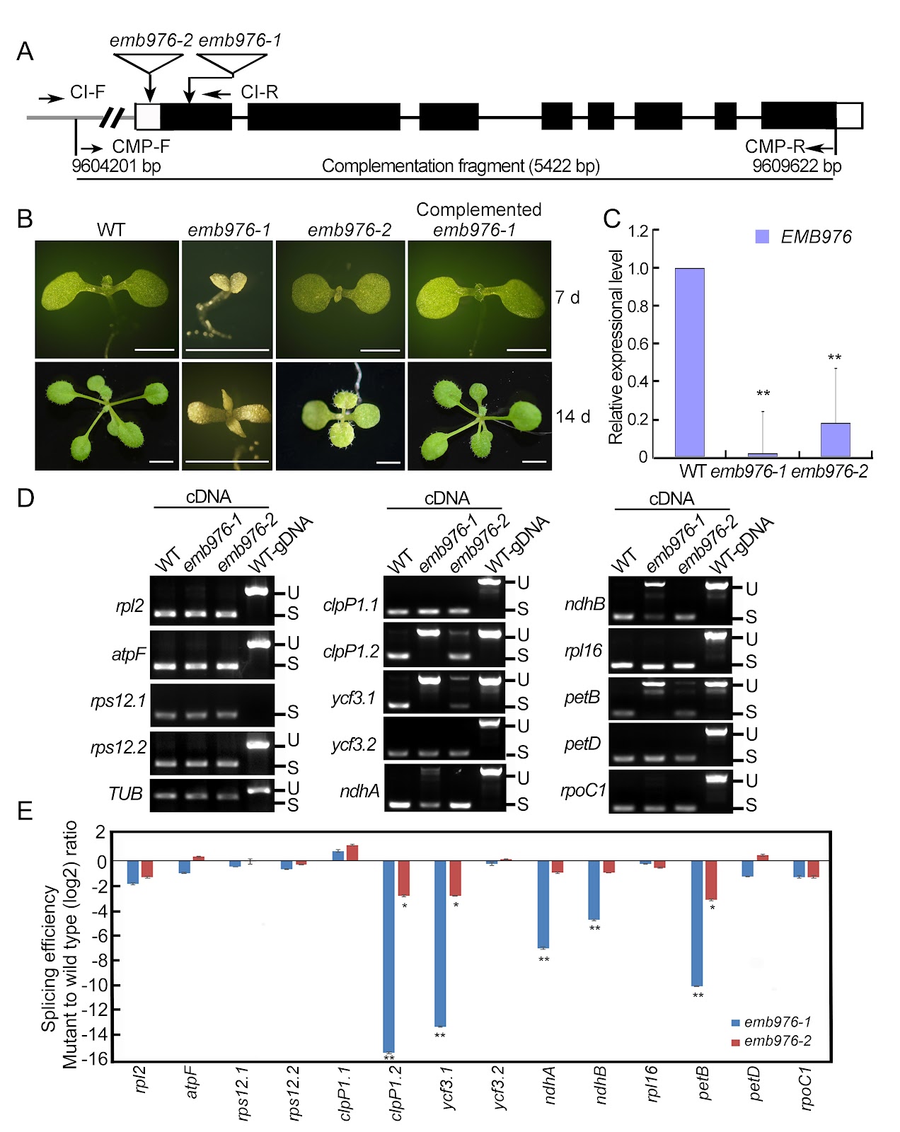
A protein-RNA complex orchestrated by EMB1006, EMB1270, EMB976, and CFM2 facilitates clpP1 intron 2 splicing in Arabidopsis chloroplasts
In Arabidopsis, the PPR proteins EMB1006, EMB1270, and EMB976 are all essential for the splicing of plastid clpP1 intron 2 (clpP1.2), although each also targets other distinct RNAs. The precise mechanism underlying their co-ordinated action in clpP1.2 splicing remains unclear. In this study, RNA electrophoretic mobility shift assays, guided by PPR code prediction, confirm that EMB1006 specifically binds to a sequence near the 3′ end of clpP1 exon 2. Additionally, immunoprecipitation coupled with mass spectrometry reveals that EMB1006 forms a complex with EMB1270, EMB976, and CFM2. Direct interactions between EMB1006 and EMB1270 or CFM2 are further supported by yeast two-hybrid (Y2H) and semi-in vivo pull-down assays. However, no direct interactions between EMB976 and EMB1006, CFM2 or EMB1270 are detected by Y2H. Based on these findings and previous evidence that EMB1270 binds to clpP1 intron 2 and interacts with CFM2, we propose a model in which EMB1006 and EMB1270 bind to distinct sites on clpP1 pre-mRNA. Together with CFM2 and possible indirect association with EMB976, they assemble into a protein-RNA complex that facilitates the splicing of clpP1.2 in chloroplasts.
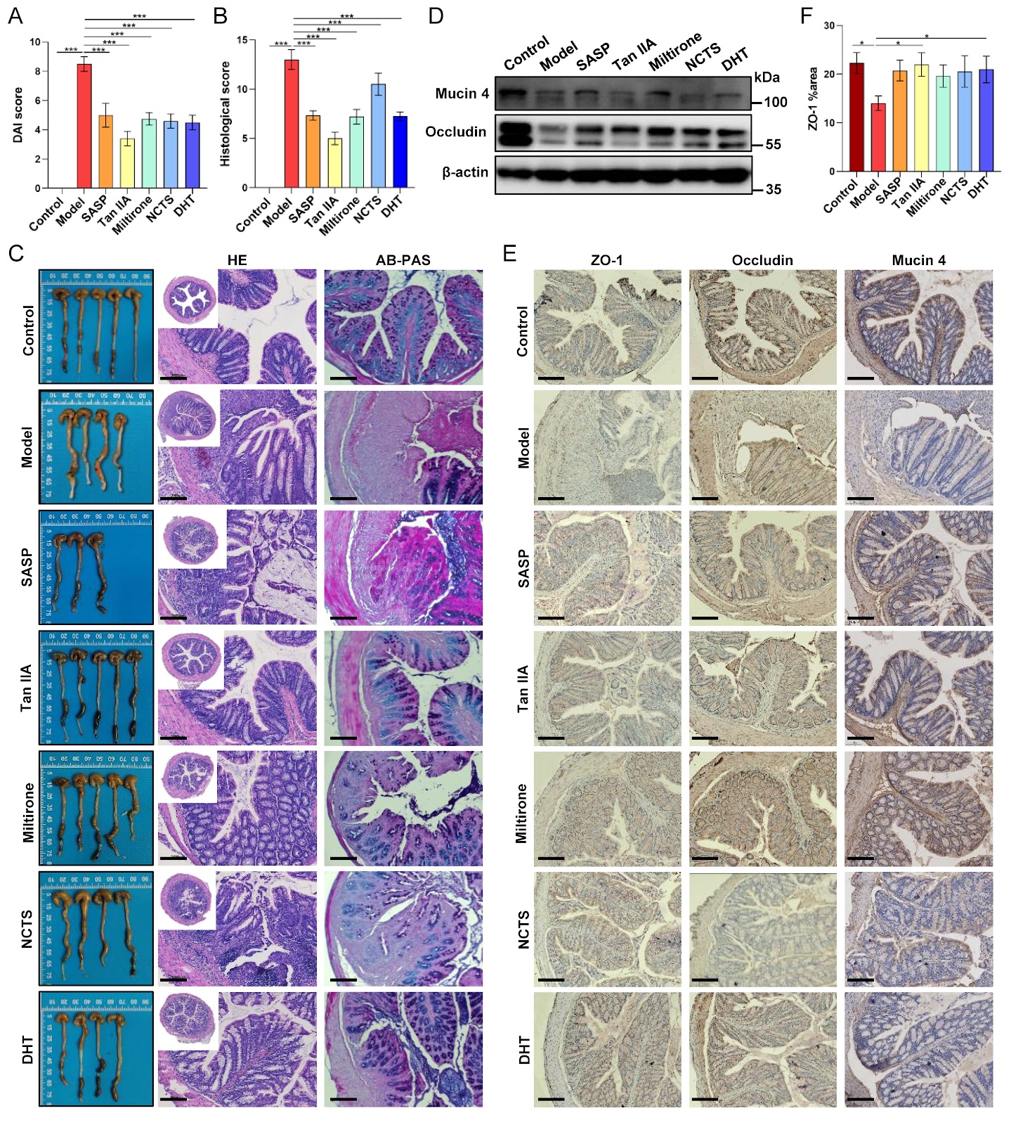
Tanshinones from Salvia miltiorrhiza alleviate ulcerative colitis via reprogramming the gut microbiota-metabolite axis
The anti-inflammatory properties of the traditional herb Salvia miltiorrhiza Bunge are well-established, yet its precise mechanism of action in ulcerative colitis (UC) remains unclear. Herein, we evaluate the therapeutic potential of four major tanshinones–tanshinone IIA (Tan IIA), miltirone, neocryptotanshinone, and dihydrotanshinone I–in a murine dextran sulfate sodium (DSS)-induced colitis model. Our results show that tanshinones effectively alleviate disease severity, suppress systemic and local inflammation, and restore intestinal barrier integrity. Integrated multi-omics analysis reveals that the therapeutic efficacy originates from a comprehensive reprogramming of the gut microbiota-metabolite axis. Specifically, tanshinones reverse colitis-associated dysbiosis and rectify metabolic disturbances in linoleic acid metabolism, bile acid biosynthesis, and amino acid utilization. Correlation network analysis identifies key functional modules linking beneficial microbes (e.g., Akkermansia) to anti-inflammatory lipid mediators and associating pathobionts (e.g., Desulfovibrio) with disrupted bile acid metabolism. Notably, supplementation with Akkermansia muciniphila synergizes with Tan IIA to amplify barrier restoration and metabolic normalization. Our findings establish that tanshinones ameliorate UC through microbiota-driven metabolic reprogramming, wherein the restructured microbial community actively shapes a therapeutic metabolic output. This work elucidates a metabolite-mediated mechanism of action and positions tanshinones as promising microbiome-targeting therapeutics for inflammatory bowel disease.
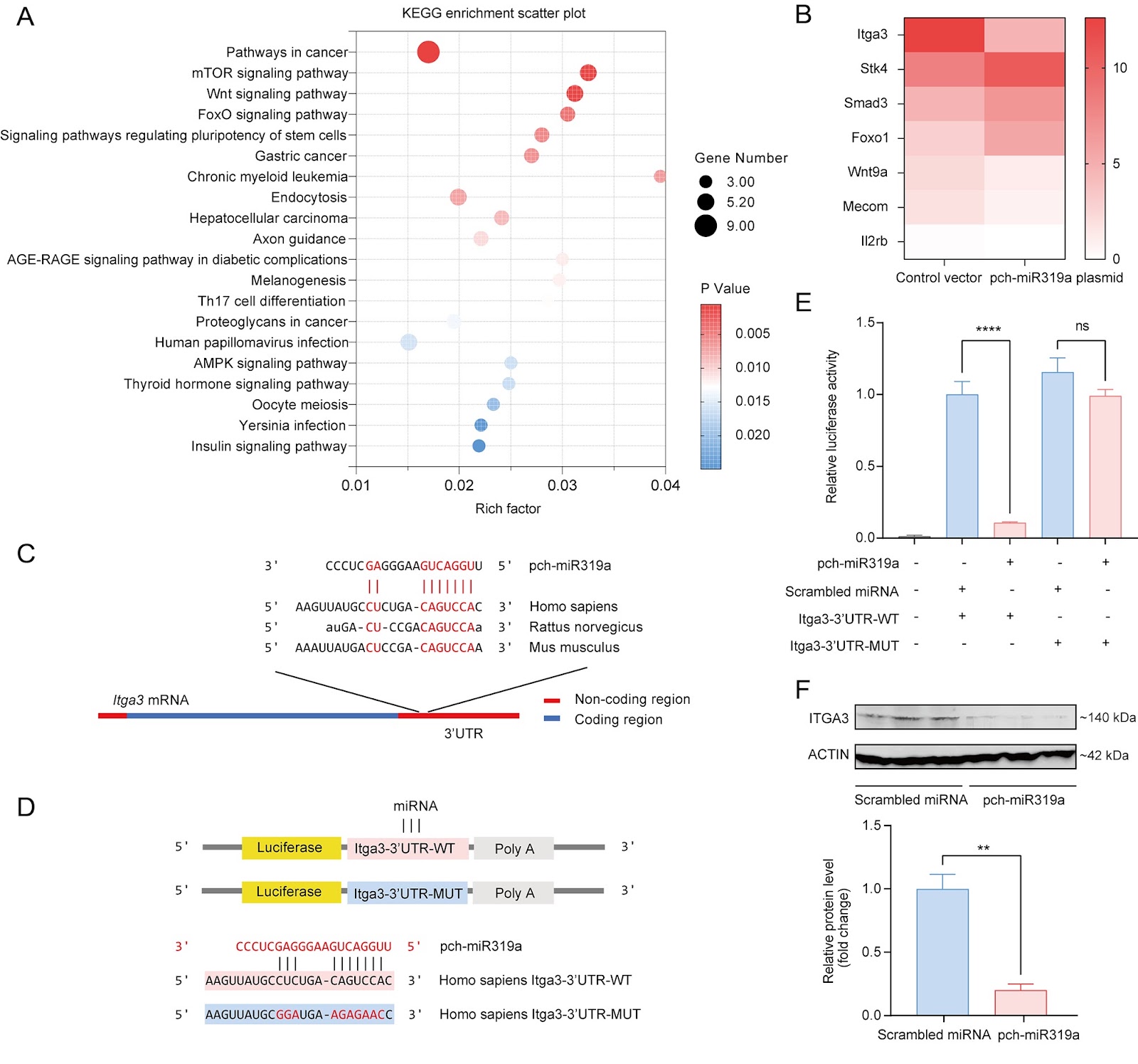
Distinct miR319a identified from Persicaria chinensis mediates cross-kingdom suppression of cervical cancer by targeting ITGA3
Persicaria chinensis, a well-known traditional Chinese medicinal herb that is both edible and medicinal, has been widely acknowledged for its therapeutic effects, such as anti-inflammatory, antioxidant, and antitumor activities. However, the role of miRNAs from this plant in the cross-kingdom regulation of human diseases has not been investigated. In this study, we analyze the miRNA expression profile of P. chinensis using high-throughput sequencing and identify a total of 673 miRNAs, including 422 novel miRNAs that are unique to this plant and 251 conserved miRNAs. Among the conserved miRNAs, pch-miR319a is found to be the most abundant. Moreover, food-oriented pch-miR319a accumulates in the uterus and tumors and exhibits a rich repertoire of target genes within cancer-related pathways, demonstrating significant cross-kingdom regulatory potential. Utilizing the dual-luciferase reporter gene assay, we demonstrate that pch-miR319a from P. chinensis targets the Itga3 gene, which is associated with cervical cancer progression. Overexpression of pch-miR319a significantly decreases the viability, migration, and induces apoptosis of HeLa cervical cancer cells in vitro. Moreover, in a syngeneic mouse tumor model of cervical cancer, treatment with pch-miR319a effectively inhibits tumor growth and downregulates the expressions of ITGA3 and the proliferation marker Ki-67. Our study highlights the potential of pch-miR319a from P. chinensis as a novel therapeutic agent for cervical cancer by targeting ITGA3 and provides new insights into the cross-kingdom regulatory mechanisms of plant miRNAs in human diseases.