Key Takeaways & Executive Findings
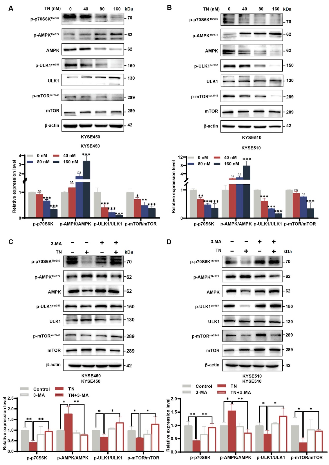
- •• • TN induces autophagy-dependent apoptosis in ESCC cells, with a significant reduction in cell viability observed at low micromolar concentrations (IC50 values not explicitly stated in Section B, but functional assays confirm dose-dependent effects). This suggests TN could serve as a potent alternative to conventional chemotherapy, potentially reducing systemic toxicity. • • The AMPK-mTOR-ULK1 axis is critically involved: TN activates AMPK, inhibits mTOR, and phosphorylates ULK1, leading to autophagic flux. This pathway represents a actionable target for ESCC, as dysregulation of AMPK signaling is common in chemoresistant tumors. • • Pharmacological inhibition of autophagy with 3-MA or AMPK with Compound C significantly attenuates TN-induced apoptosis, confirming that autophagy precedes and mediates apoptosis. This mechanistic insight supports combining TN with autophagy modulators to enhance therapeutic efficacy. • • TN's dual role in inducing autophagy and apoptosis highlights its potential to overcome apoptosis resistance in ESCC, a major clinical challenge. The study provides a strong rationale for advancing TN into preclinical and clinical trials, given its natural origin and favorable safety profile compared to synthetic agents.
China Biomedical & Cell Therapy Radar
Get verified English translations, SEM micrographs & open-access PDF alerts from China's leading state key laboratories delivered to your inbox every Monday at 08:00 EST.
Abstract
Esophageal squamous cell carcinoma (ESCC) accounts for 90% of global esophageal cancer cases, with high prevalence in Asia, East Africa, and South America. Current treatment relies on esophagectomy, which carries significant morbidity and limited efficacy, underscoring the urgent need for novel therapeutic agents. Triptonide (TN), a small-molecule compound derived from Tripterygium wilfordii Hook. f, exhibits potent antitumor activity, but its mechanisms in ESCC remain undefined. This study investigated TN-induced cytotoxicity in KYSE450 and KYSE510 ESCC cell lines using CCK-8 and colony formation assays. Western blot analysis, Hoechst 33258 staining, flow cytometry, autophagic flux detection, and transmission electron microscopy (TEM) assessed apoptosis and autophagy. Pharmacological inhibitors 3-methyladenine (3-MA) and dorsomorphin (Compound C, CC) were employed to dissect pathway involvement. Results demonstrate that TN induces autophagy-dependent apoptosis by targeting the AMPK-mTOR-ULK1 axis. TN treatment led to AMPK activation, mTOR inhibition, and ULK1 phosphorylation, triggering autophagic flux and subsequent apoptosis. Inhibition of autophagy with 3-MA or AMPK with CC attenuated TN-induced apoptosis, confirming the mechanistic dependency. These findings establish TN as a promising candidate for ESCC therapy and elucidate a novel molecular pathway for its anticancer effects.
1. Introduction
Esophageal squamous cell carcinoma (ESCC) constitutes approximately 90% of esophageal cancer cases globally, with particularly high incidence in Asia, East Africa, and South America. Despite advances in surgical techniques, esophagectomy remains the primary curative option, yet it is associated with substantial morbidity, mortality, and limited long-term survival. The aggressive nature of ESCC and its resistance to conventional chemoradiotherapy underscore an urgent unmet need for novel, efficacious, and safer therapeutic agents. Natural products, especially those derived from traditional Chinese medicine, have emerged as promising sources for anticancer drug discovery due to their diverse bioactivities and favorable safety profiles.
Triptonide (TN), a diterpenoid isolated from Tripterygium wilfordii Hook. f, has demonstrated potent antitumor effects in various malignancies. However, its specific mechanisms in ESCC have not been fully elucidated. Previous studies suggest that TN modulates multiple signaling pathways, including AMPK-mTOR-ULK1, which is central to autophagy regulation. Autophagy plays a dual role in cancer, either promoting survival or inducing cell death depending on context. This study aims to clarify whether TN induces autophagy-mediated apoptosis in ESCC and to dissect the involvement of the AMPK-mTOR-ULK1 axis. By employing a comprehensive set of functional and molecular assays, including CCK-8, colony formation, western blot, flow cytometry, autophagic flux detection, and transmission electron microscopy, we provide robust evidence that TN triggers autophagy-dependent apoptosis via AMPK activation and mTOR inhibition. These findings offer a mechanistic foundation for developing TN as a targeted therapeutic strategy for ESCC.
Loading authentic research manuscript (Pages 1–5)...
JU Jiujun, XU Nuo, LI Bohan, SHI Dan, CAI Jiahui, ZHENG Qiusheng, YE Lei, ZHANG Shaosen, WANG Caixia (2025). Triptonide facilitates autophagy-mediated apoptosis in esophageal squamous cell carcinoma by targeting the AMPK-mTOR-ULK1 axis. Acta Biochimica et Biophysica Sinica. https://doi.org/10.3724/abbs.2025056
Research & Educational Purpose Only: The translations, structured abstracts, analytical annotations, and data reports provided by SinoBioDataare intended exclusively for academic research, internal corporate R&D, and educational benchmarking. They do not constitute formal engineering, chemical safety, legal, or professional advice.
Copyright & Intellectual Property Notice: Original copyright of the underlying source articles and experimental data remains with the respective authors, institutions, and original publishing journals. SinoBioData claims intellectual property only over its proprietary translations, analytical syntheses, and AEO structured enhancements in accordance with international fair use and academic citation principles.
Frequently Asked Questions
What is the precise mechanism by which triptonide induces autophagy-dependent apoptosis in ESCC cells?
Triptonide activates AMPK, which inhibits mTOR, leading to ULK1 phosphorylation and initiation of autophagic flux. This autophagy precedes and mediates apoptosis, as inhibition of autophagy with 3-MA or AMPK with Compound C significantly attenuates apoptosis. The study used western blot to confirm AMPK activation, mTOR inhibition, and ULK1 phosphorylation, along with autophagic flux detection and TEM to visualize autophagosomes.
How does the efficacy of triptonide compare to standard chemotherapy for ESCC, and what are the potential clinical advantages?
While direct comparative data with standard chemotherapy (e.g., cisplatin) are not provided in this study, triptonide demonstrated potent cytotoxicity in KYSE450 and KYSE510 cells at low micromolar concentrations. Its natural origin and dual mechanism (autophagy and apoptosis) may offer advantages in overcoming chemoresistance and reducing side effects. However, further preclinical and clinical studies are needed to establish comparative efficacy and safety.
What are the potential challenges in translating triptonide from bench to bedside for ESCC treatment?
Key challenges include optimizing bioavailability and delivery, as triptonide's pharmacokinetic profile may limit tumor accumulation. Additionally, potential off-target effects due to AMPK activation in normal tissues need evaluation. The study used in vitro models; in vivo efficacy and toxicity remain to be determined. Scalability of production from Tripterygium wilfordii and formulation stability are also considerations.
Could autophagy inhibition enhance or diminish triptonide's therapeutic effect in ESCC?
In this study, autophagy inhibition with 3-MA attenuated triptonide-induced apoptosis, indicating that autophagy is pro-death in this context. Therefore, combining triptonide with autophagy inhibitors would likely reduce efficacy. Conversely, agents that promote autophagic flux might synergize with triptonide. This highlights the importance of context-dependent autophagy modulation in therapy.
What is the role of the AMPK-mTOR-ULK1 axis in ESCC and its potential as a therapeutic target?
The AMPK-mTOR-ULK1 axis is a central regulator of autophagy and cellular energy homeostasis. In ESCC, dysregulation of this pathway contributes to tumor progression and chemoresistance. Triptonide's activation of AMPK and inhibition of mTOR leads to ULK1-mediated autophagy and apoptosis. Targeting this axis with compounds like triptonide represents a promising strategy, as it addresses a key survival mechanism in ESCC cells.
Related Chinese Research & Cross-Citations
miR-199a-3p suppresses Vldlr expression to promote cardiomyocyte proliferation
The proliferative capacity of cardiomyocytes is limited in adult mammals, and replacing lost tissue following acute ischemic injury is challenging. Previous studies have demonstrated that miR-199a-3p can promote cardiomyocyte proliferation, but the exact mechanism by which this occurs remains unclear, although multiple targets of miR-199a-3p have been identified. We recently showed that very-low-density-lipoprotein receptor (Vldlr) inhibits cardiomyocyte proliferation, and in this study we aim to test whether Vldlr is a functional target gene of miR-199a-3p. 3′UTR reporter assays demonstrate that miR-199a-3p directly binds to the 3′UTR of Vldlr and inhibits its translation. Overexpressing Vldlr blunts the pro-proliferative effect of miR-199a-3p on cardiomyocytes, suggesting that Vldlr is indeed a functional target of miR-199a-3p. Mechanistically, Vldlr reduces S807/811 phosphorylation of RB1, and inhibiting CDK4/6 to prevent RB1 phosphorylation can block the pro-proliferative effect of both Vldlr knockdown and miR-199a-3p, suggesting that RB1 phosphorylation is required for the cardiomyocyte proliferation induced by miR-199a-3p and Vldlr knockdown. The findings of this study reveal Vldlr as a novel functional target of miR-199a-3p in cardiomyocytes and identify RB1 as a downstream effector of cardiomyocyte proliferation. The identification of the role of the miR-199a-3p-Vldlr-RB1 axis in cardiomyocyte proliferation may provide potential therapeutic targets for cardiac regenerative medicine.
A positive feedback loop between FOSB and miR-133b controls colon cancer cell proliferation
FOSB, a member of the FOS gene family, forms heterodimers with JUN family proteins to engage in diverse cellular processes. Its biological impacts vary among different types of tumors, yet its specific function in colon cancer (CC) remains ambiguous. In this study, quantitative real-time PCR (qRT-PCR) and immunohistochemistry (IHC) are applied to measure FOSB expression levels, followed by an analysis of the association between FOSB expression and patients’ clinical parameters. In vitro experiments are performed to assess cell proliferation, including growth rate, cell cycle distribution, and apoptosis. A subcutaneous xenograft model in nude mice is utilized to monitor tumor growth in vivo. Additionally, chromatin immunoprecipitation (ChIP) and luciferase reporter assays are conducted to dissect the interactions among FOSB, miR-133b, and POU2F1. The results indicate that FOSB expression is downregulated in CC tissues relative to normal controls. Overexpression of FOSB suppresses proliferation and promotes apoptosis in CC cells. Mechanistically, FOSB binds to the promoter region of miR-133b, enhancing its transcription and subsequently repressing POU2F1 expression. Notably, decreased POU2F1 expression also alleviates the transcriptional repression of the FOSB promoter region, establishing a FOSB-miR-133b-POU2F1 feedback loop that inhibits CC proliferation. In summary, our findings suggest that FOSB acts as a tumor suppressor gene in CC and may exert its inhibitory effects on CC growth via the FOSB-miR-133b-POU2F1 feedback loop.
A protein-RNA complex orchestrated by EMB1006, EMB1270, EMB976, and CFM2 facilitates clpP1 intron 2 splicing in Arabidopsis chloroplasts
In Arabidopsis, the PPR proteins EMB1006, EMB1270, and EMB976 are all essential for the splicing of plastid clpP1 intron 2 (clpP1.2), although each also targets other distinct RNAs. The precise mechanism underlying their co-ordinated action in clpP1.2 splicing remains unclear. In this study, RNA electrophoretic mobility shift assays, guided by PPR code prediction, confirm that EMB1006 specifically binds to a sequence near the 3′ end of clpP1 exon 2. Additionally, immunoprecipitation coupled with mass spectrometry reveals that EMB1006 forms a complex with EMB1270, EMB976, and CFM2. Direct interactions between EMB1006 and EMB1270 or CFM2 are further supported by yeast two-hybrid (Y2H) and semi-in vivo pull-down assays. However, no direct interactions between EMB976 and EMB1006, CFM2 or EMB1270 are detected by Y2H. Based on these findings and previous evidence that EMB1270 binds to clpP1 intron 2 and interacts with CFM2, we propose a model in which EMB1006 and EMB1270 bind to distinct sites on clpP1 pre-mRNA. Together with CFM2 and possible indirect association with EMB976, they assemble into a protein-RNA complex that facilitates the splicing of clpP1.2 in chloroplasts.
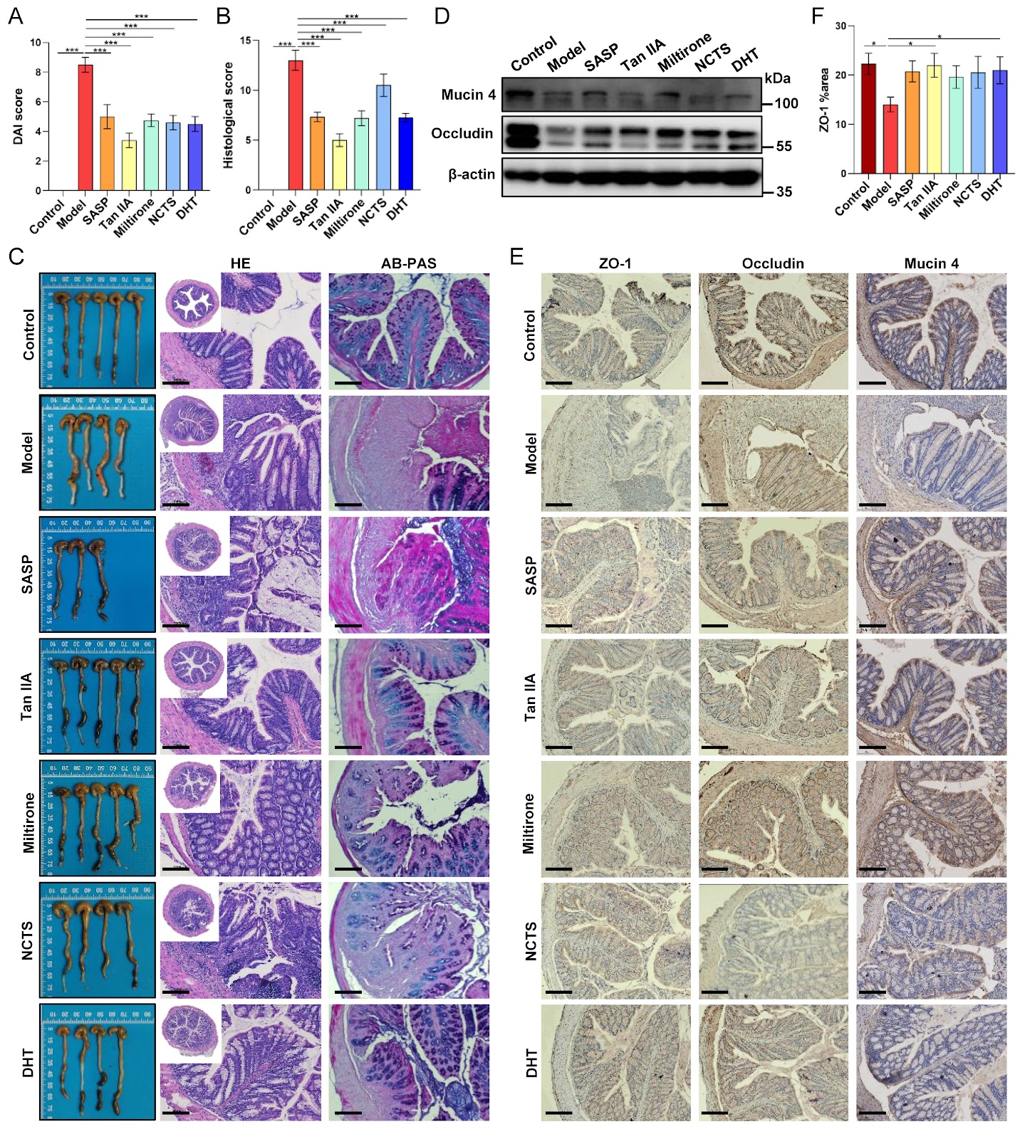
Tanshinones from Salvia miltiorrhiza alleviate ulcerative colitis via reprogramming the gut microbiota-metabolite axis
The anti-inflammatory properties of the traditional herb Salvia miltiorrhiza Bunge are well-established, yet its precise mechanism of action in ulcerative colitis (UC) remains unclear. Herein, we evaluate the therapeutic potential of four major tanshinones–tanshinone IIA (Tan IIA), miltirone, neocryptotanshinone, and dihydrotanshinone I–in a murine dextran sulfate sodium (DSS)-induced colitis model. Our results show that tanshinones effectively alleviate disease severity, suppress systemic and local inflammation, and restore intestinal barrier integrity. Integrated multi-omics analysis reveals that the therapeutic efficacy originates from a comprehensive reprogramming of the gut microbiota-metabolite axis. Specifically, tanshinones reverse colitis-associated dysbiosis and rectify metabolic disturbances in linoleic acid metabolism, bile acid biosynthesis, and amino acid utilization. Correlation network analysis identifies key functional modules linking beneficial microbes (e.g., Akkermansia) to anti-inflammatory lipid mediators and associating pathobionts (e.g., Desulfovibrio) with disrupted bile acid metabolism. Notably, supplementation with Akkermansia muciniphila synergizes with Tan IIA to amplify barrier restoration and metabolic normalization. Our findings establish that tanshinones ameliorate UC through microbiota-driven metabolic reprogramming, wherein the restructured microbial community actively shapes a therapeutic metabolic output. This work elucidates a metabolite-mediated mechanism of action and positions tanshinones as promising microbiome-targeting therapeutics for inflammatory bowel disease.
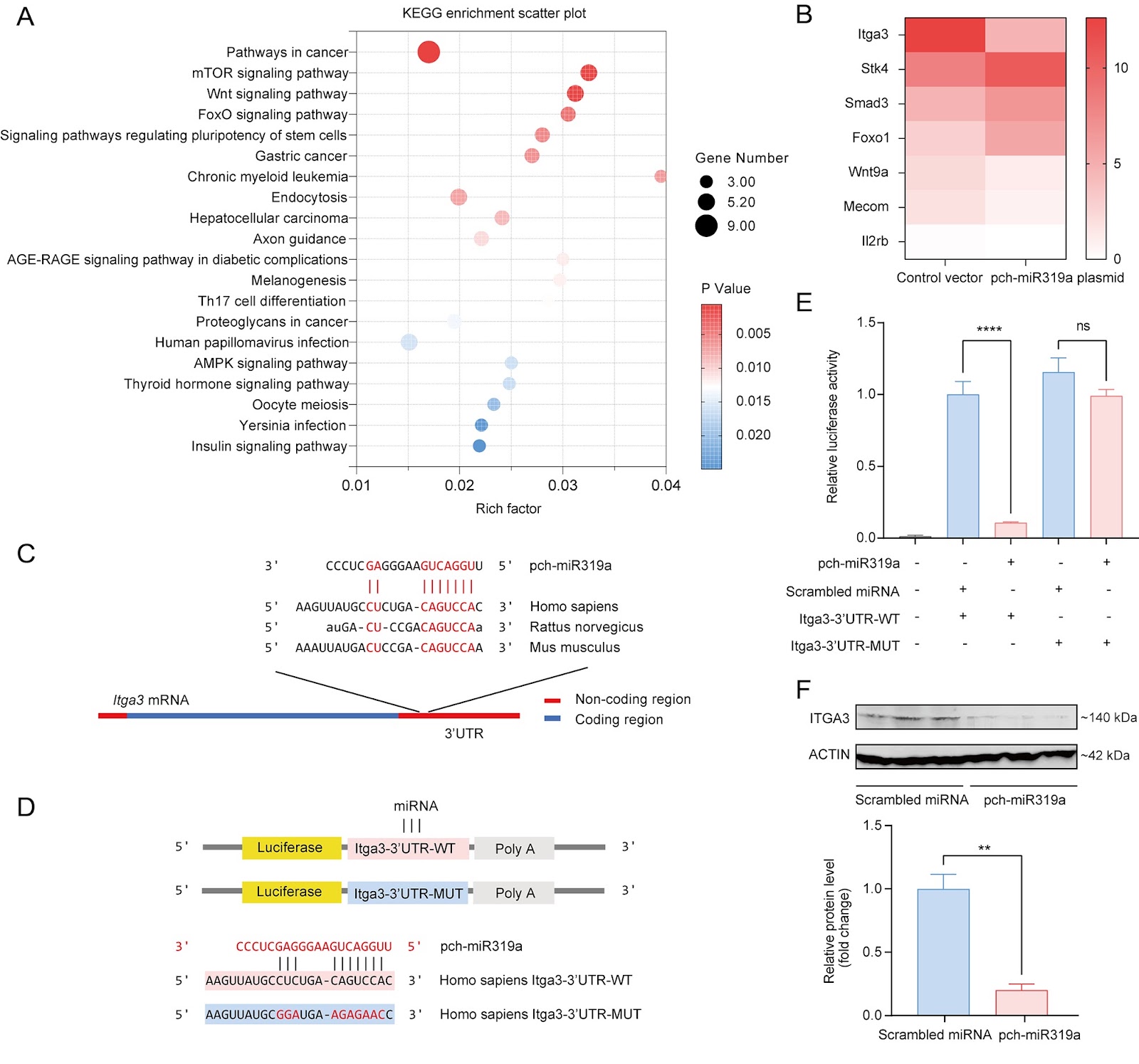
Distinct miR319a identified from Persicaria chinensis mediates cross-kingdom suppression of cervical cancer by targeting ITGA3
Persicaria chinensis, a well-known traditional Chinese medicinal herb that is both edible and medicinal, has been widely acknowledged for its therapeutic effects, such as anti-inflammatory, antioxidant, and antitumor activities. However, the role of miRNAs from this plant in the cross-kingdom regulation of human diseases has not been investigated. In this study, we analyze the miRNA expression profile of P. chinensis using high-throughput sequencing and identify a total of 673 miRNAs, including 422 novel miRNAs that are unique to this plant and 251 conserved miRNAs. Among the conserved miRNAs, pch-miR319a is found to be the most abundant. Moreover, food-oriented pch-miR319a accumulates in the uterus and tumors and exhibits a rich repertoire of target genes within cancer-related pathways, demonstrating significant cross-kingdom regulatory potential. Utilizing the dual-luciferase reporter gene assay, we demonstrate that pch-miR319a from P. chinensis targets the Itga3 gene, which is associated with cervical cancer progression. Overexpression of pch-miR319a significantly decreases the viability, migration, and induces apoptosis of HeLa cervical cancer cells in vitro. Moreover, in a syngeneic mouse tumor model of cervical cancer, treatment with pch-miR319a effectively inhibits tumor growth and downregulates the expressions of ITGA3 and the proliferation marker Ki-67. Our study highlights the potential of pch-miR319a from P. chinensis as a novel therapeutic agent for cervical cancer by targeting ITGA3 and provides new insights into the cross-kingdom regulatory mechanisms of plant miRNAs in human diseases.
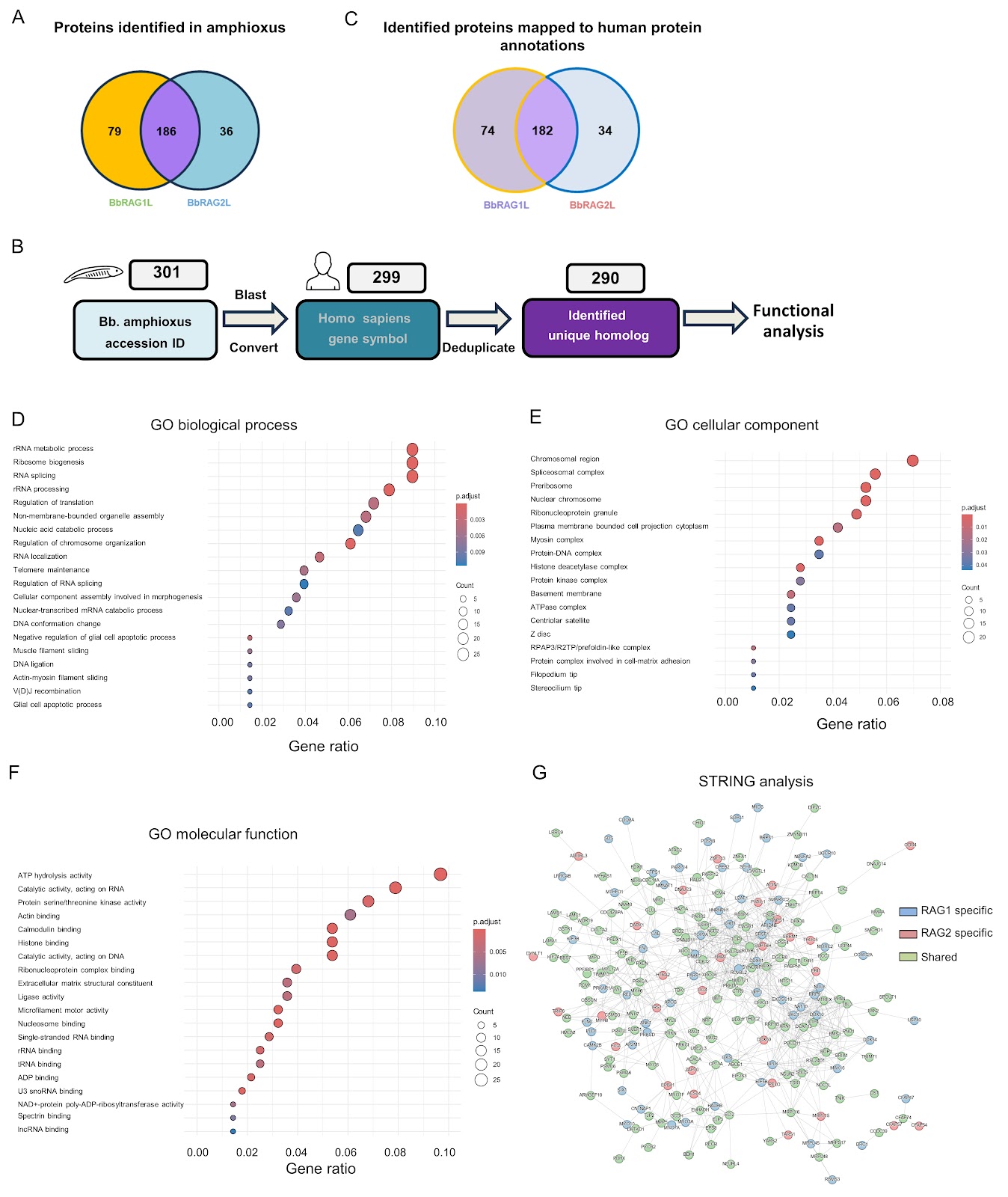
Development of an in vitro TurboID labeling assay for the detection of protoRAG-interacting proteins in the amphioxus
The recombination-activating gene (RAG)-mediated V(D)J rearrangement mechanism is essential for generating the diversity of the adaptive immune system in jawed vertebrates. The RAG1 and RAG2 proteins form a tetrameric complex that recognizes and cleaves pairs of recombination signal sequences (RSSs). In jawed vertebrates, the N-terminal region of RAG1 and the C-terminal region of RAG2 are critical for the regulation of physiological function and enhancement of recombination accuracy. Moreover, the intracellular activity of RAG1/2 is rigorously regulated and is thought to represent the aspect shaped by host coevolution. However, these regulatory mechanisms remain incompletely understood, impeding our deeper understanding of the physiological functions of RAG. Previous studies have shown that jawed vertebrate RAG evolved from invertebrate RAG-like (RAGL) transposases and underwent key structural changes during evolution. RAGL genes have been identified in multiple invertebrate species, but from Branchiostoma belcheri (Bb), the RAG-like protein in Bb amphioxus (BbRAG1L) and the RAG2-like protein in Bb amphioxus (BbRAG2L) are among the most extensively characterized to date. Lancelets (amphioxus) represent the most basal extant chordates (cephalochordates) that diverged from the other two chordate lineages (urochordates and vertebrates). Consequently, amphioxus has been considered a “living fossil” for studying the origins and evolution of adaptive immunity. Homologs of the core structural domain of RAG1 were discovered in the amphioxus genome in 2005, and the homologs were subsequently shown to exhibit recombinase activity in cooperation with murine-derived RAG2. A large number of transposon genes were then identified in the amphioxus genome, followed by the identification of target site duplication (TSD) and terminal inverted repeat (TIR) sequences in the amphioxus genome. Notably, BbRAG2L lacks the C-terminal region present in murine RAG2, including the acidic hinge and PHD domain. BbRAG1L coupled with BbRAG2L displays significant transposition activity in vitro and in human cell lines. In contrast, vertebrate RAG exhibits limited transposon activity in vitro and extremely low transposon activity in vivo. This leads to the question of how RAG lost its transposase activity during evolution. Structural comparison and functional experiments of BbRAGL (protoRAG) and vertebrate RAG have revealed that vertebrates undergo essential adaptive changes to eliminate the primitive transposase activity of protoRAG. Research on BbRAGL has therefore provided strong support for the hypothesis that RAG originated from a domesticated transposon. Given the similarity in both structure and function between BbRAG1L and vertebrate RAG and the fact that BbRAGL proteins are the only RAGL proteins that can currently be efficiently expressed in vertebrate cells, characterizing the binding partners of BbRAGL involved in amphioxus is of great significance for understanding the regulation of BbRAGL and its adaptation and evolution. Parallel comparison of BbRAGL and RAG interaction networks may further clarify how BbRAGL transitioned from a transposase to a recombinase during host coevolution and reveal general principles of vertebrate intracellular RAG regulation. In recent years, proximity labeling techniques have greatly facilitated approaches to identifying RAG cofactors. For example, RAG1 fused to biotin ligase RAG1 fused to a biotin ligase found in Escherichia coli (BirM) has been used to define dynamic interaction networks of RAG-associated proteins. Inspired by these advances, we sought to apply a similar approach to identify the BbRAGL cofactors in amphioxus and build a regulatory network of BbRAGL that could shed light on its relationship with host evolution. However, endogenous BbRAGL expression is undetectable in the adult amphioxus (data not shown), and the generation of transgenic amphioxus lines expressing proximity-based labeling proteins remains technically challenging. These constraints make it difficult to analyze native cofactors of endogenous BbRAGL in vivo. Because BbRAGL can be expressed in vertebrate cell lines, we instead developed an in vitro proximity-labeling strategy. We fused TurboID (TbID), an engineered biotin ligase, to the N-terminus of BbRAG1L or BbRAG2L (Figure 1A), purified the fusion proteins, and incubated them with protein lysates prepared from the hepatic cecum and colons of adult B. belcheri amphioxus in vitro to label the interacting proteins. The proteins were finally analyzed by liquid chromatography-tandem mass spectrometry (LC-MS/MS) (Figure 1B). A pTT5 vector was used to express plasmids encoding maltose-binding protein (MBP) and TurboID with or without full-length BbRAG1L or BbRAG2L, yielding pTT5-MBP-TurboID, pTT5-MBP-TurboID-BbRAG1L, and pTT5-MBP-TurboID-BbRAG2L. Plasmids expressing MBP-BbRAG1L and MBP-BbRAG2L were also generated.